Call Girls in Hauz Khas Delhi 💯Call Us 🔝9953322196🔝 💯Escort.
Molecular mechanisms of ovulation
1. Molecular mechanisms of ovulation: co-ordination through
the cumulus complex
Darryl L.Russell1 and Rebecca L.Robker
Research Centre for Reproductive Health, School of Paediatrics and Reproductive Health, The University of Adelaide, Adelaide, South
Australia 5005, Australia
1
Correspondence address. School of Paediatrics and Reproductive Health, The University of Adelaide, Adelaide, South Australia 5005,
Australia Tel: þ61 8 8303 4096; Fax: þ61 8 8303 4099; E-mail: darryl.russell@adelaide.edu.au
Successful ovulation requires that developmentally competent oocytes are released with appropriate timing from the
ovarian follicle. Somatic cells of the follicle sense the ovulatory stimulus and guide resumption of meiosis and release of
the oocyte, as well as structural remodelling and luteinization of the follicle. Complex intercellular communication co-
ordinates critical stages of oocyte maturation and links this process with release from the follicle. To achieve these
outcomes, ovulation is controlled through multiple inputs, including endocrine hormones, immune and metabolic
signals, as well as intrafollicular paracrine factors from the theca, mural and cumulus granulosa cells and the
oocyte itself. This review focuses on the recent advances in understanding of molecular mechanisms that commence
after the gonadotrophin surge and culminate with release of the oocyte. These mechanisms include intracellular sig-
nalling, gene regulation and remodelling of tissue structure in each of the distinct ovarian compartments. Most critical
ovulatory mediators exert effects through the cumulus cell complex that surrounds and connects with the oocyte. The
convergence of ovulatory signals through the cumulus complex co-ordinates the key mechanistic processes that
mediate and control oocyte maturation and ovulation.
Key words: ovulation/fertility/ovarian follicle/oocyte/ovary
Introduction
Successful ovulation is a complex process whereby ovarian
follicles reactivate oocyte meiosis, create a rupture pore in the
apical follicle wall and initiate tissue restructuring and differen-
tiation to form the corpus luteum. These processes are fundamen-
tal to successful establishment of pregnancy, but importantly also
impact on the developmental potential of resultant embryos. A
cascade of events drive ovulation, initiated upon receipt by the fol-
licle of a single trigger, the surge of gonadotrophins: luteinizing
hormone (LH) and follicle-stimulating hormone (FSH) from the
pituitary (Figure 1). In response, detailed changes in gene
expression and follicular structure occur with overlapping
control and interdependent consequences in the theca, granulosa,
cumulus and oocyte compartments of the ovarian follicle.
Systemic and local inputs co-ordinate with signals from the
oocyte; thus ovulation is under multipartite control facilitating
synchronization of oocyte maturation with release and permitting
the selection of oocytes with full developmental competence for
succession to the reproductive pool.
This review focuses on the dynamic tissue remodelling, cell–
cell communication, intracellular signalling and transcriptional
changes that occur in each compartment of the ovulating follicle.
Most of the recent advances in understanding of the molecular
mechanisms of ovulation have come from rodent models, with
the most complete knowledge of genes and molecular pathways
in ovulation derived from studies with mice and these are a
focus of this review. However, a considerable contribution from
studies in domestic animal and primate species is also important.
Which model species is most appropriate for understanding criti-
cal genes and processes in human ovulation is currently unclear,
making translational research in humans very important, and
available information from such research is highlighted here.
Mural granulosa cells: targets and transducers of ovulatory signals
Pre-ovulatory follicles contain two distinct sublineages of granu-
losa cells that arise during folliculogenesis as the cell populations
segregate upon formation of the fluid-filled follicular antrum. The
mural granulosa cells line the follicle wall and reside very close to
the basement membrane; and the cumulus cells are those directly
adjacent to the oocyte (Figure 1). These two cell populations
exhibit highly divergent responses during ovulation. Indeed, the
direct response to the ovulatory LH surge is predominant in
mural granulosa cells due to greater receptor levels than in
cumulus cells. Mural granulosa layers express LH-receptors
(LH-R), at levels typically an order of magnitude higher than
those in cumulus cells. Binding of human chorionic gonadotrophin
# The Author 2007. Published by Oxford University Press on behalf of the European Society of Human Reproduction and Embryology. All rights reserved. For
Permissions, please email: journals.permissions@oxfordjournals.org 289
Human Reproduction Update, Vol.13, No.3 pp. 289–312, 2007 doi:10.1093/humupd/dml062
Advance Access publication January 22, 2007
Downloaded
from
https://academic.oup.com/humupd/article/13/3/289/2457885
by
Universidade
Federal
da
Santa
Maria
user
on
13
May
2021
2. (hCG) within intact follicles is consistently reported to be at least
9-fold higher in mural granulosa cells than in cumulus of rat
(Amsterdam et al., 1975; Bortolussi et al., 1979; Lawrence
et al., 1980), pig (Channing et al., 1981), hamster (Oxberry and
Greenwald, 1982) and mouse (Wang and Greenwald, 1993b).
The earliest studies utilized in vivo and in vitro binding of radio-
labelled hCG to detect sites of action within the ovary and found
that cumulus cells bound little or no hCG in contrast to mural gran-
ulosa cells within the same proestrus follicles (Amsterdam et al.,
1975; Bortolussi et al., 1979; Channing et al., 1981; Oxberry and
Greenwald, 1982; Wang and Greenwald, 1993a). Isolated
cumulus–oocyte complex (COC) were similarly reported to have
a 10-fold less hCG-binding capacity than isolated mural
granulosa cells (Lawrence et al., 1980; Channing et al., 1981).
Subsequent studies using in-situ hybridization showed LH-R
mRNA expression is stratified within the rat pre-ovulatory follicle,
with mural granulosa cells expressing the highest, levels and
cumulus cells and oocytes having low to undetectable levels
(Peng et al., 1991). An immunohistochemical study in rats
supported mRNA analyses, demonstrating far greater LH/hCG
receptor protein in theca, stroma and luteal tissue than mural or
cumulus, but low levels of cell surface receptor were detected on
cumulus cells after ovulation (Bukovsky et al., 1993). Confirming
these in vivo observations of follicular expression, LH-R mRNA
was low or undetectable in freshly isolated murine, bovine
and equine cumulus cells but highly expressed in mural cells
(Eppig et al., 1997; Goudet et al., 1999; Robert et al., 2003).
Postovulatory human cumulus cells undergo structural changes
consistent with luteinization (Motta et al., 1995; Familiari et al.,
2006), and this process includes the induction of LH-R. In a
recent study, the relative levels of LH-R mRNA 36 h after hCG
treatment in human granulosa versus cumulus cells were reported
to be similar (Foong et al., 2006). However, LH-R mRNA levels
prior to LH treatment or the responsiveness of human COC to
FSH versus LH was not reported. This is essential information
for understanding whether human, similar to animal models,
have higher LH-R in the mural granulosa of pre-ovulatory
follicles.
Numerous studies indicate that pre-ovulatory cumulus cells
respond poorly, if at all, to direct LH exposure. Highly purified
LH alone could not cause cumulus expansion in isolated mouse
COCs (Eppig, 1979a, 1979b) in contrast to highly purified FSH.
Furthermore, although cumulus expansion in vivo is induced by
pure hCG treatment, this effect required the presence of other fol-
licular compartments indicating the response in cumulus is indir-
ect (Eppig, 1980). In rats, Hillensjo et al. (1981) reported that
COC mucification is an FSH-specific effect and that FSH was
10-fold more potent than LH in stimulating progesterone (P)
secretion by cumulus cells (Hillensjo et al., 1981). Subsequent
experiments in mouse showed that FSH and LH can sequentially
stimulate cumulus matrix synthesis, with FSH required prior to
LH responsiveness, and that FSH or FSH plus estradiol induced
LH-binding activity on cumulus cells, peaking after 8 h when
cumulus expansion is well advanced in the mouse (Chen et al.,
1994). Indeed most studies demonstrating LH action on COCs
or maturation of oocytes in vitro include prior treatment or
co-culture in the presence of FSH (Shimada et al., 2003; Foong
et al., 2006). The preponderance of current evidence indicates
that the ovulatory LH signal is received and responded to in the
mural granulosa and theca cell layers and these may transmit a sec-
ondary signal to cumulus cells inducing myriad changes including
the up-regulation of LH-R expression.
LH-induced intracellular signals
The LH surge triggers the activity of multiple intracellular signal-
ling pathways in granulosa cells that culminate in altered tran-
scriptional complexes mediating expression of ovulatory genes.
The LH-R, a classical Gas G-protein-coupled receptor (GPCR)
activates adenylate cyclase, resulting in a large intracellular
cAMP increase that activates the cAMP-dependent serine kinase
protein kinase A (PKA), a reaction which has been intensively
studied (Marsh, 1976; Richards, 1994). Downstream of PKA,
the cAMP regulatory element-binding protein (CREB) is phos-
phorylated at serine 133 (Mukherjee et al., 1996; Gonzalez-
Robayna et al., 1999; Salvador et al., 2002; Russell et al.,
2003b) and then recruits the CBP/p300 transcriptional coactivator
(Arias et al., 1994). Phosphodiesterases are required to maintain
tonic cAMP levels in responsive cells and PDE4D, in particular,
regulates cAMP levels in granulosa cells (Tsafriri et al., 1996).
The LH surge is thought to stimulate additional signalling path-
ways, since LH treatment increased inositol triphosphate pro-
duction in rat granulosa cells (Davis et al., 1986) as well as
increased phospholipase C activity and intracellular calcium in
non-ovarian cells (Gudermann et al., 1992; Zhu et al., 1994). Fur-
thermore, cAMP and PKC activators, such as phorbol ester, are
both required to emulate LH responses in cultured granulosa
cells (Morris and Richards, 1995; Sriraman et al., 2003). Paradoxi-
cally studies have shown that LH-R activation (by hCG) does not
activate PKC nor is a PKC inhibitor able to block hCG-mediated
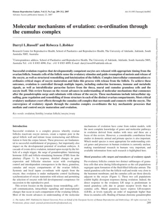
Figure 1. Intraovarian signalling cascade that mediates ovulation in the
mouse. The ovulatory LH surge from the hypothalamus initiates secondary
signals within the ovarian follicle, which converge on the COC. In the thecal
compartment, LH acts on thecal cells or leukocytes to induce Insl-3 and
IL-1 secretion. In mural granulosa layers, Egf-L: (amphiregulin, epiregulin
and betacellulin) are produced and transduce the ovulatory signal to the
cumulus cells. FSH acts directly on receptors expressed by mural as well as
cumulus cells. IaI enters the cumulus complex from circulation. Within the
COC, growth and differentiation factor-9 (Gdf-9) and/or bone morphogenetic
protein-15 (BMP-15) from the oocyte exert paracrine effects on cumulus cells
through the Alk5/BMPRII receptor dimer. Prostaglandin E2 (PGE2) acts in an
autocrine fashion via the PGE2 receptor (EP2). LH-R, luteinizing hormone
receptor; FSH-R, follicle-stimulating hormone receptor; ErbB, Egf receptor
family; Alk5, activin receptor-like kinase-5; BMPRII, BMP receptor type-II;
LGR8, Insl3 receptor; IL-1R, interleukin-1 receptor.
D.L.Russell and R.L.Robker
290
Downloaded
from
https://academic.oup.com/humupd/article/13/3/289/2457885
by
Universidade
Federal
da
Santa
Maria
user
on
13
May
2021
3. effects on rat granulosa cells (Salvador et al., 2002). Thus,
although LH signalling likely involves activation of multiple
parallel signal transduction pathways, PKC may not be one.
It is agreed that LH action on granulosa cells stimulates the
extracellular regulated kinase (Erk1/2 or MAPK) pathway (Das
et al., 1996; Maizels et al., 2001; Seger et al., 2001; Salvador
et al., 2002; Choi et al., 2005), with activation of Erk being very
rapid (Sela-Abramovich et al., 2005) and dependent on PKA
(Salvador et al., 2002; Russell et al., 2003b). It is not yet entirely
clear whether Erks are activated via direct phosphorylation by
PKA or through some intermediate step such as inactivation of
Erk phosphatases (Cottom et al., 2003), nor is the downstream
function of Erks in granulosa cells, particularly in relation to ovu-
lation, well understood; reports to date have primarily focused on
their role in progesterone synthesis as cells luteinize (Seger et al.,
2001; Dewi et al., 2002; Tajima et al., 2003). Recently, the
transcription factor Rhox5 was identified as a transcriptional
target of LH via Erks in ovulating follicles (MacLean et al.,
2005). Ovulatory hCG also activates AP1 transcription factors of
the Jun, Fos and Fra family (Sharma and Richards, 2000), consist-
ent with general observations that Erk1/2 typically phosphorylate
Fos, Myc, Elk-1 and Stat3 (Roux and Blenis, 2004). The effectors
of Erk-mediated transcriptional regulation of granulosa cell gene
expression during ovulation remain to be determined.
Follicular cGMP levels are increased during the course of ovu-
lation, and at least one cGMP effector, the cGMP-dependent
kinase cGKII, is also up-regulated at the time of ovulation by
cAMP and Erk1/2 via progesterone receptor (PR) and epidermal
growth factor (Egf) mediated pathways (Sriraman et al., 2005).
Guanylyl cyclase receptors which generate cGMP, have been loca-
lized within the ovary (LaPolt et al., 2003). Expression of guanylyl
cyclase-A receptor, and its ligand atrial natriuretic peptide (ANP),
is up-regulated in mouse granulosa cells in response to ovulatory
hCG (Sriraman et al., 2005); whereas guanylyl cyclase isoforms
GC-B, GC-C and sGC are expressed at low levels and not hormon-
ally regulated. Elevated production and activity of cGMP during
the course of ovulation may alternatively occur via guanylyl
cyclase activation by nitric oxide (NO), since nitric oxide synthase
(NOS) in the theca produces NO during ovulation (Zackrisson
et al., 1996). Clear effects of cGMP on granulosa cell gene
expression, steroidogenesis and oocyte maturation are reported,
but how this cascade impacts ovulation in particular is not
known (LaPolt et al., 2003).
Studies using LH- or hCG-stimulated granulosa-lutein cells
obtained from IVF patients confirm that PKA and PKC pathways
are active in human granulosa cells (Freimann et al., 2004, 2005).
More work is needed, however, to understand, as in rodents, the
complex array of intracellular signalling pathways activated in
response to the simple LH surge.
Periovulatory regulation of gene transcription
Signalling pathways induced by LH rapidly modify the transcrip-
tional machinery of mural granulosa cells, reprogramming cellular
function towards ovulation concurrent with luteinization. Exten-
sive reprogramming of gene expression after the LH surge is
achieved through modulation of a suite of transcriptional regula-
tors, which in turn mediate effector gene transcription. A cohort
of periovulatory genes with key roles in ovulation exhibits a
common pattern of transcriptional induction involving binding
of Sp1/Sp3 transcription factors to GC-box promoter elements.
Sp1 and Sp3 are constitutively present in granulosa cells but
exhibit sequence-specific, LH-regulated binding to gene promo-
ters (Russell et al., 2003b). The signal transduction mechanism
that modifies Sp1 binding activity in granulosa cells is uncertain,
but in most tissues involves post-translational phosphorylation or
glycosylation (Li et al., 2004).
Activity of Sp1/Sp3 complexes induces expression of
additional transcription factors that further contribute to a
cascade of ovulatory gene expression (Figure 2). Induction of
PR in mouse and rat mural granulosa cells occurs in response to
the LH surge through a cAMP/PKA-dependent mechanism,
whereas phorbol ester (PKC activation) has a synergistic effect
(Clemens et al., 1998; Sriraman et al., 2003; Sriraman and
Richards, 2004). The cAMP-mediated induction of PR requires
Sp1/Sp3 binding to multiple GC-box elements in the PR promoter
(Sriraman et al., 2003) and consequent recruitment of additional
transcription factors. In the rhesus monkey, PR is similarly
induced in the periovulatory period (Stouffer, 2002). PR is
present in human pre-ovulatory granulosa cells (Suzuki et al.,
1994; Revelli et al., 1996; Chang et al., 2005). Two isoforms of
PR are produced from distinct promoters on the same gene;
PR-A is predominant in rat granulosa cells (Natraj and Richards,
1993) and is the most critical for ovulation, since mice lacking
only PR-A have severely reduced ovulation (Mulac-Jericevic
et al., 2000). Human ovaries express PR-A and PR-B isoforms,
with PR-A most consistently found to predominate (Stouffer,
2003), however, the regulation of each isoform in ovulating
follicles is not yet determined.
Early growth response-1 (Egr-1) is a transcription factor
induced through cAMP/PKA and Erk signalling. Transcriptional
induction of Egr-1 requires Sp1/Sp3 and phosphorylated-CREB
binding to the proximal promoter. This is thought to lead to
recruitment of a combinatorial transcription initiator complex
involving CBP and other cofactors (Russell et al., 2003b).
Another transcription regulator Rhox5 is also regulated by the
combination of Ets/GABP, Sp1 and Sp3 (MacLean et al., 2005).
These LH-induced transcription factors in turn co-ordinately
activate transcription of a further suite of ovulatory effector
genes. For instance, the protease cathepsin L is induced through
Sp1/Sp3, PR, Egr-1 and CREB-mediated transactivation (Jaffe
et al., 1989; Sriraman and Richards, 2004), and another ovulatory
protease ADAMTS-1 is induced through Sp1/Sp3 and PR in com-
bination with C/EBPb, and NF-1-like factor (Doyle et al., 2004).
The Egf-like factor epiregulin is also regulated by Sp1/Sp3
binding its promoter in rat granulosa cells (Sekiguchi et al., 2002).
Thus, the cascade of diverse periovulatory gene expression in
mural granulosa cells involves the induction and recruitment of
a cohort of transcription factors that subsequently induce effector
gene products, including ADAMTS-1 and cathepsin L proteases,
Egf-like ligands (Egf-L) and others. These ovulatory genes
exhibit a signature pattern of mRNA and protein expression: a
rapid, transient increase after the LH surge followed by down-
regulation. Recurring modifications of the transcriptional machin-
ery interacting with these gene promoters mediate this character-
istic expression. The common denominator is Sp1/Sp3
transcription factor binding, suggesting that these are universal
mediators of periovulatory gene expression. The Sp factors
recruit chromatin-remodelling complexes that can switch in
Molecular mechanisms of ovulation
291
Downloaded
from
https://academic.oup.com/humupd/article/13/3/289/2457885
by
Universidade
Federal
da
Santa
Maria
user
on
13
May
2021
4. response to changing cell context to modify transcriptional
complex assembly, or alter promoter occupancy at neighbouring
enhancer sites (Li et al., 2004) (Figure 2). This process has been
well described for LH-R transactivation by Sp and nuclear
receptor transcription factors in granulosa cells (Zhang and
Dufau, 2003). Histone deacetylases recruited to Sp1/Sp3-bound
promoters silence transcription, but can be rapidly exchanged
for histone-acetylating complexes that open local chromatin
structure to RNA polymerase access where Sp1/Sp3 are present.
More detail is needed to better understand how LH-induced
intracellular signals impact specific transcriptional complexes
that distinctly regulate ovulation versus luteinization events and
how this model relates to human periovulatory gene regulation.
Essential effectors of ovulation in mural granulosa cells
A number of mural granulosa cell genes play essential roles in ovu-
lation. Mouse models, in particular, have yielded a great deal of
information about the role of key genes in specific aspects of the
ovulatory process. Of those identified to date, a few are expressed
prior to the LH surge, such as the transcription co-regulator,
RIP140, and the phosphodiesterase, PDE4D; however, the majority
are transiently expressed during the discrete ovulatory process.
Many are transcriptional regulators, including PR, Egr-1 and
CAAT enhancer binding protein (C/EBPb). These co-ordinately
mediate expression of ovulatory effector genes.
Transcriptional regulators: PR, RIP140, Egr-1 and C/EBPb. Pro-
gesterone and PR are very specific key regulators of follicular
rupture. Antagonists of PR, including RU486 (Tsafriri et al.,
1987; Loutradis et al., 1991), or inhibitors of P synthesis each
reduces or completely blocks ovulation in several species
including rats (Espey, 1998), sheep (Murdoch et al., 1986) and
humans (Baird et al., 2003). Pre-ovulatory granulosa cells begin
expressing progesterone in the days prior to the LH surge (Gore-
Langton and Armstrong, 1994; Richards, 1994), whereas PR is
acutely induced after the LH surge and is immediately activated
by the high local concentration of ligand, before its expression is
down-regulated. Mice with a targeted deletion of the PR gene
(PRKO) exhibit normal follicle growth and luteinization, but a
complete and specific block in ovulation (Lydon et al., 1996;
Robker et al., 2000). Key PR-dependent genes expressed in the
periovulatory period, but absent in PRKO mice and most likely
contributing to their anovulation, include proteases ADAMTS-1
and cathepsin L (Robker et al., 2000).
The nuclear receptor co-regulator RIP140 is most highly
expressed in pre-ovulatory granulosa cells and is rapidly down-
regulated after the LH surge in the periovulatory period (White
et al., 2000). RIP140 plays a broad role in the regulation of
genes that are involved in follicle rupture, suggesting that this tran-
scription cofactor is part of a cell-specific transcriptional complex
formed in the granulosa compartment of ovulating follicles. Mice
null for RIP140 exhibit apparently normal follicular growth and
luteinization, but a block in ovulation due to the failed induction
of numerous critical mural and cumulus cell genes, such as
ADAMTS-1, epiregulin, amphiregulin, betacellulin, hyaluronan
synthase-2(HAS-2), cyclooxygenase-2(COX-2), pentraxin-
3(PTX-3) and tumour necrosis factor stimulator gene-6(TSG-6)
(Tullet et al., 2005). Thus, RIP140 may regulate differentiation
of follicles to pre-ovulatory maturity or mediate a single master
event in the periovulatory response to LH. The close similarity
of the PRKO phenotype to that of RIP140 null mice suggests
that these two transcriptional regulators are in the same essential
pathway.
The LH surge also initiates rapid and transient induction of tran-
scription factors Egr-1 (Russell et al., 2003b) and C/EBPb (Sirois
and Richards, 1993) in mural granulosa cells. Egr-1 null mice are
Figure 2. Signal transduction in mural granulosa cells in the mouse periovulatory phase. LH interacts with its G-protein (Gas)-coupled receptor activating AC and
hence intracellular cAMP production as well as iP3/Caþþ
mobilization. Consequently, PKA and Erk activation result in phosphorylation (line arrows) of transcrip-
tion factors including cAMP response element-binding protein (CREB) and stimulatory proteins 1 and 3 (Sp1/3). Transcriptional complexes assembled on the pro-
moters of periovulatory genes expressed in mural granulosa cells include Sp1/3 and additional factors including CREB, reproductive homeobox 5 (Rhox5) or other
induced transcription factors. Transcriptional complexes modify chromatin structure and stabilize transcriptional machinery to transiently activate high gene
expression. Genes expressed in this rapid transient fashion via this signalling cascade include PR, a disintegrin and metalloproteinase with thrombospondin
repeats (ADAMTS-1), Egr-1 cathepsin L and versican.
D.L.Russell and R.L.Robker
292
Downloaded
from
https://academic.oup.com/humupd/article/13/3/289/2457885
by
Universidade
Federal
da
Santa
Maria
user
on
13
May
2021
5. infertile, primarily due to loss of LH secretion due to its role in
LHb gene synthesis in the pituitary (Lee et al., 1996; Topilko
et al., 1998). Thus, fertility may be controlled at multiple levels
by Egr-1, including both pituitary and ovarian roles in the ovula-
tory process. It has been reported that ovarian LH-R expression
is activated by Egr-1 and is disrupted in Egr-1 null mouse
ovaries (Topilko et al., 1998; Yoshino et al., 2002). However,
closer analysis of these mice indicates that after exogenous
gonadotrophin replacement, ovulation remains impaired but that
ovarian LH-R is normally expressed (Russell 2004). Null
mutants for C/EBPb show oocytes entrapped in CL; however,
evidence suggests these are among the few models with affected
luteinization in addition to ovulation (Sterneck et al., 1997). Con-
sistent with this it was shown that, C/EBPb may regulate steroi-
dogenesis through the StAR gene (Christenson et al., 1999).
cAMP metabolism. The phosphodiesterase PDE4D, an important
regulator of LH-mediated cAMP accumulation in granulosa cells
(Tsafriri et al., 1996), is required for normal ovulation. PDE4D
null mice exhibit dramatically reduced ovulation rates and litter
size due to altered responsiveness of granulosa cells to the LH
surge and reduced expression of ovulatory genes, including PR,
cathepsin L and COX-2 (Jin et al., 1999; Park et al., 2003).
Many follicles fail to reach pre-ovulatory stage due to premature
luteinization. However, by in-situ hybridization, it was shown
that the morphologically normal pre-ovulatory follicles, although
reduced in number, still express ovulatory genes such as the
Egf-L (Park et al., 2004). Thus, rather than rendering the granulosa
cells unable to respond to ovulatory LH, the lack of PDE4D pri-
marily permits accumulation of cAMP such that follicles exhibit
premature luteinization and entrapment of oocytes in immature
luteinized follicles.
Egf-L: amphiregulin, betacellulin and epiregulin. The Egf-L, epir-
egulin, amphiregulin and betacellulin, are very rapidly induced
specifically in mural granulosa cells within 1–3 h after the LH
surge and act as major secondary signals that transmit the ovula-
tory signal to the cumulus complex (Park et al., 2004)
(Figure 1). Epiregulin and amphiregulin are also LH inducible in
human granulosa cells (Freimann et al., 2004). Owing presumably
to functional redundancy between epiregulin, amphiregulin and
betacellulin, null mutant mice for each of these genes are fertile
(Luetteke et al., 1999; Lee et al., 2004). Each of the Egf-L
factors are synthesized as integral membrane proteins, and clea-
vage of the precursor forms is required for their interaction with
cumulus-expressed receptors since protease inhibitors can block
transmission of the LH signal to cumulus cells, whereas co-
treatment with epiregulin reverses this block (Ashkenazi et al.,
2005). Identification of the processing enzyme will reveal a key
regulatory event in intrafollicular co-ordination of the ovulatory
processes.
Extracellular matrix protease ADAMTS-1. The extracellular pro-
tease ADAMTS-1 was identified as LH- and PR-induced gene in
mural granulosa cells in the rat (Espey et al., 2000) and mouse
(Robker et al., 2000). This gene has been identified in bovine
(Madan et al., 2003), equine (Boerboom et al., 2003), primate
(Young et al., 2004) and human ovaries (Freimann et al., 2005)
and may be associated with the aetiology or symptoms of anovu-
latory human polycystic ovarian syndrome (Jansen et al., 2004),
but has yet to be studied in human normal ovulation. The latent
pro-form of ADAMTS-1 is synthesized by the mural granulosa
cells, but the secreted, mature form is selectively localized to
the extracelullar matrix (ECM) and cell surfaces of the expanded
cumulus complex (Russell et al., 2003a), suggesting this is where
it mediates its principal effects. Two lines of mice with null
mutation of the ADAMTS-1 gene have 70–90% reduced ovu-
lation rates and 75% reduction in litter size (Shindo et al., 2000;
Mittaz et al., 2004). From these studies, it is clear that
ADAMTS-1 is an important mediator of LH and progesterone
effects in ovulation across species. Substrates for ADAMTS-1
include versican, which is also produced by rodent mural granu-
losa cells and stimulated by the LH surge (Russell et al., 2003a)
and has been identified in human ovaries (Eriksen et al., 1999;
Relucenti et al., 2005). As with ADAMTS-1, versican is produced
in mural granulosa cells, but the majority of protein in the perio-
vulatory period is incorporated into the cumulus cell matrix.
Clearly, many mural granulosa cell gene products are required
for successful and optimal ovulation. Many of these genes
encode proteins that sense and respond to systemic inputs, includ-
ing the LH surge. A cohort of essential ovulatory mediators are
transcription factors that are up- and down-regulated by the LH
surge and function to translate the ovulatory trigger into global
reprogramming of gene transcription and ultimately cell function.
Interestingly, a new group of effector gene products is emerging,
including the Egf-L, versican and ADAMTS-1, which are pro-
duced by mural granulosa cells, but specifically translocate to
and act within the COC. This combined endocrine and paracrine
intrafollicular communication enables the co-ordination of
maternal receptivity with oocyte maturation and ovulation.
Cumulus cells: conduit of ovulatory signals
The pre-ovulatory oocyte is surrounded by several cell layers,
known as the cumulus granulosa cells, which are tightly connected
to each other and the oocyte through intercellular membrane pro-
cesses and gap junctions that facilitate exchange of glucose metab-
olites, small signalling molecules and ions. Oocytes depend on the
cumulus cells for metabolism of glucose and supply of pyruvate
for energy production (Biggers et al., 1967; Gardner et al.,
1996; Preis et al., 2005). The cumulus cells exhibit hormone
responsiveness and gene expression profiles distinct from those
of the mural granulosa layers. In preovulatory follicles, cumulus
and mural cells present abundant cell surface FSH receptors, but
the cumulus mass and granulosa cells very close to the antrum
show the greatest proliferative response to FSH during follicle
growth (Robker and Richards, 1998). The cumulus are the cells
most directly exposed to mitogenic factors secreted by the
oocyte (Erickson and Shimasaki, 2000). Oocyte-secreted factors
(OSF) belonging to the Tgfb superfamily establish a morphogen
gradient that promotes the cumulus cell characteristics including
enhanced cell proliferation, and repression of LH-R and PR
gene transcription, and progesterone biosynthesis (Eppig et al.,
1998; Elvin et al., 1999a; Gilchrist et al., 2004). The rate of pro-
liferation of these cells has been suggested to correlate with
implantation potential in human assisted reproduction (Gregory,
1998). Following the ovulatory LH surge, cumulus cells respond
with a unique pattern of gene induction leading to production
and stabilization of a local ECM that envelopes the COC. This
Molecular mechanisms of ovulation
293
Downloaded
from
https://academic.oup.com/humupd/article/13/3/289/2457885
by
Universidade
Federal
da
Santa
Maria
user
on
13
May
2021
6. process, known as cumulus expansion or mucification, is depen-
dent on a specific cascade of intracellular signals and ECM gene
expression within the COC and plays a key role in ovulation
(Chen et al., 1993; Hess et al., 1999; Fulop et al., 2003; Salustri
et al., 2004) and subsequent fertility success (Tanghe et al.,
2003; Somfai et al., 2004; Yang et al., 2005).
Intracellular signalling in periovulatory cumulus cells
Cumulus cells respond rapidly and change dramatically after the
gonadotrophin surge. The program of gene induction in the
cumulus cells responding to the gonadotrophin surge is potentially
mediated, at least in part, by high FSH that accompanies the LH
surge. In women, as in other mammalian species, FSH levels
surge with similar timing kinetics to LH at the midcycle (Hoff
et al., 1983). Local paracrine effectors including Egf-L from
granulosa cells, interleukin-1 from the thecal compartment and
oocyte-derived signals also contribute to cumulus cell responses
(Figure 1).
Cumulus response to endocrine and paracrine stimuli
Follicle stimulating hormone. In isolated COCs in vitro, FSH
induces periovulatory changes in cumulus cells and oocytes (Su
et al., 2002) and in vivo very high concentrations of pure FSH
have even been found to initiate oocyte maturation and ovulation
in rodents (Tsafriri et al., 1976b; Galway et al., 1990; Wang and
Greenwald, 1993a) and primates (Zelinski-Wooten et al., 1998).
However, the levels of FSH used to activate COC expansion in
vitro are supraphysiological, and in animal models or human
IVF cycles, levels of FSH, which reflect physiological circulating
levels during the ovulatory surge, do not enhance molecular events
associated with ovulation or oocyte development (Tsafriri et al.,
1976b; Vermeiden et al., 1997). Although periovulatory gene
expression is induced by FSH in the cumulus, it has been widely
shown that ovulation and oocyte maturation are efficiently
induced in vivo by pure exogenous LH preparations that mimic
the levels of an endogenous LH surge. These observations indicate
that FSH may play a minor role, but is not essential, whereas intra-
follicular responses to the LH surge mediate ovulation.
Since FSH is effective in stimulating ovulatory cumulus gene
expression, it is widely used to study molecular aspects of this
process. The FSH receptor is a GPCR closely related to LH-R
that activates adenylate cyclase mediating cAMP synthesis and
resulting in PKA-activated intracellular signalling (Figure 3). In
addition, FSH independently, activates the PI3K/Akt pathway
(Gonzalez-Robayna et al., 2000; Zeleznik et al., 2003) and recruits
the 14-3-3 adaptor protein (Cohen et al., 2004) to mediate cell sur-
vival and proliferation in granulosa cells. Through the Akt
pathway, FSH also increases transcription of the hypoxia inducible
factor-1a (Alam et al., 2004) leading to the induction of
expression from genes such as vascular endothelial growth
factor (VEGF), a critical mediator of angiogenesis during ovu-
lation/luteinization (see below). Most importantly, FSH activates
ERK signalling in granulosa (Das et al., 1996) and in cumulus
cells (Su et al., 2002) and Erk activity is requisite in cumulus
cells but not in oocytes for COC expansion and oocyte meiotic
maturation (Su et al., 2002). Inhibitors of Erk, p38 kinase or Akt
block COC expansion induced by FSH or cAMP treatment
(Ochsner et al., 2003a), indicating that each signalling kinase
pathway converges to mediate important periovulatory cumulus
gene expression.
Prostaglandins. Cumulus cells in ovulating COC produce pros-
taglandins via induction of COX-2 and other prostaglandin syn-
thetic enzymes. Induction of COX-2 in COC can be stimulated
by Egf in cows (Nuttinck et al., 2002) and mice (Park et al.,
2004) or by FSH through cAMP/PKA activation (Joyce et al.,
2001). Pharmacological activators of cAMP synthesis induce
COX-2, but this is sensitive to inhibition of p38-MAPK or
Erk1/2 (Ochsner et al., 2003a), indicating that Erk-mediated tran-
scriptional induction, distinct from or in conjunction with the clas-
sical cAMP pathway, is required for COX-2 expression. The
COX-2 gene promoter has been studied in detail and shown in
mouse granulosa cells to be dependent on oocyte-derived Gdf-9
(Elvin et al., 2000) and induced through an E-box element that
interacts with the upstream stimulatory factor (USF) transcription
factor in a manner regulated by PKA (Morris and Richards, 1996;
Sayasith et al., 2004; Sayasith et al., 2005). The predominant pros-
taglandin produced is prostaglandin (PGE2) which interacts with
the PGE2-receptor (EP-2) to induce changes in cumulus gene
expression (Segi et al., 2003) critical for COC expansion and ovu-
lation (Hizaki et al., 1999). Prostaglandins made by the cumulus
cells potentially mediate important signalling effects through
other receptors also, since disruption to fertility in EP-2 null
mice is far less severe than in COX-2 nulls that produce no pros-
taglandins (Table I). Very recently, the PGE2 receptor, EP-4, was
also shown to be induced in periovulatory cumulus cells and to
participate with EP-2 in oocyte maturation, cumulus cell survival
and matrix production (Takahashi et al., 2006). Both PGE2 recep-
tors are GPCR that stimulate cAMP production; thus in cumulus
cells, prostaglandin synthesis establishes an autocrine signalling
loop synergizing with FSH and reinforcing cAMP/PKA and Erk-
dependent gene induction in cumulus cells (Figure 3). This loop of
cAMP-mediated PKA activation in cumulus cells may be con-
trolled by the regulator of G-protein signalling (RGS-2), which
has a temporal and spatial pattern of expression very similar to
COX-2 and EP-2 in periovulatory COCs (Ujioka et al., 2000).
Adenylate cyclase activity can be either augmented or inhibited
by RGS-2 (Kehrl and Sinnarajah, 2002), and its role in COC has
yet to be more fully investigated. Human granulosa-lutein cells
express EP-2 (Harris et al., 2001)and secrete PGE2 (Chandras
et al., 2004), indicating that a similar signalling loop likely
exists in human ovulation.
Epidermal growth factor-like ligands. Egf-L (amphiregulin, beta-
cellulin and epiregulin) from LH-stimulated mural granulosa cells
also activate periovulatory COC gene expression (Espey and
Richards, 2002; Park et al., 2004; Rimon et al., 2004). These
ligands activate the Egf-receptor tyrosine kinase and subsequently
the Erk activity (Kansra et al., 2004) (Figure 3). Cumulus cell
expression of ECM genes, COC expansion and ovulation are
blocked by pharmacological inhibition of the Egf-receptor tyro-
sine kinase or downstream Erk function (Lorenzo et al., 2001;
Su et al., 2002; Park et al., 2004; Tsafriri et al., 2005). Ovulation
induction by hCG is also blocked by inhibition of Egf-receptor
activity; oocytes are entrapped in follicles surrounded by
compact cumulus cells and cumulus matrix gene induction in cul-
tured follicles is inhibited (Ashkenazi et al., 2005). Thus, both
FSH and Egf-L signals promote cumulus periovulatory gene
expression through activation of Erk signalling.
D.L.Russell and R.L.Robker
294
Downloaded
from
https://academic.oup.com/humupd/article/13/3/289/2457885
by
Universidade
Federal
da
Santa
Maria
user
on
13
May
2021
7. Androgen. Cumulus cells of several species, including rat (Pelle-
tier et al., 2000; Szoltys et al., 2003) and human (Hickey et al.,
2005), express the androgen receptor and thus are thought to
respond directly to androgens produced by theca cells. There is
limited evidence for the consequences of androgen action in ovu-
lation; however, in female mice lacking androgen receptor a
minor, albeit significant reduction in ovulation rate was reported
(Hu et al., 2004). Furthermore, ovulatory genes, including PR,
HAS2 and TSG-6, were not induced, and the cumulus complexes
ovulated were reported to have a disorganized structure,
suggesting a role for androgen receptor in COC expansion and
the ovulatory process (Hu et al., 2004).
Interleukin-1b. Other cells within the theca interna, possibly leu-
kocytes surrounding periovulatory follicles, may also contribute to
cumulus expansion in vivo (Figure 1). These cells produce IL-1b
(Jasper and Norman, 1995; Wang et al., 1997; Minge et al., 2005)
in response to LH (Kol et al., 1999). The IL-1b receptor activates a
receptor-associated kinase, again resulting in Erk activation (Eder,
1997). Thus, in vivo, paracrine signals from the theca also transmit
signals to the cumulus and co-ordinate the cumulus response to the
LH surge. Hyaluronan (HA) synthesis and cumulus expansion are
induced by IL-1b (Kokia et al., 1993; Singh and Armstrong,
1997), and cumulus expansion and ovulation can be blocked by
IL-1 antagonism in rodents (Simon et al., 1994). In humans,
IL-1b and IL-1a have been found to be induced by gonado-
trophins and serum levels correlate with successful fertilization
(Mendoza et al., 1999) or implantation in assisted reproductive
cycles (Karagouni et al., 1998). However, subsequent studies evi-
denced either no correlation (Barrionuevo et al., 2000) or reverse
correlation (Mendoza et al., 2002).
Extracellular matrix. The COC matrix itself may have important
effects in mediating or modulating cumulus intracellular signal-
ling. The HA receptor, CD44 is expressed on cumulus cells,
induced after the LH surge in many species including humans
(Ohta et al., 1999; Yokoo et al., 2002; Schoenfelder and Einspa-
nier, 2003) and can transduce multiple intracellular signals
(Bourguignon, 2001) via cytoskeletal changes, which transactivate
the Egf-R family member ErbB2 and activate Erk. Proteoglycan
components of the COC matrix, such as CD44 and versican,
likely bind growth factors and modulate signalling. However,
CD44 null mice have normal fertility (Schmits et al., 1997;
Protin et al., 1999), suggesting the involvement of redundant
HA receptors such as RHAMM, which is present in bovine COC
(Turley et al., 2002). Actions of the proteoglycan inter-alpha
trypsin inhibitor (IaI) found in the COC matrix include modulat-
ing kinases Erk and Akt to mediate gene expression changes
(Kobayashi et al., 2001). Microarray analysis identified 29 genes
up-regulated and 5 genes down-regulated 12 h after hCG in
Figure 3. Signal transduction in cumulus cells in the mouse periovulatory phase. FSH binds FSH receptors on cumulus cells to activate AC, cAMP production and
PKA. This pathway activates Erk as do Egf receptors in response to Egf-L amphiregulin (AR), betacellulin (BC) and epiregulin (ER). Transcription factors regulated
by these kinase cascades include AP-1 factors, Elk-1 and cAMP response element-binding protein (CREB). In growing follicles, cAMP is translocated to the oocyte
via gap junctions until these are inactivated through phosphorylation by Erk and cells separate from the oocyte through cumulus expansion. Consequently, cAMP
levels in the oocyte fall. In a separate pathway, oocyte-secreted growth and differentiation factor-9 (Gdf-9) and bone morphogenetic protein-15 (BMP-15) activate
integral receptor kinase activity on cumulus cells, which results in phosphorylation of SMAD2/3, which translocate to the nucleus in dimers with SMAD4. These
transcription factors promote expression of key cumulus genes required for specification of the cumulus-specific response to the ovulatory surge. Genes induced via
these two signal transduction mechanisms include HAS-2, TSG-6, PTX-3, COX-2, PGE2 receptor (EP2) and regulator of GPCR-2 (RGS-2). PGE2 activates the EP2
receptor to mediate a signal transduction pathway similar to FSH, and RGS-2 may control the activation of G-proteins.
Molecular mechanisms of ovulation
295
Downloaded
from
https://academic.oup.com/humupd/article/13/3/289/2457885
by
Universidade
Federal
da
Santa
Maria
user
on
13
May
2021
8. Table I: Female Anovulatory Infertility Phenotypes Reported in Null Mutant Mouse Models
Gene Ovulation rate
% null versus controls
Fertilization rate
in vivo (%)
Pregnancy rate
C pregnant
% control
Litter size of
pregnant C (%)
Comment Reference
Transcription regulators or cell signalling
PR 0 0 0 — Normal morphological COC
expansion described
Lydon et al. (1995) Robker
et al. (2000)
AR 62 N/D 69 51 COC expansion disrupted Hu et al. (2004)
RIP140 0 — 0 — COC expansion disrupted
COC gene expression
disrupted
White et al. (2000)
EP2 67 8.5 20 N/D COC expansion disrupted Hizaki et al. (1999)
COX-2 40 0% natural cycle 2%
superovulation
0 — No first polar body extrusion Lim et al. (1997)
COX-2 5 N/D N/D N/D COC expansion disrupted Davis et al. (1999)
C/EBPb 12 N/D N/D N/D Cumulus not assessed Sterneck et al. (1997)
PDE4D 22 N/D N/D 50 COC gene expression
disrupted Premature
luteinization
Jin et al. (1999) Park et al.
(2003)
Extracellular proteins
ADAMTS-1 N/D N/D 13 24 Shindo et al. (2000)
ADAMTS-1 33 N/D 50 50 COC expansion observed Mittaz et al. (2004)
TSG-6 48 0 0 — COC expansion disrupted Fulop et al. (2003)
Bikunin 43 ,10 40 9 COC expansion disrupted Zhuo et al. (2001)
Bikunin 14 N/D 19 N/D d3 embryos lack zona
pellucida
Sato et al. (2001)
PTX-3 45 2 ,50 27 COC expansion disrupted Varani et al. (2002)
PTX-3 Not differenta
0 0 — Oocyte transport defect Salustri et al. (2004)
N/D, not determined.
a
Oocytes collected at 14 versus 20 h hCG may explain different results in Salustri versus Varani PTX-3 null ovulation rates.
D.L.Russell
and
R.L.Robker
296
Downloaded
from
https://academic.oup.com/humupd/article/13/3/289/2457885
by
Universidade
Federal
da
Santa
Maria
user
on
13
May
2021
9. COC of mice that lack IaI relative to wild-type controls (Suzuki
et al., 2004), suggesting that it regulates gene expression
changes in addition to being a key matrix component.
To date, transcription regulators modulating periovulatory
cumulus cell gene expression have not been studied in detail due
to lack of an appropriate in vitro model for such studies. Repeat-
edly it has been reported that signals that activate periovulatory
cumulus gene expression act through the Erk kinase family. The
transcription factors most commonly phosphorylated by Erk
kinases include AP-1 factors, signal transducer and activator of
transcription (STAT) and serum-responsive transcription factors.
However, in addition, cumulus-cell-specific gene expression
requires oocyte-derived signals.
Cumulus cell response to signals from the oocyte
Mature oocytes, although transcriptionally inactive, secrete
signals that play a key permissive role in periovulatory cumulus
gene expression in mice (Eppig et al., 2002). This oocyte-derived
activity, originally referred to as cumulus-expansion-enabling
factor (CEEF), is secreted by mouse, pig and cow oocytes
(Eppig et al., 1993; Singh et al., 1993; Vanderhyden et al.,
1993; Gilchrist et al., 2004). This ability is linked to the
oocytes’ developmental competence (Eppig et al., 1993). A
range of experiments seeking to identify the CEEF in mice
suggests that Gdf-9 as well as other Tgfb superfamily members
can support cumulus ECM gene expression (Vanderhyden et al.,
2003; Dragovic et al., 2005). However, the identity of the true
oocyte-secreted factor(s) facilitating COC expansion remains con-
troversial, since antibody neutralization of Gdf-9 secreted by
oocytes only partially blocks COC expansion (Dragovic et al.,
2005), whereas suppression of Gdf9 mRNA in mouse oocytes
was more highly effective (Gui and Joyce, 2005).
The specific roles for Gdf-9, BMP-15 and perhaps other Tgfb
family members such as BMP-6, in ovulation have also been
difficult to fully interpret because of species variations and their
critical role early in folliculogenesis. In pigs and cows, the
oocyte does not need to be present at the time of cumulus expan-
sion, but in cows, BMP-15 is a necessary cumulus cell survival
factor during oocyte maturation and COC expansion (Hussein
et al., 2005). The impact of growth factor signals from the
human oocyte has not been directly tested due to obvious con-
straints on available COCs for analysis. Thus, the model system
that best represents the human is unknown.
In all species, the initiator of ovulatory cumulus gene expression
is clearly not Gdf-9 and/or BMP-15, since these factors are con-
stitutively synthesized by oocytes and affect follicular somatic
cell development throughout folliculogenesis (Elvin et al.,
1999b; Juengel et al., 2002), including in humans (Di Pasquale
et al., 2004), without activating matrix production. It is thought
that in all species, oocyte signals specify the cumulus cell
lineage which orchestrates the cumulus-specific responses after
the LH surge (Erickson and Shimasaki, 2000; McNatty et al.,
2005). Therefore, by producing Gdf-9 and BMP-15, the oocyte
controls the differentiation of its immediate environment and
hence its ovulation.
Signalling by Gdf-9 in cumulus cells is achieved by binding to
the BMP type-II receptor (BMPRII), followed by recruitment and
activation of the type-I receptor, Alk5 (Mazerbourg et al., 2004)
(Figure 3). Thus Gdf-9 uses similar intracellular signalling to
Tgfb. Activated Alk5 in human granulosa-lutein cells phosphory-
lates transcription factors SMAD2 and SMAD3, which dimerize
with SMAD4, translocate to the nucleus and bind to
SMAD-response elements in gene promoters containing the core
nucleotide sequence CAGA, such as the inhibin-B promoter
(Kaivo-Oja et al., 2005). Not surprisingly, factors that share the
capacity to activate SMAD2/3 in cumulus cells are those
previously shown to share the CEEF activity.
The mRNA and protein for SMAD2 and 4 are induced in whole
mouse ovary by FSH (Gueripel et al., 2004), indicating that the
pre-ovulatory follicle is primed for a robust response to Gdf-9
and other Tgfb-related ligands in the periovulatory phase. Knock-
out of the SMAD3 gene results in anovulatory infertility primarily
due to defects in follicle growth (Tomic et al., 2004), a similar but
not identical phenotype to Gdf-92/2
. This may indicate that the
Gdf-9 functions partially but not exclusively through SMAD3.
SMAD2 and SMAD4 knockout mice die in embryonic develop-
ment. Whether SMADs bind directly to promoters of each
cumulus-expressed periovulatory gene or regulate these genes
through indirect means is not known. It has been suggested that
Gdf-9 also activates Erk signalling in cumulus (Su et al., 2003),
but since other Erk activators such as Egf cannot substitute for
the signal from oocytes, SMAD 2/3 activation is more likely to
be the requisite mechanism of Gdf-9-mediated cumulus gene
expression.
Essential cumulus gene expression and regulation
The set of genes expressed in periovulatory cumulus cells are criti-
cal for normal rates of ovulation and fertility (Table I). Among the
earliest induced genes is COX-2, the rate-limiting enzyme in
prostaglandin production (Sirois and Richards, 1992; Wong and
Richards, 1992). COX-2 production is rapidly induced within
2 h of an hCG stimulus in rodent mural and cumulus cells
(Sirois et al., 1992), but expression is higher and persists longer
in the COC (Elvin et al., 1999a; Joyce et al., 2001; Segi et al.,
2003). In larger species with longer periovulatory periods, this
induction happens later and COX-2 has been proposed to be part
of a molecular clock that sets the species-specific timing of ovu-
lation (Sirois et al., 2004). This suggestion is supported by
studies in the macaque monkey, but in this model, the timing of
prostaglandin secretion is dependent on induction of COX-2,
microsomal prostaglandin-E synthase and most particularly on
expression and post-translational activation of phospholipase A2
in granulosa cells (Duffy et al., 2005). This mechanism places
more stringent control on the process required for prostaglandin
synthesis and the timing of ovulation. It is not known whether a
similar complex regulatory mechanism operates in human
ovulation.
Null mutation of the mouse COX-2 gene results in defective
cumulus expansion, reduced ovulation rate and infertility
(Dinchuk et al., 1995; Lim et al., 1997; Davis et al., 1999;
Ochsner et al., 2003b). This is explained by the reduced expression
of PGE2/EP-2-dependent genes such as TSG-6 in COX-22/2
as
well as EP-22/2
mice (Ochsner et al., 2003a). Likewise, treatment
of mice and rats with COX-2 inhibitors indomethacin or NS398
also blocks ovulation and TSG-6 expression (Mikuni et al.,
1998; Yoshioka et al., 2000; Espey and Richards, 2002). Indo-
methacin treatment also caused delayed ovulation by 2–12 days
in women, (Athanasiou et al., 1996) and another COX-2 inhibitor,
Molecular mechanisms of ovulation
297
Downloaded
from
https://academic.oup.com/humupd/article/13/3/289/2457885
by
Universidade
Federal
da
Santa
Maria
user
on
13
May
2021
10. rofecoxib, had a similar effect (Pall et al., 2001). These studies
collectively demonstrate that prostaglandin production via
COX-2 enzyme is a key event in the cumulus gene expression
cascade and vital to ovulation across mammalian species.
Genes encoding COC matrix components are also induced in
cumulus cells by FSH or Egf in conjunction with oocyte signals
including HAS-2, the enzyme that synthesizes the HA backbone
of the cumulus matrix, (Eppig, 1979a, 1981; Tirone et al., 1997;
Elvin et al., 1999a; Dragovic et al., 2005) and HA binding proteins
TSG-6 (Fulop et al., 1997; Yoshioka et al., 2000) and pentraxin-3
(PTX-3) (Varani et al., 2002; Salustri et al., 2004). The inhibitor of
BMP signalling Gremlin is expressed in pre-ovulatory mural and
cumulus cells, but maintained only in cumulus cells after ovulation
induction through Gdf-9 action (Pangas et al., 2004). The need for
oocyte factors as well as FSH or Egf for induction of these
cumulus specific genes has been characterized, but the full regulat-
ory mechanism in cumulus cells has not been studied in detail.
Interestingly, induction of TSG-6 in cumulus cells is dependent
on autocrine/paracrine prostaglandin action, yet HAS-2
expression is not (Ochsner et al., 2003b). However, PGE2 treat-
ment of isolated COC can induce HAS-2 mRNA (Eppig, 1981).
These observations indicate that HAS-2 induction requires
cAMP-PKA and/or Erk signalling in addition to Gdf-9/BMP-15
and that diverse regulatory mechanisms control different periovu-
latory genes in cumulus cells in vivo.
Cumulus matrix function
Appropriate formation of the expanded cumulus matrix is critical
for ovulation. In fact, the success of follicular rupture as well as
fertilization is exquisitely sensitive to perturbations in the compo-
sition and hence functional capacity of the cumulus matrix. This
has been clearly established in mice by gene ablation of COC
matrix proteins (Sato et al., 2001; Zhuo et al., 2001; Varani
et al., 2002; Fulop et al., 2003; Salustri et al., 2004) (Table I).
Other approaches that inhibit the expansion, organization or func-
tion of cumulus matrix proteins support the observations from
knockout studies (Chen et al., 1993; Hess et al., 1999; Ochsner
et al., 2003a).
Composition of expanded cumulus matrix. The backbone of the
expanded cumulus matrix is HA, a large disaccharide chain
common to many extracellular matrices including cartilage
where it provides strong osmotic force and viscoelastic properties.
Synthesis of HA requires glucose as a substrate for the hexosamine
pathway, which generates the disaccharide subunits of HA.
Glucose uptake and glycolytic activity in cumulus cells is mark-
edly stimulated by the LH surge in rodents, cows and humans
(Tsafriri et al., 1976a; Zuelke and Brackett, 1992; Roy and
Terada, 1999; Sutton et al., 2003). Glucose flux through the hex-
osamine pathway in bovine COC increases during oocyte matu-
ration (Sutton-McDowall et al., 2004). Strands of HA matrix are
stabilized and organized into a specific functional structure by
cross-linking proteins that bind HA through canonical
link-protein-like modules. Protein cross-linkers play a fundamen-
tal role of stabilization of the HA matrix, as demonstrated by its
dissolution by treatment with protease (Cherr et al., 1990).
Organizers of the COC matrix, which bind HA directly, include
TSG-6 (Carrette et al., 2001; Mukhopadhyay et al., 2001;
Ochsner et al., 2003a), synthesized by cumulus cells and versican
synthesized by the mural granulosa cells (Russell et al., 2003c) and
the heavy chains (HCs) of IaI, a serum protein secreted by hepato-
cytes (Steinbauch and Loeb 1961). Circulating IaI is a trimeric
protein complex comprising two HC subunits joined through chon-
droitin sulphate bridges to bikunin (Bost et al., 1998). It circulates
in high concentrations and enters the ovulating ovary in blood
plasma as a result of increased follicular vascular permeability
during ovulation (Castillo and Templeton, 1993). The HC binds
HA in the expanding COC ECM, whereas the bikunin subunit
remains in solution and takes no further part in the COC matrix for-
mation. Cartilage link protein may also be a product of FSH or
IGF-1-stimulated mural granulosa cells (Sun et al., 2003) and is
found in pre-ovulatory follicles of rodents and humans (Kobayashi
et al., 1999). Exogenous link protein enhances volumetric COC
expansion in vitro, but is not an essential matrix component (Sun
et al., 2002). PTX-3 is an immune protective and anti-
inflammatory protein that does not possess an HA-binding motif,
but localizes intensely to the cumulus ECM in rodents and
human. In addition, fibronectin, tenascin-C and laminin have
been reported in the human COC matrix (Familiari et al., 1996).
Function of HA cross-linking proteins in cumulus matrix. Evidence
suggests that the function of the cumulus matrix is dependent on a
specific structure produced through the binding of HC and
HC-TSG-6 complexes to HA (Carrette et al., 2001; Mukhopad-
hyay et al., 2001; Fulop et al., 2003; Ochsner et al., 2003a)
(Figure 4). Formation of the appropriate covalent complex of
IaI-HC to HA requires an intermediate interaction with TSG-6
(Chen et al., 1996; Carrette et al., 2001; Jessen and Odum,
2003). Loss of either TSG-6 or IaI synthesis results in almost iden-
tical phenotypes: HA is produced by cumulus cells but not stabil-
ized, resulting neither in matrix aggregation nor morphological
COC expansion in vitro or in vivo. As a result, fewer (,50%)
oocytes are ovulated (Sato et al., 2001; Zhuo et al., 2001; Fulop
et al., 2003). Of the few oocytes that are ovulated in TSG-62/2
mice, no fertilization or implantation has been observed in
natural matings, but fertilization under IVF conditions has not
been assessed (Fulop et al., 2003). A TSG-6 neutralising antibody
can prevent cumulus matrix assembly in vitro (Ochsner et al.,
2003a). Treatment of mice with inhibitors of the HA synthetic
pathway (Chen et al., 1993) or short HA oligosaccharides that
adsorb TSG-6 (Hess et al., 1999) also block COC expansion and
ovulation. These data combined with TSG-62/2
studies indicate
that TSG-6 as well as IaI interactions with HA are required for
the formation of the COC matrix.
The role of PTX-3 is different; mice with PTX-3 null mutation
achieve COC matrix expansion in vivo, but the cumulus cells
appear abnormally distributed. Oocytes are ovulated but fail to fer-
tilize because of the rapid degradation and loss of the COC matrix
during or after release from the follicle (Varani et al., 2002;
Salustri et al., 2004). Thus, PTX-3 appears to be required for
matrix stability and retention. In vivo fertilization was ,2% of
the normal rate in one PTX-32/2
mouse line (Varani et al.,
2002) and completely deficient in another (Salustri et al., 2004).
Under IVF conditions, fertilization of PTX-32/2
oocytes occurred
at the normal rate. This is expected since it is known that the need
for the COC matrix can be circumvented in vitro by the use of high
sperm concentrations; it also indicates that there is no profound
oocyte deficiency in the absence of PTX-3 (Salustri et al.,
D.L.Russell and R.L.Robker
298
Downloaded
from
https://academic.oup.com/humupd/article/13/3/289/2457885
by
Universidade
Federal
da
Santa
Maria
user
on
13
May
2021
11. 2004). PTX-3 protein forms homo-decameric complexes that may
interact with HA-bound TSG-6 molecules to form anchoring com-
plexes that cross-link multiple independent HA molecules (Salu-
stri et al., 2004). The above observations together demonstrate
that distinct functional aspects of the COC matrix composition
and structure are involved in release of the oocyte and its protec-
tion during passage through the oviduct.
Versican, a chondroitin-sulphate-substituted large aggregating
proteoglycan of the lectican family, is closely related to aggrecan
that cross-links HA providing strength and elasticity to cartilage.
Versican is produced primarily by mural granulosa cells and
rapidly incorporated into the cumulus matrix as it forms,
suggesting that it binds HA through its link module and is
another organizer of COC matrix structure (Russell et al.,
2003c). Whether versican is obligatory for COC matrix expansion
or ovulation has not been directly confirmed. Mutation of the ver-
sican gene in mice is uninformative since these mice die before
embryonic day 7 because of defective heart development
(Mjaatvedt et al., 1998). However, an important role for versican
protein synthesis and proteolytic processing may be implied from
other studies. The protease ADAMTS-1 selectively accumulates
in the expanding COC matrix possibly through direct interaction
with versican and specifically cleaves versican within the COC
matrix (Russell et al., 2003a). Ovaries of PRKO mice have 10%
of normal ADAMTS-1 protein abundance and 50% lower versican
processing, but morphologically normal COC expansion occurs.
PRKO mice are anovulatory due in part to reduced cleavage of
versican resulting in altered COC matrix function (Russell et al.,
2003a). ADAMTS-1 null mutation also results in reduced ovu-
lation rate and subfertility in two independent null lines (Shindo
et al., 2000; Mittaz et al., 2004). These observations suggest that
cross-linking of COC HA by versican occurs prior to ovulation,
but rapid cleavage of versican is requisite for normal matrix func-
tion and ovulation of COC.
Oocyte release and COC matrix expansion seem inseparable,
co-dependent processes, yet how the cumulus matrix affects or
participates in ovulation remains unknown. The composition and
structure of the expanded cumulus matrix is of clear importance
for fertility in rodents. A matrix of similar composition forms
around human oocytes (Relucenti et al., 2005), and the degree
of cumulus expansion and expression of mRNA for the HAS-2
enzyme have been correlated with human oocyte quality (McKen-
zie et al., 2004), but composition and function have been less well
studied.
Additional key properties of the cumulus matrix. In all the dis-
rupted cumulus matrix models thus far tested, in vivo fertilization
rate is the most dramatically reduced fertility parameter (Table I).
Disrupted cumulus matrix may alter oviductal transport of the
COC. After ovulation, oocyte capture requires selective adhesion
between the COC matrix and ciliated infundibular epithelium of
the oviduct (Lam et al., 2000). Matrix enclosed COC is efficiently
transported through the oviductal lumen (Lam et al., 2000; Talbot
et al., 2003), whereas cumulus-denuded oocytes are not (Odor and
Blandau, 1973; Mahi-Brown and Yanagimachi, 1983). Impaired
oocyte capture or transport in women can cause infertility and/
or ectopic pregnancy (Harper, 1994). However, animal models
of cumulus matrix disruption and reduced ovulation frequently
show entrapment of oocytes in luteinized follicles (Davis et al.,
1999; Robker et al., 2000; Shindo et al., 2000; Varani et al.,
2002; Mittaz et al., 2004), consistent with specific effects on
oocyte release rather than capture or transport of the oocyte.
Cumulus matrix also plays a role in capacitation of sperm (Van
Soom et al., 2002) and may present a selective barrier that screens
for sperm with high fertilization potential (Hong et al., 2004). A
proteoglycan component of the COC consistent in size and compo-
sition to versican accelerates human sperm acrosome reaction
(Tesarik et al., 1988; Drahorad et al., 1991). PTX-3 binds sperm
and may either anchor or attract sperm to the COC in the
oviduct in vivo (Salustri et al., 2004). Furthermore, a form of
glycodelin in the COC matrix appears to have the effect of promot-
ing sperm binding to the oocyte (Yeung et al., 2006). Thus com-
ponents of the COC matrix may be an active sperm attractant
and capacitator.
The presence of cumulus has also been found to reduce
oxidative stress on the oocyte (Fatehi et al., 2005); this may be
an important role for cumulus and one explanation for the reported
beneficial effect of cumulus inclusion in bovine IVF (Luciano
et al., 2005). Pericellular versican is known to be a potent inhibitor
of oxidative damage in cells (Wu et al., 2005), and hence versican
in the COC matrix may protect the oocyte during the events of
tissue hyperaemia and inflammation, which increase oxidative
free radicals during ovulation.
The synthesis of COC matrix and cumulus gene expression has
been linked to the determination of oocyte quality and develop-
mental capacity. Degree of cumulus expansion has been used as
a criterion for selection of oocytes for IVF for many years (Ball
et al., 1983; Laufer et al., 1984; Foote, 1987). More recently,
PTX-3 expression was correlated with oocyte quality in one
study (Zhang et al., 2005); however, in another study, PTX-3
expression was not significantly correlated with blastocyst grade,
Figure 4. Molecular structure of the expanded COC matrix. The intercellular
matrix between cumulus cells is composed of HA bound to cell surface recep-
tors (CD44 and Rhamm). HA is stabilised and organized into a specific struc-
ture by cross-linking proteins. Versican, Tsg6 and the inter-alpha-inhibitor HC
each bind HA, whereas PTX3 decamers may interact with multiple Tsg6 mol-
ecules. These protein complexes result in the assembly of a network of HA
strands cross-linked at globular intersections, as has been observed by electron
microscopy observation of this matrix. Versican also interacts with cell surface
proteins including integrins through its C-terminal region and may further
anchor the HA matrix to cumulus cells. The protease ADAMTS-1 (a disintegrin
and metalloproteinase with thrombospondin motifs) cleaves versican in this
matrix, possibly modulating the COC matrix structure and function and its
role during ovulation.
Molecular mechanisms of ovulation
299
Downloaded
from
https://academic.oup.com/humupd/article/13/3/289/2457885
by
Universidade
Federal
da
Santa
Maria
user
on
13
May
2021
12. whereas COX-2 and HAS-2 were (McKenzie et al., 2004). In each
case, these parameters most likely serve as surrogate measures of
the level of endocrine, paracrine and OSF action on cumulus cells.
The resulting cumulus matrix gene expression and structure is thus
indicative of follicular maturity and oocyte quality, which are pre-
dictive of developmental competence.
The oocyte: arbiter of its own release
Throughout follicle growth, oocytes are arrested at meiotic
prophase-I, but progress through a series of maturational stages,
both cytoplasmic and nuclear, which determine its eventual devel-
opmental competence. The timing whereby the oocyte reaches its
fully grown state is determined by bi-directional feedback signal-
ling between the oocyte and its companion somatic cells and is
critical to its post-fertilization developmental success (De La
Fuente and Eppig, 2001). By the pre-ovulatory stage, mature
oocytes are transcriptionally silent until activation of the embryo-
nic genome occurs after fertilization. The mechanism of transcrip-
tion silencing involves sequestering of transcription factors and
RNA polymerases into inactive complexes as well as condensation
of chromatin structure (De La Fuente et al., 2004) and histone dea-
cetylation (Borsuk and Milik, 2005). This specific chromatin
organization is a key aspect of oocyte developmental competence
and meiotic progression after ovulatory initiation. Thus, the
follicle-oocyte dialogue synchronizes the activation of oocyte
growth and developmental capacity with its release, and this
process is co-ordinated by signals received and transmitted
through ovarian somatic cells, in particular the cumulus cells.
Follicular control of oocyte developmental competence
Cyclic AMP transport. Meiotic arrest is maintained prior to ovu-
lation through communication of a suppressive effect from the sur-
rounding somatic cells; purines in follicular fluid (Eppig et al.,
1985) and high levels of cAMP in the oocyte are particularly
important (Schultz et al., 1983; Bornslaeger et al., 1986).
During folliculogenesis, cumulus cells synthesize cAMP in
response to FSH and deliver cAMP through gap junctions
shared with oocytes (Webb et al., 2002). Intra-oocyte cAMP
levels are balanced by a range of factors, including the rate of syn-
thesis and metabolism in each compartment and the maintenance
of gap junctional transport from cumulus to oocyte. The gonado-
trophin surge elicits a strong increase in cAMP in cumulus cells;
yet in the oocyte, cAMP levels fall, promoting the resumption of
meiosis. This partitioning of responses is in part achieved by ces-
sation of oocyte and somatic cell gap junctional communication
(Dekel et al., 1981) via phosphorylation of gap junction proteins
and down-regulation of connexins that assemble gap junctions
(Granot and Dekel, 1994; Kalma et al., 2004; Sela-Abramovich
et al., 2005). Physical dissociation of cumulus cells from the
oocyte also occurs as a result of COC expansion (Larsen, 1996).
Partitioning of cAMP metabolizing phosphodiesterases also
accounts for the disparity in cAMP levels in oocytes and
cumulus. Oocytes metabolize cAMP via a specific PDE3
isoform (Wiersma et al., 1998; Richard et al., 2001; Shitsukawa
et al., 2001), and selective pharmacological inhibition of PDE3
blocks meiotic resumption without affecting ovulation in rats
(Wiersma et al., 1998). Likewise, PDE3 inhibition delays spon-
taneous or gonadotrophin-induced meiotic resumption in the
rhesus monkey (Jensen et al., 2002; Jensen et al., 2005) and spon-
taneous maturation of denuded human oocytes (Nogueira et al.,
2006).
Oocytes enter a critical stage of development in response to
diminishing cAMP after the LH surge, resuming meiosis and pro-
gressing through precisely synchronized nuclear and cytoplasmic
maturation, to achieve full developmental competence. Nuclear
maturation involves breakdown of the oocyte nuclear membrane,
formation of the meiotic spindle and extrusion of half the oocyte
chromosomes into the first polar body to complete metaphase-I.
Meiosis-activating sterol. Progression of oocytes through
metaphase-I involves accumulation of a meiosis-activating sterol
(MAS; 4,4-dimethyl-5a-cholest-8,14,24,triene-3b-ol) after the
LH-surge (Byskov et al., 1995; Marin Bivens et al., 2004a;
Marin Bivens et al., 2004b), which may contribute to synchroniza-
tion of oocyte developmental competence with follicular develop-
ment and ovulation. However, whether MAS is a critical mediator
of meiotic progression remains controversial (Tsafriri et al., 2005),
and control of meiotic resumption is a complex process certainly
modulated by numerous input signals.
Paracrine control of oocyte maturation by follicular compart-
ments. Signals produced by cumulus cells appear to influence
oocyte maturation even after loss of gap junctions, since bovine-
denuded oocytes matured in culture have minimal developmental
potential unless intact COCs are co-cultured (Luciano et al.,
2005). Cytoplasmic maturation of human oocytes is also improved
by maintaining cumulus cells in the IVF culture (Goud et al.,
1998). Maturation of bovine oocytes in co-culture with isolated
cumulus cell monolayers or conditioned media induced no
similar effect (Ikeda et al., 2000; Tanghe et al., 2003; Luciano
et al., 2005), suggesting that the appropriate organization and
communication between the cumulus cells, oocyte and/or the
associated matrix is important for promoting oocyte developmen-
tal competence. Calcium mobilization by Egf has also been
suggested to depend upon the organization of cumulus cells in
intact mouse COC (O’Donnell et al., 2004). Human immature
oocytes matured in vitro without cumulus cells also exhibit a
lack of co-ordination of maturation processes (Combelles et al.,
2002) and co-culture with cumulus cells in a 3D matrix is
coming under investigation as a means to more successfully gen-
erate mature, MII-arrested oocytes in vitro (Combelles et al.,
2005). Co-culture of human embryos with cumulus cells has
been also suggested to increase fertilization rate (Tanghe et al.,
2003), embryo grade (Saito et al., 1994) and clinical pregnancy
rates (Carrell et al., 1999). These results suggest that cumulus
cells support both oocyte and embryo development.
Three recently described paracrine signals from follicular
somatic compartments may enhance oocyte maturation. Thecal
cells produce insulin-like factor-3 (Insl-3), a member of the
relaxin family of insulin-related peptides, in response to the LH-
surge (Bathgate et al., 1999; Kawamura et al., 2004) (Figure 1).
The Insl-3 receptor (LGR-8) present on oocytes is a Gai-coupled
GPCR. Oocytes (and male germ cells) undergo a reduction in
intracellular cAMP production via LGR8/Gai-mediated
inhibition of adenylate cyclase activity. The reduction in cAMP
initiates meiotic resumption and germinal vesicle breakdown
(Kawamura et al., 2004). Insl-3 null mice exhibit female subferti-
lity and disrupted estrous cyclicity (Nef and Parada, 1999), but
D.L.Russell and R.L.Robker
300
Downloaded
from
https://academic.oup.com/humupd/article/13/3/289/2457885
by
Universidade
Federal
da
Santa
Maria
user
on
13
May
2021
13. oocyte competence has not been analysed in detail. Leydig cells of
the human testis produce Insl-3, but its expression has not been
investigated in women.
Secondly, brain-derived neurotropic factor (BDNF) is
expressed by mural and cumulus granulosa cells after hCG
treatment and its receptor TrkB is exclusively expressed in the
oocyte. Recombinant BDNF enhanced the progression of in vitro
fertilized mouse embryos to blastocysts, whereas treatment of
mice with a Trk receptor antagonist in vivo during the periovulatory
period reduced the progression of fertilized embryos to blastocyst
(Kawamura et al., 2005).
Finally, meiotic resumption and oocyte developmental compe-
tence are also increased by Egf-L signals from granulosa cells.
Egf itself has long been known to be an effective inducer of
oocyte meiotic resumption in mice (Downs et al., 1998; Smitz
et al., 1998; Su et al., 2002), cows (Lorenzo et al., 2001), pigs
(Grupen et al., 1997; Procházka, 2000; Prochazka et al., 2003)
and rats (Dekel and Sherizly, 1985). The Egf-L amphiregulin,
epiregulin and betacellulin are also efficient inducers of meiosis
in follicle enclosed oocytes from mice (Park et al., 2004) or rats
(Ashkenazi et al., 2005; Tsafriri et al., 2005). Signalling through
the Egf pathway is superior to FSH in promoting oocyte polarity
and developmental competence (De La Fuente et al., 1999;
Sanfins et al., 2003; Sanfins et al., 2004; Rossi et al., 2006).
This effect is specifically mediated through cumulus cells
(O’Donnell et al., 2004; Jamnongjit et al., 2005). Furthermore,
Egf-L signals appear to require synthesis of Egf-L followed by
proteolytic cleavage and changes in cumulus cell gene expression,
which potently mediate the LH effect on meiotic resumption
in vivo (Ashkenazi et al., 2005). Protease activators of Egf-L,
though as yet unidentified, may place a further level of control
on oocyte maturation and ovulation via somatic follicular cells.
The multiple overlapping mechanisms of follicular control of
oocyte maturation testify to the complexity and fine balance of
this process. Fertilization and especially the development of ferti-
lized oocytes to healthy blastocyst is limited by the quality of the
oocyte, and cumulus cells play a critical role in determining oocyte
developmental potential both before and after ovulation.
Oocyte control of ovulation rate
Genetic studies have confirmed that oocytes produce signals that
can regulate the ovulation quota of humans, sheep and mice.
The source of this effect is difficult to confirm because of differing
regulation and activity of Gdf-9 and BMP-15 in species with
different ovulation rates. Mice lacking Gdf-9 exhibit a block in
folliculogenesis at the early primary (type-IV) stage (Dong
et al., 1996; Elvin et al., 1999a; Elvin et al., 1999b), and thus
no pre-ovulatory follicles occur. Mutations in the related
BMP-15 gene in mice produce subtle alterations of follicle
growth and ovulation, but BMP-15 appears to play a more import-
ant role in combination with Gdf-9 (Yan et al., 2001). It is thought
that a reduced role for BMP-15 has evolved in mice because of
alteration of the pro-domain sequence that perturbs secretion of
mouse BMP-15, leading to reduced function (Hashimoto et al.,
2005). In sheep, BMP-15 is a critical, but complex mediator
of folliculogenesis since ewes with a single copy mutant gene
have an increased ovulation rate, whereas homozygosity for
null BMP-15 mutations results in complete ovulatory failure
(Galloway et al., 2000). The same relationship between gene
dosage and ovulation rate has been observed for one mutation of
Gdf-9 in sheep (Hanrahan et al., 2004). Short-term immunoneutra-
lization of Gdf-9 or BMP-15 can also raise ovulation rates in sheep
without overtly harming the developmental trajectories of the
resulting embryos and fetuses (Juengel et al., 2004), but impor-
tantly, longer term neutralization perturbs follicular and oocyte
function (Juengel et al., 2002). Humans carrying a heterozygous
mutation in BMP-15 suffer ovarian failure and infertility (Di Pas-
quale et al., 2004), whereas ovulation rate may be enhanced by a
mutation of Gdf-9 (Montgomery et al., 2004). Oocyte health, fol-
licle growth and ovulation are dependent on some level of Gdf-9
and BMP-15 signalling, and mildly reducing their activity elicits
an intraovarian response increasing ovulatory follicle develop-
ment. Thus, in most species including humans Gdf-9 and
BMP-15 are oocyte-specific genes that influence ovulation via
responses elicited in the follicular somatic cells. This oocyte/
somatic cell regulatory loop guides follicle growth and sets the
ovulation quota limit (Moore et al., 2004; McNatty et al., 2005).
Appropriate promotion of periovulatory cumulus gene
expression can be achieved only by matured oocytes (Joyce
et al., 2001; Latham et al., 2004; Eppig et al., 2005), but this
effect also requires an interaction with unknown products of gran-
ulosa cells from mature follicles (Joyce et al., 2001). Thus, stage-
specific feedback communication between oocytes and somatic
cells co-ordinates control of meiotic resumption and ovulation,
whereas immature or incompetent oocytes fail to interact appropri-
ately in this communication loop and are not released (Albertini,
1992; Albertini et al., 2003; Plancha et al., 2005). Follicular and
oocyte signals converge through the cumulus cells that transmit
maturation-promoting effects to the oocyte. It can be supposed
that a good quality follicular/oocyte unit generates a signature
gene expression profile in cumulus cells. Indeed, correlative
relationships between follicular gene expression levels and
oocyte developmental trajectory have been reported in humans
(McKenzie et al., 2004; Hasegawa et al., 2005; Zhang et al.,
2005). The dependence on this fine internal balance is demon-
strated when deviations are introduced in the normal regulation
of follicle and oocyte maturation such as through exogenous
ovarian hyperstimulation which overrides the normal control
permitting ovulation of oocytes with inferior developmental
competence (Sato and Marrs, 1986; Yun et al., 1987; Greve and
Callesen, 2001).
Structural remodelling and rupture of the follicle
Prior to ovulation, the ovarian and follicular walls present a multi-
laminar barrier that must be breached for release of the COC into
the oviduct. High tensile strength of these tissues is derived from
the granulosa layers, the follicular basement membrane, the theca
interna and externa layers with associated collagenous ECM, the
type-I collagen-rich tunica albuginea and the ovarian surface epi-
thelium with its underlying basal lamina. After the LH surge, cel-
lular and ECM layers at the follicular apex become thinned and the
circumferential basement membrane is degraded through proteo-
lytic actions. At the same time, the entire follicular structure is
remodelled through rapid angiogenesis and infiltration of the neo-
vasculature, thecal cells and immune cells into the antral space,
accompanying differentiation of granulosa cells to luteal cells.
Thus, remodelling of the follicle wall structure is an important
Molecular mechanisms of ovulation
301
Downloaded
from
https://academic.oup.com/humupd/article/13/3/289/2457885
by
Universidade
Federal
da
Santa
Maria
user
on
13
May
2021
14. process for oocyte release as well as CL formation, requiring basal
lamina degradation and driven by rapid angiogenic morphogenesis
of the thecal layers.
Thecal and vascular remodelling
Several proteases have been shown to increase in expression and
activity in the periovulatory period. These include matrix metallo-
proteinases, MMP-2 (Russell et al., 1995; Curry et al., 2001;
Jo et al., 2004; Smith et al., 2005), MMP-9 (Hagglund et al.,
1999; Robker et al., 2000), MT1-MMP (MMP-14) and MMP-19
(Hagglund et al., 1999; Jo and Curry, 2004; Jo et al., 2004),
ADAMTS-1 (Espey et al., 2000), cathepsin L (Robker et al.,
2000) and kallikreins (Holland et al., 2001). Type-1 collagenase,
MMP-1, is induced in primate granulosa cells by LH (Stouffer
and Duffy, 2003). The role of MMP, their inhibitors (TIMP) and
other proteases in ovarian function has been reviewed elsewhere
(Curry and Osteen 2003; Ohnishi et al., 2005) and few have
emerged as obligatory for ovulatory follicle remodelling. This is,
at least in part, due to redundant actions of multiple proteases
with overlapping substrate specificity being co-ordinately
involved in matrix degradation of the various follicular compart-
ments. Thus, although protease activities are certainly required
for the remodelling processes, the identification of the key individ-
ual enzymes is ongoing.
Pharmacological inhibition, which blocks activity of specific
enzyme families, is likely to be a better experimental approach
than targeted genetic deletions because it can account for redun-
dant functionality as well as compensatory enzyme expression.
Reduced ovulation has been reported using broad spectrum
MMP inhibitors administered into the ovarian bursa (Reich
et al., 1985) or in vitro perfused rat ovaries (Brannstrom et al.,
1988; Butler et al., 1991). Furthermore, specific intrafollicular
inhibition of MMP-2 in ewes blocked ovulation and the normal
thecal vascular projections associated with CL morphogenesis
(Gottsch et al., 2002). This result was very similar to the effect
seen in earlier studies actively immunising ewes against the N-
terminal peptide of inhibin-a, which reduced MMP-2 activation,
thecal/vascular invagination and ovulation (Russell et al., 1995).
Simultaneous with follicular basement membrane remodelling,
very rapid angiogenesis is initiated in the follicular vasculature
(Hazzard and Stouffer, 2000). This process is required for neovas-
cularization of the forming corpus luteum; however, several
models of ovulatory insufficiency show a link between disruption
of angiogenesis and ovulatory failure. Angiogenesis in ovulating
follicles appears to be primarily regulated through induction of
VEGF expression (Neulen et al., 1995; Gomez et al., 2003).
Several approaches that block the action of VEGF and hence
angiogenesis result in a severe failure of follicle wall remodelling
and ovulation, resulting in entrapment of oocytes in corpora
lutea and infertility. These studies include immuno-inhibition
of VEGF-A (Fraser et al., 2000; Zimmermann et al., 2001)
or VEGF receptor-2 in primates (Zimmermann et al., 2002;
Zimmermann et al., 2003), antagonism with soluble VEGFR
decoy in rats (Ferrara et al., 1998) and primates (Wulff et al.,
2001; Hazzard et al., 2002; Wulff et al., 2002) or inhibition of
angiogenesis through angiopoietin antagonism in monkeys (Xu
et al., 2005). Thus, VEGF mediated angiogenic remodelling of
the follicle wall is necessary for efficient follicle rupture.
Contribution of leukocytes
Considerable evidence indicates that immune cells contribute to
the ECM remodelling of follicular rupture (Wu et al., 2004). In
particular macrophages and neutrophils accumulate around perio-
vulatory follicles, potentially through binding to the ICAM-1 leu-
kocyte receptor induced in ovarian vascular endothelium of
ovulating follicles (Bonello et al., 2004). The important role for
immune cells in ovulation is demonstrated through enhanced ovu-
lation rate after their addition to in vitro ovarian perfusion medium
(Hellberg et al., 1991).
Macrophages gather in the follicular thecal layers during follicle
growth of many species (Suzuki et al., 1998; Tamura et al., 1998)
and may secrete factors involved in follicular growth and develop-
ment (Matsuura et al., 2002; Wang et al., 2005). Macrophages are
also specifically recruited in greater numbers after the LH surge
and mediate aspects of ovulation since depletion of macrophages
in perfused mouse ovaries results in reduced ovulation rate and
delayed estrous cycle progression (Van der Hoek et al., 2000).
Neutrophil invasion of the perifollicular thecal tissues after the
LH surge has been demonstrated in humans (Brannstrom et al.,
1994), rats (Brannstrom et al., 1993), sheep (Cavender and
Murdoch, 1988) and pigs (Standaert et al., 1991), particularly
near the follicular apex (Brannstrom et al., 1993). Neutrophils
are important mediators of tissue remodelling (Vaday et al.,
2001) and may secrete neutrophil elastase (MMP-8) and other
matrix-degrading proteases in ovulating ovaries. Acute neutrophil
depletion using a cytotoxic antibody reduced ovulation rate in rats
demonstrating their role in facilitating ovulation (Brannstrom
et al., 1995).
Leukocytes release cytokines and free radicals, such as NO
which are likely to mediate the inflammatory-like aspects of ovu-
lation and contribute to processes of tissue destruction
(Brannstrom and Enskog 2002; Minge et al., 2005). Studies inhi-
biting NOS in vivo or ex vivo in rodents demonstrate that NO is
required for ovulation (Shukovski and Tsafriri 1994; Bonello
et al., 1996). Both inducible NOS and endothelial NOS isoforms
have been identified in the ovary, predominantly in the thecal com-
partment, and are hormonally regulated (Jablonka-Shariff and
Olson, 1997), but their specific roles are still unclear. It is likely
that NO acts at several levels in the follicle to optimally control
blood vessel dilation and permeability, and leukocyte recruitment
(Bonello et al., 1996), in addition to the stimulation of
nitrosylation-sensitive signalling cascades in neighbouring
ovarian compartments.
Platelet activating factor (PAF), a mediator of inflammatory
processes, may also mediate aspects of ovulation. Inhibition of
PAF blocked ovulation in rats (Abisogun et al., 1989; Kikukawa
et al., 1991), and PAF secretion was found to increase in ovulating
sheep ovaries (Alexander et al., 1990); however, in rat ovaries,
PAF mRNA decreased after ovulatory hCG (Espey et al., 1989)
and the PAF receptor was found to be the lowest in proestrous
mouse ovaries (Lash and Legge, 2001). Thus, PAF may play a
role in the inflammatory aspects of ovulation; however, this role
appears complex or may vary among species.
Basement membrane degradation
The basement membrane, which surrounds the granulosa layers of
every follicle, separates these epithelial-like cells from the
D.L.Russell and R.L.Robker
302
Downloaded
from
https://academic.oup.com/humupd/article/13/3/289/2457885
by
Universidade
Federal
da
Santa
Maria
user
on
13
May
2021
15. mesenchyme-derived thecal cells. Laminin, collagen type-IV,
fibronectin and proteoglycans are the major ECM components of
this basement membrane found in most species (Bortolussi
et al., 1989). Versican is also abundant either within or adjacent
to the basement membrane of preovulatory follicles (Rodgers
et al., 2003; Russell et al., 2003c). An intriguing, but unusual
study that induced precocious ovulation in 13-day-old mice by
blocking the interaction between laminin and its integrin receptor
(a6b1) (Nakamura et al., 1997) suggests that disruption of cellular
interactions with basement membrane components can promote
follicular rupture. After the LH surge, the basement membrane
ECM undergoes rapid dissolution early in the periovulatory
period (Palotie et al., 1984). This is essential both for apical per-
foration and passage of the COC, as well as for the basal infiltra-
tion of theca and vascular components during the formation of the
corpus luteum structure (Rodgers et al., 2003).
Structural components of basement membranes, such as col-
lagen type-IV and laminin, are targets for proteolysis by specific
enzymes including the gelatinases (MMP-2 and -9), plasmin and
the plasminogen activators (tPA and uPA). Gelatinases and uPA
degrade and remodel the basal lamina of the ovarian surface
epithelium (Yang et al., 2004), and thus it is likely they target
the follicular basal lamina as well. Interestingly, a cohort of infer-
tile women undergoing IVF showed significantly lower follicular
fluid MMP-2 and -9 as well as increased tissue inhibitor of metal-
loproteinases (TIMP-1) suggestive of impaired basement mem-
brane remodelling in these infertile women (D’Ascenzo et al.,
2004). The gelatinases, MMP-2 and -9, are found in growing fol-
licles of most species and MMP-9, in particular, is induced during
ovulation (Robker et al., 2000; Curry et al., 2001; D’Ascenzo
et al., 2004). However, null mutation of each of these genes has
not yielded any evidence that they are essential for basement
membrane degradation or follicular rupture (Itoh et al., 1997;
Vu et al., 1998). Similarly, inhibitor studies and null mutation in
mice indicate that the PAs and plasmin are not individually or
jointly essential for ovulation (Ny et al., 1999). Furthermore,
among the many anovulatory experimental models, including
those with altered thecal/vascular remodelling (Russell et al.,
1995; Gottsch et al., 2002), there are no experimental reports
with demonstrated persistence of the basement membrane struc-
ture after induction of the ovulatory cascade. Thus, a critical pro-
tease or enzyme system has not yet been elucidated in this process.
This indicates that basement membrane degrading proteases are
highly redundant, but also suggests that the basal lamina does
not present a key barrier to COC release in ovulatory failure.
Rupture at the follicular apex
At the follicular apex, the cellular and ECM layers are first thinned
and then breached by a combination of proteolytic action as well
as cell death and migration of thecal fibroblasts away from the
apical site (Espey, 1994, 1998). The surface epithelial cells die
through apoptosis and are sloughed from the follicle apex region
(Murdoch et al., 1999). Fibrillar collagenase activity is presum-
ably required to degrade the thick collagen fibres found in the
theca and tunica albuginea (Espey and Rondell, 1968; Espey,
1970; Tsafriri, 1995). In the rabbit, pro-MMP-1 (interstitial
type-I collagenase) was identified in increased concentration at
the follicle apex around the time of ovulation (Tadakuma et al.,
1993). However, as with basement membrane degradation,
enzymes directly involved have not been clearly identified and
there is little strong evidence to implicate MMP-1 or other specific
proteases in follicle rupture. Gene deletions of MMPs with inter-
stitial collagenase activity in mice have yielded no follicular or
ovulatory defects. These include MMP-3 (Alexander et al.,
2001), MMP-7 (Wilson et al., 1997) and MMP-13 (Stickens
et al., 2004). The Medaka Teleost (bony fish) provides a more
primitive and perhaps simplified model of rupture of the follicle
apex in which combined actions of membrane-bound matrix
metalloproteinases MT1-MMP and MT2-MMP along with
MMP-2 were shown to be necessary and sufficient (Ogiwara
et al., 2005). However, the degree to which this species model
reflects the mammalian system is not known. Inactivation of the
MT1-MMP (MMP-14) gene in mice results in quite severe
growth and connective tissue defects, yet ovulation was not appar-
ently influenced (Holmbeck et al., 1999). Thus, it can be con-
cluded that ovulation is assured through redundant and
overlapping activities of proteases that rupture the follicular apex.
A few studies have reported treatments that encourage follicle
rupture at regions other than the apex. Inhibition of COX-2
using indomethacin disrupted the guidance of follicle rupture,
causing random rupture into the interstitium or vascular spaces
at the basal side of some follicles (Osman and Dullaart, 1976;
Gaytan et al., 2003). But surprisingly, this treatment did not
appear to block cumulus expansion, which is an expected conse-
quence of blocking COX-2 action (Eppig, 1981; Downs and
Longo, 1983; Davis et al., 1999; Ochsner et al., 2003b). Basal
rupture of follicles was also reported after inhibition of the plasmi-
nogen activator system by antibodies or antiplasmin administered
intrabursally (Tsafriri et al., 1989). This may result from surround-
ing the ovary in a high dose of antiprotease, since in other studies,
particularly in plasmin or plasminogen-activator-deficient mice,
misguided follicle rupture location has not been reported.
To date, the only individual protease that has been demonstrated
to be essential for follicle rupture is ADAMTS-1. Aspects of fol-
licle matrix dissolution appear to be dependent on ADAMTS-1,
since ADAMTS-1 null mice have minimal ovulation rates and
exhibit oocytes retained within luteinized follicles (Mittaz et al.,
2004). The incidence of pregnancy and the litter size of females
that do become pregnant are each reduced .50% (Table I)
(Shindo et al., 2000; Mittaz et al., 2004). Functionally related
family members are also expressed in the bovine ovary (Madan
et al., 2003), and in mice, ADAMTS-4 is induced by ovulatory
LH in mural and cumulus cells and ADAMTS-5 is expressed in
granulosa cells of all healthy follicles (Richards et al., 2005).
Although these related ADAMTS proteases likely rescue aspects
of ADAMTS-1 action in ADAMTS-12/2
ovaries, reduced ferti-
lity is still observed. Mice deficient in ADAMTS-4 or
ADAMTS-5 show no fertility defects (Stanton et al., 2005), indi-
cating that ADAMTS-1 uniquely plays a critical and non-
redundant role in ovulation.
Reduced expression of ADAMTS-1 was also identified in
PRKO mice (Robker et al., 2000) and RIP140 null mice (White
et al., 2000), both models with complete, and specific, defects in
follicle rupture. The anovulation of PRKO and RIP140 null
mice is more severe than that of ADAMTS-1 null mice and
likely due to the loss of co-operative proteases. ADAMTS-4 and
-5 are not dysregulated in PRKO follicles (Richards et al.,
2005), but other ADAMTS family members have not been
Molecular mechanisms of ovulation
303
Downloaded
from
https://academic.oup.com/humupd/article/13/3/289/2457885
by
Universidade
Federal
da
Santa
Maria
user
on
13
May
2021