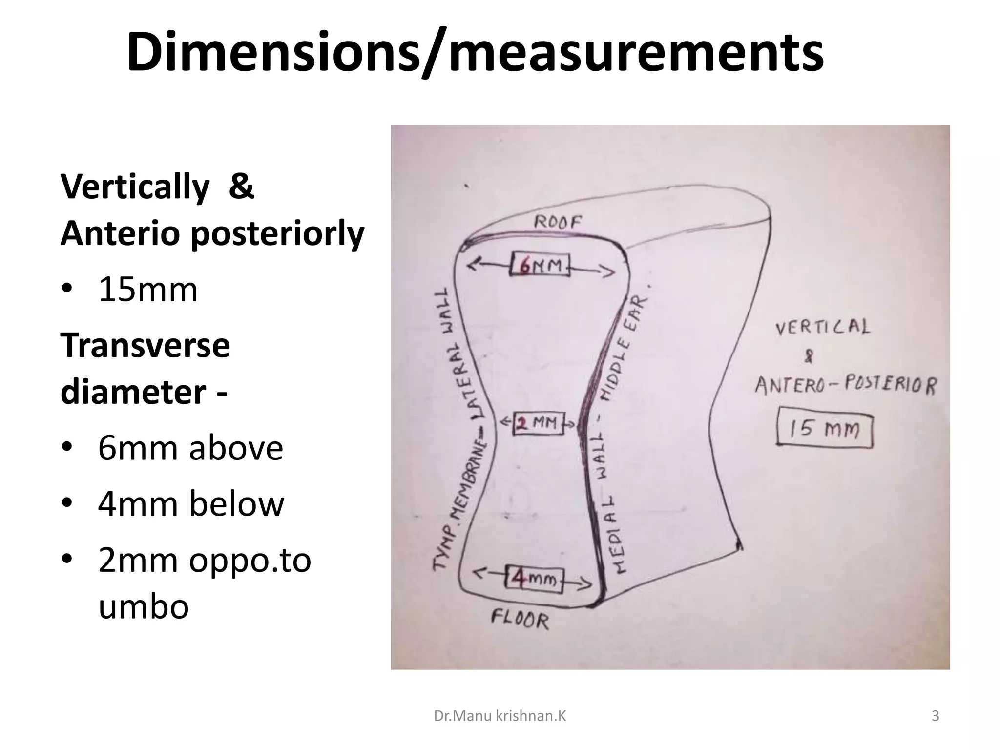
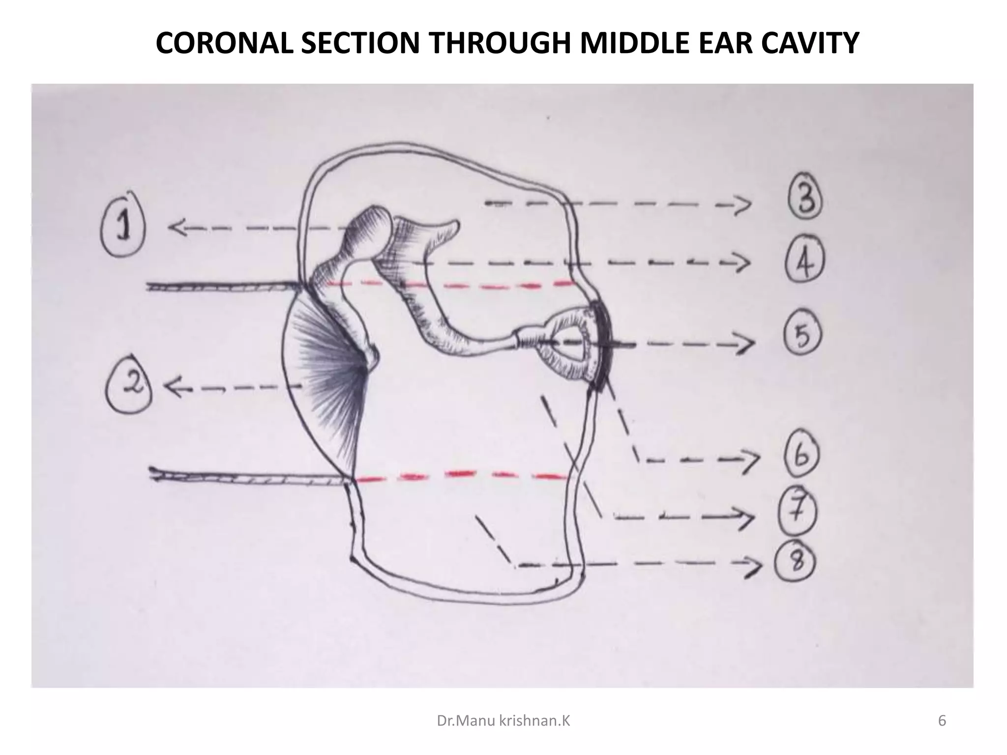
Middle ear
Dr. Manu krishnan.K
Tympanic cavity/tympanum
• Narrow air filled space situated in the petrous
part of the temporal bone b/w external ear
& internal ear.
• Resembles a six sided box ( like a match box)
Dr.Manu krishnan.K 2
Dimensions/measurements
Vertically &
Anterio posteriorly
• 15mm
Transverse
diameter -
• 6mm above
• 4mm below
• 2mm oppo.to
umbo
Dr.Manu krishnan.K 3
PARTS
• Tympanic cavity proper – opposite to the
Tympanic membrane
• Epitympanic recess (attic)– above the level of
Tympanic membrane(TM)
Dr.Manu krishnan.K 4
Middle ear cavity is further divided :
Portion of middle ear lies
1. Epitympanum-above the level of TM
2. Mesotympanum –at the level opposite to TM
3. Hypotympanum-below the level of TM
4. Protympanum- around the eustachian tube
opening
Dr.Manu krishnan.K 5
Dr.Manu krishnan.K 6
CORONAL SECTION THROUGH MIDDLE EAR CAVITY
PARTS
1. MALLEUS
2. TYMPANIC MEMBRANE
3. EPITYMPANUM
4. INCUS
5. STAPES
6. OVAL WINDOW
7. MESOTYMPANUM
8. HYPOTYMPANUM
Dr.Manu krishnan.K 7
COMMUNICATIONS & CONTENTS
COMMUNICATIONS
• Anteriorly-nasopharynx with eustachian tube
• Posteriorly –mastoid antrum &mastoid air cells
CONTENTS
• 3 small bones-Malleus, Incus, Stapes(ear ossicles)
• Ligment of ear ossicles
• Tensor tympani & stapedius muscle
• Vessels supplying & draining the middle ear
• Nerves –Chorda tympani & tympanic plexus
Dr.Manu krishnan.K 8
WALLS OF MIDDLE EAR CAVITY
Six walls:
1. Roof/tegmental
wall
2. Floor/jugular wall
3. Anterior
wall/carotid wall
4. Posterior/mastoid
wall
5. Lateral/membran
ous wall
6. Medial/labrynthi
ne wall
Dr.Manu krishnan.K 9
The Roof/ Tegmental wall
• Separates the middle ear from the middle cranial
fossa
• Formed by thin plate of bone called Tegmen
tympani.
• Plate is prolonged backwards as the roof of the
canal for the tensor tympani.
• Pierced by greater and lesser petrosal nerves.
Dr.Manu krishnan.K 10
Dr.Manu krishnan.K 11
Floor / jugular Wall
• Formed by thin plate of bone
• separates middle ear from the superior bulb
of the internal jugular vein.
• Aperture for Jacobson nerve
Dr.Manu krishnan.K 12
Dr.Manu krishnan.K 13
Anterior / carotid Wall
• Narrow ,separates middle ear from internal
carotid artery.
• Upper part - the opening of the canal for the
tensor tympani and opening of auditory tube
• lower part- superior & Inferior carotico
tympanic vessels &nerves
Dr.Manu krishnan.K 14
Posterior/mastoid wall
• Superiorly-aditus through which epitympanic
recess communicate with the mastoid antrum.
• Fossa incudis-depression which lodges the short
process of the incus
• Pyramid-a conical projection ,opening at the apex
for the passage of tendon of stapedius muscle
• Posterior canaliculus-for the chorda tympani
Dr.Manu krishnan.K 15
Dr.Manu krishnan.K 16
Lateral / Membranous wall
Separates middle ear from External auditory
meatus
Formed by
• Tympanic membrane
• Tympanic ring & sulcus
• Partially by squamous temporal bone
Apertures
• Petro tympanic fissure-upper end of the bony rim
• Anterior canaliculus-for chorda tymapani nerve
Dr.Manu krishnan.K 17
Dr.Manu krishnan.K 18
Medial wall / labryinthine wall
Separates middle ear from inner ear
Promontory –rounded bulging produced by basal
turn of cochlea
Fenestra vestibuli (Oval window)
-Oval opening posterio superior to promontory
-Measures 3.25x1.75mm
Fenestra cochlea(round window)
- Lies postero inferior to promontory
-Opens into floor scala tympani
-Measures 1.5x1.3mm
Dr.Manu krishnan.K 19
Dr.Manu krishnan.K 20
Blood supply
Arterial supply
• Anterior tympanic branch of maxillary artery
• Super.Tympanic branch of middle meningeal
artery
• branch of internal carotid artery
• Petrosal branch of middle meningeal artery.
Venous drainage
• Superior petrosal sinus & pterigoid plexus
Dr.Manu krishnan.K 21
Nerve supply
• Tympanic branch of glossopharyngeal nerve
• Superior & inferior Carotico tympanic nerves
Dr.Manu krishnan.K 22

Middle ear cavity(with handmade illustrations)

  • 1.
  • 2.
    Tympanic cavity/tympanum • Narrowair filled space situated in the petrous part of the temporal bone b/w external ear & internal ear. • Resembles a six sided box ( like a match box) Dr.Manu krishnan.K 2
  • 3.
    Dimensions/measurements Vertically & Anterio posteriorly •15mm Transverse diameter - • 6mm above • 4mm below • 2mm oppo.to umbo Dr.Manu krishnan.K 3
  • 4.
    PARTS • Tympanic cavityproper – opposite to the Tympanic membrane • Epitympanic recess (attic)– above the level of Tympanic membrane(TM) Dr.Manu krishnan.K 4
  • 5.
    Middle ear cavityis further divided : Portion of middle ear lies 1. Epitympanum-above the level of TM 2. Mesotympanum –at the level opposite to TM 3. Hypotympanum-below the level of TM 4. Protympanum- around the eustachian tube opening Dr.Manu krishnan.K 5
  • 6.
    Dr.Manu krishnan.K 6 CORONALSECTION THROUGH MIDDLE EAR CAVITY
  • 7.
    PARTS 1. MALLEUS 2. TYMPANICMEMBRANE 3. EPITYMPANUM 4. INCUS 5. STAPES 6. OVAL WINDOW 7. MESOTYMPANUM 8. HYPOTYMPANUM Dr.Manu krishnan.K 7
  • 8.
    COMMUNICATIONS & CONTENTS COMMUNICATIONS •Anteriorly-nasopharynx with eustachian tube • Posteriorly –mastoid antrum &mastoid air cells CONTENTS • 3 small bones-Malleus, Incus, Stapes(ear ossicles) • Ligment of ear ossicles • Tensor tympani & stapedius muscle • Vessels supplying & draining the middle ear • Nerves –Chorda tympani & tympanic plexus Dr.Manu krishnan.K 8
  • 9.
    WALLS OF MIDDLEEAR CAVITY Six walls: 1. Roof/tegmental wall 2. Floor/jugular wall 3. Anterior wall/carotid wall 4. Posterior/mastoid wall 5. Lateral/membran ous wall 6. Medial/labrynthi ne wall Dr.Manu krishnan.K 9
  • 10.
    The Roof/ Tegmentalwall • Separates the middle ear from the middle cranial fossa • Formed by thin plate of bone called Tegmen tympani. • Plate is prolonged backwards as the roof of the canal for the tensor tympani. • Pierced by greater and lesser petrosal nerves. Dr.Manu krishnan.K 10
  • 11.
  • 12.
    Floor / jugularWall • Formed by thin plate of bone • separates middle ear from the superior bulb of the internal jugular vein. • Aperture for Jacobson nerve Dr.Manu krishnan.K 12
  • 13.
  • 14.
    Anterior / carotidWall • Narrow ,separates middle ear from internal carotid artery. • Upper part - the opening of the canal for the tensor tympani and opening of auditory tube • lower part- superior & Inferior carotico tympanic vessels &nerves Dr.Manu krishnan.K 14
  • 15.
    Posterior/mastoid wall • Superiorly-aditusthrough which epitympanic recess communicate with the mastoid antrum. • Fossa incudis-depression which lodges the short process of the incus • Pyramid-a conical projection ,opening at the apex for the passage of tendon of stapedius muscle • Posterior canaliculus-for the chorda tympani Dr.Manu krishnan.K 15
  • 16.
  • 17.
    Lateral / Membranouswall Separates middle ear from External auditory meatus Formed by • Tympanic membrane • Tympanic ring & sulcus • Partially by squamous temporal bone Apertures • Petro tympanic fissure-upper end of the bony rim • Anterior canaliculus-for chorda tymapani nerve Dr.Manu krishnan.K 17
  • 18.
  • 19.
    Medial wall /labryinthine wall Separates middle ear from inner ear Promontory –rounded bulging produced by basal turn of cochlea Fenestra vestibuli (Oval window) -Oval opening posterio superior to promontory -Measures 3.25x1.75mm Fenestra cochlea(round window) - Lies postero inferior to promontory -Opens into floor scala tympani -Measures 1.5x1.3mm Dr.Manu krishnan.K 19
  • 20.
  • 21.
    Blood supply Arterial supply •Anterior tympanic branch of maxillary artery • Super.Tympanic branch of middle meningeal artery • branch of internal carotid artery • Petrosal branch of middle meningeal artery. Venous drainage • Superior petrosal sinus & pterigoid plexus Dr.Manu krishnan.K 21
  • 22.
    Nerve supply • Tympanicbranch of glossopharyngeal nerve • Superior & inferior Carotico tympanic nerves Dr.Manu krishnan.K 22