The transmission electron microscope is a very powerful tool for material science. A high energy beam of electrons is shone through a very thin sample, and the interactions between the electrons and the atoms can be used to observe features such as the crystal structure and features in the structure like dislocations and grain boundaries. Chemical analysis can also be performed. TEM can be used to study the growth of layers, their composition and defects in semiconductors. High resolution can be used to analyze the quality, shape, size and density of quantum wells, wires and dots.
In the late 16th century several Dutch lens makers designed devices that magnified objects, but in 1609 Galileo Galilei perfected the first device known as a microscope. Dutch spectacle makers Zaccharias Janssen and Hans Lipperhey are noted as the first men to develop the concept of the compound microscope.
Transmission electron microscope, high resolution tem and selected area elect...Nano Encryption
The transmission electron microscope is a very powerful tool for material science. A high energy beam of electrons is shone through a very thin sample, and the interactions between the electrons and the atoms can be used to observe features such as the crystal structure and features in the structure like dislocations and grain boundaries. Chemical analysis can also be performed. TEM can be used to study the growth of layers, their composition and defects in semiconductors. High resolution can be used to analyze the quality, shape, size and density of quantum wells, wires and dots.
I am John G. I am a Chemistry Assignment Expert at eduassignmenthelp.com. I hold a Ph.D. in Chemistry, from London, UK. I have been helping students with their homework for the past 6 years. I solve assignments related to Chemistry.
Visit eduassignmenthelp.com or email info@eduassignmenthelp.com.
You can also call on +1 678 648 4277 for any assistance with Chemistry Assignments.
Poster of my master\'s research presented at the Physics@FOM conference at Veldhoven on 20 januari 2010. There\'s one error in the equations, can you find it?
Earliest Galaxies in the JADES Origins Field: Luminosity Function and Cosmic ...Sérgio Sacani
We characterize the earliest galaxy population in the JADES Origins Field (JOF), the deepest
imaging field observed with JWST. We make use of the ancillary Hubble optical images (5 filters
spanning 0.4−0.9µm) and novel JWST images with 14 filters spanning 0.8−5µm, including 7 mediumband filters, and reaching total exposure times of up to 46 hours per filter. We combine all our data
at > 2.3µm to construct an ultradeep image, reaching as deep as ≈ 31.4 AB mag in the stack and
30.3-31.0 AB mag (5σ, r = 0.1” circular aperture) in individual filters. We measure photometric
redshifts and use robust selection criteria to identify a sample of eight galaxy candidates at redshifts
z = 11.5 − 15. These objects show compact half-light radii of R1/2 ∼ 50 − 200pc, stellar masses of
M⋆ ∼ 107−108M⊙, and star-formation rates of SFR ∼ 0.1−1 M⊙ yr−1
. Our search finds no candidates
at 15 < z < 20, placing upper limits at these redshifts. We develop a forward modeling approach to
infer the properties of the evolving luminosity function without binning in redshift or luminosity that
marginalizes over the photometric redshift uncertainty of our candidate galaxies and incorporates the
impact of non-detections. We find a z = 12 luminosity function in good agreement with prior results,
and that the luminosity function normalization and UV luminosity density decline by a factor of ∼ 2.5
from z = 12 to z = 14. We discuss the possible implications of our results in the context of theoretical
models for evolution of the dark matter halo mass function.
Toxic effects of heavy metals : Lead and Arsenicsanjana502982
Heavy metals are naturally occuring metallic chemical elements that have relatively high density, and are toxic at even low concentrations. All toxic metals are termed as heavy metals irrespective of their atomic mass and density, eg. arsenic, lead, mercury, cadmium, thallium, chromium, etc.
ESR spectroscopy in liquid food and beverages.pptxPRIYANKA PATEL
With increasing population, people need to rely on packaged food stuffs. Packaging of food materials requires the preservation of food. There are various methods for the treatment of food to preserve them and irradiation treatment of food is one of them. It is the most common and the most harmless method for the food preservation as it does not alter the necessary micronutrients of food materials. Although irradiated food doesn’t cause any harm to the human health but still the quality assessment of food is required to provide consumers with necessary information about the food. ESR spectroscopy is the most sophisticated way to investigate the quality of the food and the free radicals induced during the processing of the food. ESR spin trapping technique is useful for the detection of highly unstable radicals in the food. The antioxidant capability of liquid food and beverages in mainly performed by spin trapping technique.
ANAMOLOUS SECONDARY GROWTH IN DICOT ROOTS.pptxRASHMI M G
Abnormal or anomalous secondary growth in plants. It defines secondary growth as an increase in plant girth due to vascular cambium or cork cambium. Anomalous secondary growth does not follow the normal pattern of a single vascular cambium producing xylem internally and phloem externally.
This presentation explores a brief idea about the structural and functional attributes of nucleotides, the structure and function of genetic materials along with the impact of UV rays and pH upon them.
Nutraceutical market, scope and growth: Herbal drug technologyLokesh Patil
As consumer awareness of health and wellness rises, the nutraceutical market—which includes goods like functional meals, drinks, and dietary supplements that provide health advantages beyond basic nutrition—is growing significantly. As healthcare expenses rise, the population ages, and people want natural and preventative health solutions more and more, this industry is increasing quickly. Further driving market expansion are product formulation innovations and the use of cutting-edge technology for customized nutrition. With its worldwide reach, the nutraceutical industry is expected to keep growing and provide significant chances for research and investment in a number of categories, including vitamins, minerals, probiotics, and herbal supplements.
The ability to recreate computational results with minimal effort and actionable metrics provides a solid foundation for scientific research and software development. When people can replicate an analysis at the touch of a button using open-source software, open data, and methods to assess and compare proposals, it significantly eases verification of results, engagement with a diverse range of contributors, and progress. However, we have yet to fully achieve this; there are still many sociotechnical frictions.
Inspired by David Donoho's vision, this talk aims to revisit the three crucial pillars of frictionless reproducibility (data sharing, code sharing, and competitive challenges) with the perspective of deep software variability.
Our observation is that multiple layers — hardware, operating systems, third-party libraries, software versions, input data, compile-time options, and parameters — are subject to variability that exacerbates frictions but is also essential for achieving robust, generalizable results and fostering innovation. I will first review the literature, providing evidence of how the complex variability interactions across these layers affect qualitative and quantitative software properties, thereby complicating the reproduction and replication of scientific studies in various fields.
I will then present some software engineering and AI techniques that can support the strategic exploration of variability spaces. These include the use of abstractions and models (e.g., feature models), sampling strategies (e.g., uniform, random), cost-effective measurements (e.g., incremental build of software configurations), and dimensionality reduction methods (e.g., transfer learning, feature selection, software debloating).
I will finally argue that deep variability is both the problem and solution of frictionless reproducibility, calling the software science community to develop new methods and tools to manage variability and foster reproducibility in software systems.
Exposé invité Journées Nationales du GDR GPL 2024
DERIVATION OF MODIFIED BERNOULLI EQUATION WITH VISCOUS EFFECTS AND TERMINAL V...Wasswaderrick3
In this book, we use conservation of energy techniques on a fluid element to derive the Modified Bernoulli equation of flow with viscous or friction effects. We derive the general equation of flow/ velocity and then from this we derive the Pouiselle flow equation, the transition flow equation and the turbulent flow equation. In the situations where there are no viscous effects , the equation reduces to the Bernoulli equation. From experimental results, we are able to include other terms in the Bernoulli equation. We also look at cases where pressure gradients exist. We use the Modified Bernoulli equation to derive equations of flow rate for pipes of different cross sectional areas connected together. We also extend our techniques of energy conservation to a sphere falling in a viscous medium under the effect of gravity. We demonstrate Stokes equation of terminal velocity and turbulent flow equation. We look at a way of calculating the time taken for a body to fall in a viscous medium. We also look at the general equation of terminal velocity.
Lateral Ventricles.pdf very easy good diagrams comprehensive
Microscopy_&_Resolution.ppt
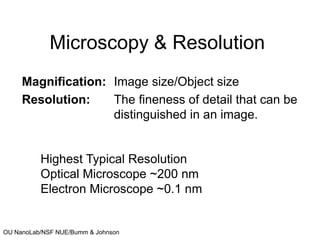
1. OU NanoLab/NSF NUE/Bumm & Johnson
Microscopy & Resolution
Magnification: Image size/Object size
Resolution: The fineness of detail that can be
distinguished in an image.
Highest Typical Resolution
Optical Microscope ~200 nm
Electron Microscope ~0.1 nm
2. OU NanoLab/NSF NUE/Bumm & Johnson
Definitions
• Acceptance angle θ
• Numerical Aperture
NA = n sinθ
• Rayleigh resolution criterion for a circular
aperture Δx = 0.61 λ/NA
θ
3. OU NanoLab/NSF NUE/Bumm & Johnson
OPTICAL MICROSCOPES
Image construction for a simple biconvex lens
4. OU NanoLab/NSF NUE/Bumm & Johnson
Rayleigh criterion for resolution
www.microscopy.fsu.edu ; www.imb-jena.de
See more interactive tutorials at www.microscopy.fsu.edu
Numerical Aperature Resolution Rayleigh Criterion
5. OU NanoLab/NSF NUE/Bumm & Johnson
Comparison
Bright-
Field
Dark-
Field
• Full
aperture
is
illuminated
• A central
obstruction blocks
the central cone.
6. OU NanoLab/NSF NUE/Bumm & Johnson www.microscopy.fsu.edu
Dark-Field
Optical Microscopy
•A central obstruction
blocks the central cone.
•The sample is only
illuminated by the
marginal rays.
•These marginal rays must
be at angles too large for
the objective lens to
collect.
•Only light scattered by the
object is collected by the
lens.
8. OU NanoLab/NSF NUE/Bumm & Johnson
THE ELECTRON MICROSCOPE
The wavelength of the electron can be tuned by changing the
accelerating voltage.
de Broglie : λ = h/mv
λ: wavelength associated with the particle
h: Plank’s constant 6.63×10⁻³⁴ Js;
mv: momentum of the particle
me= 9.1×10⁻³¹ kg; e = 1.6×10⁻¹⁹ coulomb
P.E eV = ½mv2 λ = h/(2meV) = 12.3/V (for V in KV, λ in Å)
V of 60 kV, λ = 0.05 Å Δx ~ 2.5 Å
Microscopes using electrons as illuminating radiation
TEM & SEM
10. OU NanoLab/NSF NUE/Bumm & Johnson
Components of the TEM
1. Electron Gun: Filament, Anode/Cathode
2. Condenser lens system and its apertures
3. Specimen chamber
4. Objective lens and apertures
5. Projective lens system and apertures
6. Correctional facilities (Chromatic, Spherical, Astigmatism)
7. Desk consol with CRTs and camera
Transformers: 20-100 kV; Vacuum pumps: 10-6 – 10-10 Torr
11. OU NanoLab/NSF NUE/Bumm & Johnson
Schematic of E Gun & EM lens
Magnification: 10,000 – 100,000; Resolution: 1 - 0.2 nm
www.udel.edu
12. OU NanoLab/NSF NUE/Bumm & Johnson
TEM IMAGES
www.udel.edu ; www.nano-lab. com ; www.thermo.com