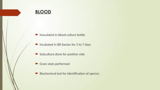
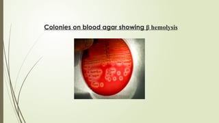
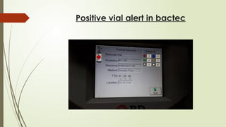
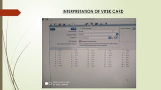
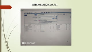
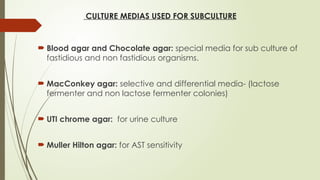
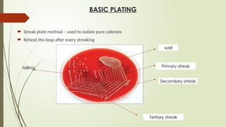
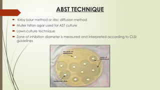
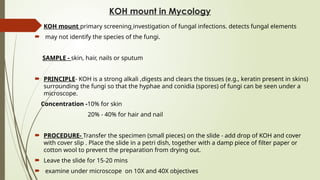
MICROBIOLOGY POSTING
26th
April – 1st
May
OBJECTIVES
Blood, Urine, Pus, CSF collection for culture and processing of specimen
Basic plating and ABST techniques.
Bactec and Vitec
KOH mount in Mycology
ZN staining and processing of sputum samples
Molecular techniques in infectious disease
BLOOD
 Inoculated in blood culture bottle
 Incubated in BD bactec for 5 to 7 days
 Subculture done for positive vials
 Gram stain performed
 Biochemical test for identification of species
Colonies on blood agar showing β hemolysis
URINE
 Gross- colour, amount
 Wet mount –pus cell ,organism
 Culture plating on UTI chrome AGAR
 Growth on whole culture plate - >105
cfu/ml- significant
Colonies on UTI chrome agar
Ecoli(purple)
Klebsiella(bluish green)
Pus
 Appearance, amount and site of the sample
 Gram stain
 Cultured on blood and MacConkey agar
 Incubate for 48 hrs
 If growth present, various biochemical test done for identification of species
like coagulase test , catalase test
IMVIC for Enterobacteriaceae
 Antibiotic sensitivity test done
CSF
 gross
 wet mount
 india ink – cryptococcus
 culture 48 hrs to see growth
 subculture if growth is present –for identification of organism
STOOL
 gross
 microscopy- to detect trophozoites , cyst and eggs of helminthes
- wet mount-motility of trophozoites
- iodine mount- cyst-E.coli, E.histolytica, G. lamblia
Gram staining
 to differentiate between gram positive (purple) and gram negative bacteria(red).
 Principle: the differences in cell wall composition of gram-positive and gram-negative bacteria
 gram-positive cell wall contains a thick layer of peptidoglycan with numerous teichoic acid cross-linking which
resists the decolorization where
 gram negative bacteria having double layer thin cell wall loses the primary stain on decolourization ,hence takes
the secondary stain.
 Reagents: Crystal violet( primary stain )
Grams iodine( mordant )
alcohol or acetone alcohol( decolourizing agent )
saffranin ( Secondary stain )
INTERPRETATION OF GRAMS STAIN
GRAM POSITIVE COCCI GRAM NEGATIVE COCCI
Bactec
 Automated blood culture systems
 based on fluorescent technology in detecting the growth of organisms in the
blood culture bottles.
 detects increase CO2 concentration in culture
 Positive vials will be determined by the BD BACTEC
 Subcultured on blood , MacConkey and chocolate agar
 colony morphology identified and further ABS Test done.
anaerobic
culture vial
Aerobic
culture vial
Positive vial alert in bactec
Vitek
 It is an automated microorganism identification and antimicrobial susceptibility system
 It uses a colorimetric methodology to determine individual biochemical reactions for organism identification and a
turbidimetric method for susceptibility testing using a 64 well barcoded card.
• The ID card(reagent card) has 64 wells, each containing individual test substrates.
• Substrates measure metabolic activities such as acidification, alkalinisation, enzyme hydrolysis and growth in
presence of inhibitory substances
• These cards have barcodes that contain information on product type, expiry date and a unique identifier that can be
linked to the sample.
• Each card also has a pre-inserted transfer tube for inoculation of the microorganisms from the test suspension tube
Vitek card
VITEK2
INTERPRETATION OF VITEK CARD
INTERPRETATION OF AST
CULTURE MEDIAS USED FOR SUBCULTURE
 Blood agar and Chocolate agar: special media for sub culture of
fastidious and non fastidious organisms.
 MacConkey agar: selective and differential media- (lactose
fermenter and non lactose fermenter colonies)
 UTI chrome agar: for urine culture
 Muller Hilton agar: for AST sensitivity
BASIC PLATING
 Streak plate method – used to isolate pure colonies
 Reheat the loop after every streaking
well
Primary streak
Secondary streak
Tertiary streak
tailing
ABST TECHNIQUE
 Kirby baur method or disc diffusion method
 Muller hilton agar used for AST culture
 Lawn culture technique
 Zone of inhibition diameter is measured and interpreted according to CLSI
guidelines
KOH mount in Mycology
 KOH mount primary screening investigation of fungal infections. detects fungal elements
 may not identify the species of the fungi.
SAMPLE - skin, hair, nails or sputum
 PRINCIPLE- KOH is a strong alkali ,digests and clears the tissues (e.g., keratin present in skins)
surrounding the fungi so that the hyphae and conidia (spores) of fungi can be seen under a
microscope.
Concentration -10% for skin
20% - 40% for hair and nail
 PROCEDURE- Transfer the specimen (small pieces) on the slide - add drop of KOH and cover
with cover slip . Place the slide in a petri dish, together with a damp piece of filter paper or
cotton wool to prevent the preparation from drying out.
 Leave the slide for 15-20 mins
 examine under microscope on 10X and 40X objectives
 INTERPRETATION - Translucent hyaline hyphae are oserved , branching and
septation are seen
Hyalin hyphae
MYCOLOGY CULTURE
 Done on SDA (Sabourd Dextrose Agar)
Additives
 Gentamicin –to inhibit growth of bacteria( blue)
 Cyclohexamide-to inhibit growth of commensal fungal(red)
 Observation- culture tube is examined as -
obverse-(front) -for morphology of colony
reverse-(back) - for pigment
Ziehl Neelsen staining
 PRINCIPLE- The procedure is used to stain acid fast bacilli, mycobacterium tuberculosis and
mycobacterium leprae. These bacteria stain with carbol fuschin, which is a red dye. They retain
the dye when treated with acid, which is because of the presence of mycolic acid in their cell wall.
 REAGENTS
1. Carbol fuschin (basic dye)
2. Mordant (heat)
3. 20% sulphuric acid (decolorizer)
4. Methylene blue (counter stain) or Malachite green
 RESULT
 Acid fast bacilli stain pink, straight or slightly curved rods, may have beaded appearance. The
background appears blue due to methylene blue.
INTERPRETATIONS
 If definite bacilli are seen, reported as AFB positive.
• > 10 AFB/high power field –>+++
• 1-10 AFB/high power field –> ++
• 10-100 AFB/100 high power fields –> +
• 1-9 AFB/100 high power fields –> exact number has to be reported
if no AFB is seen, result reported as ‘no AFB seen’ and not negative.
• Share
ZN STAINING SHOWING ACID FAST BACILLI
Molecular techniques in infectious disease
LINE PROBE ASSAY:
 rapid drug susceptibility test
 Identifies drug resistant M. tuberculosis
 Resistant to rifampicin and isoniazid
 Can be used directly on sputum
 Done for smear positive samples
 Results are available within 1 to 2 days
OTHER MOLECULAR TECHNIQUES
 PCR - being used for DNA viruses like hepatitis B, CMV
 RT PCR - currently being used for COVID19
THANK YOU

MICROBIOLOGY PRESENTATION.laboratorypptx

  • 1.
  • 2.
    OBJECTIVES Blood, Urine, Pus,CSF collection for culture and processing of specimen Basic plating and ABST techniques. Bactec and Vitec KOH mount in Mycology ZN staining and processing of sputum samples Molecular techniques in infectious disease
  • 3.
    BLOOD  Inoculated inblood culture bottle  Incubated in BD bactec for 5 to 7 days  Subculture done for positive vials  Gram stain performed  Biochemical test for identification of species
  • 4.
    Colonies on bloodagar showing β hemolysis
  • 5.
    URINE  Gross- colour,amount  Wet mount –pus cell ,organism  Culture plating on UTI chrome AGAR  Growth on whole culture plate - >105 cfu/ml- significant
  • 6.
    Colonies on UTIchrome agar Ecoli(purple) Klebsiella(bluish green)
  • 7.
    Pus  Appearance, amountand site of the sample  Gram stain  Cultured on blood and MacConkey agar  Incubate for 48 hrs  If growth present, various biochemical test done for identification of species like coagulase test , catalase test IMVIC for Enterobacteriaceae  Antibiotic sensitivity test done
  • 8.
    CSF  gross  wetmount  india ink – cryptococcus  culture 48 hrs to see growth  subculture if growth is present –for identification of organism
  • 9.
    STOOL  gross  microscopy-to detect trophozoites , cyst and eggs of helminthes - wet mount-motility of trophozoites - iodine mount- cyst-E.coli, E.histolytica, G. lamblia
  • 10.
    Gram staining  todifferentiate between gram positive (purple) and gram negative bacteria(red).  Principle: the differences in cell wall composition of gram-positive and gram-negative bacteria  gram-positive cell wall contains a thick layer of peptidoglycan with numerous teichoic acid cross-linking which resists the decolorization where  gram negative bacteria having double layer thin cell wall loses the primary stain on decolourization ,hence takes the secondary stain.  Reagents: Crystal violet( primary stain ) Grams iodine( mordant ) alcohol or acetone alcohol( decolourizing agent ) saffranin ( Secondary stain )
  • 11.
    INTERPRETATION OF GRAMSSTAIN GRAM POSITIVE COCCI GRAM NEGATIVE COCCI
  • 12.
    Bactec  Automated bloodculture systems  based on fluorescent technology in detecting the growth of organisms in the blood culture bottles.  detects increase CO2 concentration in culture  Positive vials will be determined by the BD BACTEC  Subcultured on blood , MacConkey and chocolate agar  colony morphology identified and further ABS Test done.
  • 13.
  • 14.
  • 15.
    Vitek  It isan automated microorganism identification and antimicrobial susceptibility system  It uses a colorimetric methodology to determine individual biochemical reactions for organism identification and a turbidimetric method for susceptibility testing using a 64 well barcoded card. • The ID card(reagent card) has 64 wells, each containing individual test substrates. • Substrates measure metabolic activities such as acidification, alkalinisation, enzyme hydrolysis and growth in presence of inhibitory substances • These cards have barcodes that contain information on product type, expiry date and a unique identifier that can be linked to the sample. • Each card also has a pre-inserted transfer tube for inoculation of the microorganisms from the test suspension tube
  • 16.
  • 17.
  • 18.
  • 19.
  • 20.
    CULTURE MEDIAS USEDFOR SUBCULTURE  Blood agar and Chocolate agar: special media for sub culture of fastidious and non fastidious organisms.  MacConkey agar: selective and differential media- (lactose fermenter and non lactose fermenter colonies)  UTI chrome agar: for urine culture  Muller Hilton agar: for AST sensitivity
  • 21.
    BASIC PLATING  Streakplate method – used to isolate pure colonies  Reheat the loop after every streaking well Primary streak Secondary streak Tertiary streak tailing
  • 22.
    ABST TECHNIQUE  Kirbybaur method or disc diffusion method  Muller hilton agar used for AST culture  Lawn culture technique  Zone of inhibition diameter is measured and interpreted according to CLSI guidelines
  • 23.
    KOH mount inMycology  KOH mount primary screening investigation of fungal infections. detects fungal elements  may not identify the species of the fungi. SAMPLE - skin, hair, nails or sputum  PRINCIPLE- KOH is a strong alkali ,digests and clears the tissues (e.g., keratin present in skins) surrounding the fungi so that the hyphae and conidia (spores) of fungi can be seen under a microscope. Concentration -10% for skin 20% - 40% for hair and nail  PROCEDURE- Transfer the specimen (small pieces) on the slide - add drop of KOH and cover with cover slip . Place the slide in a petri dish, together with a damp piece of filter paper or cotton wool to prevent the preparation from drying out.  Leave the slide for 15-20 mins  examine under microscope on 10X and 40X objectives
  • 24.
     INTERPRETATION -Translucent hyaline hyphae are oserved , branching and septation are seen Hyalin hyphae
  • 25.
    MYCOLOGY CULTURE  Doneon SDA (Sabourd Dextrose Agar) Additives  Gentamicin –to inhibit growth of bacteria( blue)  Cyclohexamide-to inhibit growth of commensal fungal(red)  Observation- culture tube is examined as - obverse-(front) -for morphology of colony reverse-(back) - for pigment
  • 26.
    Ziehl Neelsen staining PRINCIPLE- The procedure is used to stain acid fast bacilli, mycobacterium tuberculosis and mycobacterium leprae. These bacteria stain with carbol fuschin, which is a red dye. They retain the dye when treated with acid, which is because of the presence of mycolic acid in their cell wall.  REAGENTS 1. Carbol fuschin (basic dye) 2. Mordant (heat) 3. 20% sulphuric acid (decolorizer) 4. Methylene blue (counter stain) or Malachite green  RESULT  Acid fast bacilli stain pink, straight or slightly curved rods, may have beaded appearance. The background appears blue due to methylene blue.
  • 27.
    INTERPRETATIONS  If definitebacilli are seen, reported as AFB positive. • > 10 AFB/high power field –>+++ • 1-10 AFB/high power field –> ++ • 10-100 AFB/100 high power fields –> + • 1-9 AFB/100 high power fields –> exact number has to be reported if no AFB is seen, result reported as ‘no AFB seen’ and not negative. • Share
  • 28.
    ZN STAINING SHOWINGACID FAST BACILLI
  • 29.
    Molecular techniques ininfectious disease LINE PROBE ASSAY:  rapid drug susceptibility test  Identifies drug resistant M. tuberculosis  Resistant to rifampicin and isoniazid  Can be used directly on sputum  Done for smear positive samples  Results are available within 1 to 2 days
  • 30.
    OTHER MOLECULAR TECHNIQUES PCR - being used for DNA viruses like hepatitis B, CMV  RT PCR - currently being used for COVID19
  • 31.