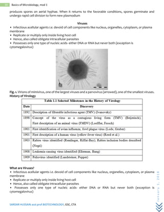
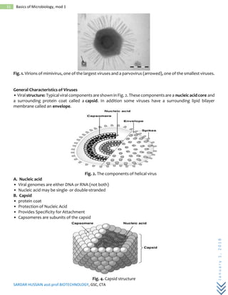
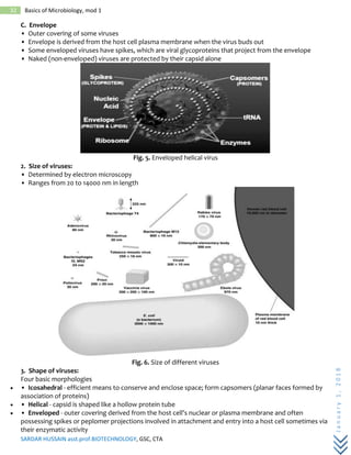
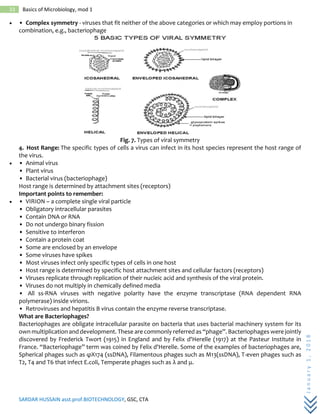
SlideShare a Scribd company logo
1 of 37
Download to read offline
SARDAR HUSSAIN asst.prof.BIOTECHNOLOGY, GSC, CTA
January1,2018
0 Basics of Microbiology, mod 1
1/1/2018 Basics of
Microbiology, mod
1
History and scope, Microbial world
classification, Taxonomy, General
features
By
SARDAR HUSSAIN
GSC, CTA
SARDAR HUSSAIN asst.prof.BIOTECHNOLOGY, GSC, CTA
January1,2018
1 Basics of Microbiology, mod 1
History and scope of Microbiology:
The environment:
• Microbes are responsible for the cycling of carbon, nitrogen phosphorus (geochemical cycles)
• Maintain ecological balance on earth
• They are found in association with plants in symbiotic relationships, maintain soil fertility and may
also be used to clean up the environment of toxic compounds (bio-remediation).
• Some are devasting plant pathogens, but others act as biological control agents against these
diseases.
Medicine:
• Disease causing ability of some microbes such as
• Small Pox (Variola virus)
• Cholera ( Vibrio cholera )
• Malaria ( Plasmodium , protozoa) etc.
• They have also provided us with the means of their control in the form of antibiotics and other
medically important drugs.
Food:
• Microorganisms have been used to produce food, from brewing and wine making, through cheese
production and bread making, to manufacture of soy sauce.
• Microbes are also responsible for food spoilage.
Biotechnology:
• Commercial applications include the synthesis of acetone, organic acids, enzymes, alcohols and
many drugs.
• Genetic engineering – bacteria can produce important therapeutic substances such as insulin,
human growth hormone, and interferon.
Research:
• Because of their simple structure they are easier to study most life processes in simple unicellular
organisms than in complex multicellular ones.
• Millions of copies of the same single cell can be produced in large numbers very quickly and at low
cost to give plenty of homogenous experimental material.
• Because they reproduce very quickly, they are useful for studies involving the transfer of genetic
information.
Brief history of microbiology
• Robert Hook (1665) – reported that life's smallest structural units were ‘little boxes' or ‘cells'. This
marked the beginning of cell theory – that all living things are composed of cells.
• Van Leuwenhoek (1673) – discovered the ‘invisible' world of microorganisms ‘animalcules'.
• Until second half of nineteenth century many believed that some forms of life could arise
spontaneously from non-living matter – spontaneous generation.
• Francesco Redi (1668) – Strong opponent of spontaneous generation. He demonstrated that
maggots appear on decaying meat only when flies are able to lay eggs on the meat.
• John Needham (1745) – claimed that microorganisms could arise spontaneously from heated
nutrient broth.
• Lazzaro Spallanzani (1765) – repeated Needhams experiments and suggested that Needham's
results were due to microorganisms in the air entering the broth.
• Rudolf Virchow (1858) – concept of biogenesis – living cells can arise only from preexisting cells.
• Louis Pasteur (1822-1895) – Pasteur's experiments on swan shaped necks resolved the controversy
of spontaneous generation. His discoveries led to the development of aseptic techniques used in the
laboratory and medical procedure to prevent contamination by microorganisms that are in the air.
Golden age of microbiology:
SARDAR HUSSAIN asst.prof.BIOTECHNOLOGY, GSC, CTA
January1,2018
2 Basics of Microbiology, mod 1
• Rapid advances in the science of microbiology were made between 1857 and 1914.
Fermentation and Pasteurization:
• Pasteur found that yeast ferments sugars to alcohols and that bacterium can oxidize the alcohol
to acetic acid.
• Heating processes called pasteurization is used to kill bacteria in some alcoholic beverages and
milk.
The Germ theory of disease:
• Agostino Bassi (1934) and Pasteur (1865) – showed a casual relationship between microorganisms
and disease.
• Joseph Lister (1860s) – introduced the use of disinfectant to clean surgical dressings in order to
control infection in humans
• Robert Koch (1876) – proved that microorganisms transmit disease – Koch's postulates which are
used today to prove that a particular microorganism causes a particular disease.
• Introduced pure cultures
• Koch's postulates (Henle-Koch's Postulates) are,
1. A specific organism should be found constantly in association with the disease.
2. The organism should be isolated and grown in a pure culture in the laboratory.
3. The pure culture when inoculated into a healthy susceptible animal should produce
symptoms/lesions of the same disease
4. From the inoculated animal, the microorganism should be isolated in pure culture.
5. An additional criterion introduced is that specific antibodies to the causative organism
shouldbe demonstrable in patient's serum.
• Angelina – American wife of Koch's assistant suggested solidifying broths with agar as an aid to
obtaining pure cultures.
• Koch also developed techniques for isolating organisms. Identified the bacillus that causes
tuberculosis and anthrax, developed tuberculin and studied various diseases in Africa and Asia. His
studies on Tuberculosis won him Nobel prize for philosophy and medicine in 1905.
Vaccination:
• Immunity is conferred by inoculation with a vaccine.
• Edward Jenner(1798 ) – demonstrated that inoculations with cowpox material provides humans
with immunity from small pox
• Pasteur (1880) – discovered that avirulent bacteria could be used as a vaccine for chicken cholera;
he coined the word vaccine
• Modern vaccines are prepared from living avirulent microorganisms or killed pathogens, from
isolated components of pathogens, and by recombinant DNA techniques.
Emergence of special fields of Microbiology:
Immunology:
• Immunization was first used against small pox. Edward Jenner used fluid from cowpox blisters to
immunize against it.
• Pasteur developed techniques to weaken organisms so they would produce immunity without
producing disease.
• Elie Metchnikoff discovered that certain cells in the body would ingest microbes and named them
as phagocytes.
Industrial Microbiology and Microbial ecology:
• Pasteur – fermentation technology and pasteurization. One of his most important discoveries was
that some fermentative microorganisms were anaerobic and others were able to live either aerobically or
anaerobically.
Microbial ecology – Two pioneers –
SARDAR HUSSAIN asst.prof.BIOTECHNOLOGY, GSC, CTA
January1,2018
3 Basics of Microbiology, mod 1
• Sergei N. Winogradsky (1856-1953) – Soil microbiology – discovered that soil bacteria could oxidize
iron, sulfur and ammonia to obtain energy and many bacteria incorporate CO2 into organic matter.
He also isolated anaerobic nitrogen fixing soil bacteria and studied the decomposition of cellulose.
• Martinus Beijerinck (1851-1931) – He isolated aerobic nitrogen fixing bacterium Azotobacte r, a
root nodule bacterium also capable of fixing nitrogen (later renamed as Rhizobium ); and sulfate
reducing bacteria. Both of them developed enrichment culture technique and use of selective media,
which have been of great importance in microbiology.
Virology:
• Beijerinck characterized viruses as pathogenic molecules that could take over a host cells
mechanisms for their own use
• Wendell Stanley (1935) – crystallized TMV and crystals consisted of protein and RNA.
• Viruses were first observed with an EM in 1939.
• Alfred Hershey and Martha Chase (1952) – demonstrated that the genetic material of some viruses
is DNA
• James Watson and Francis Crick (1953) -determined the structure of DNA
Chemotherapy:
• There are two types of chemotherapeutic agents: synthetic drugs and antibiotics.
• Elrlich (1910) introduced an arsenic containing chemical called Salvarsan to treat Syphilis.
• Alexander Fleming (1928) – observed that the mold Penicillium inhibited the growth of bacteria
and named the active ingredient as penicillin. Penicillin has been used clinically as an antibiotic since
the 1940s. Domagk and others developed sulfa drugs.
• Waksman and others developed Streptomycin and other antibiotics derived from soil organisms.
• Researchers are tackling the problem of drug-resistant microbes.
Genetics and Molecular Biology:
• 1900 – Modern genetics began with the rediscovery of Gregor Mendel's principles of genetics.
• Frederick Griffith (1928) - discovered that previously harmless bacteria could change their nature
and become capable of causing disease
• Avery, McCarty and MacLeod (1940's) – showed that this genetic change was due to DNA. After
this finding came the crucial discovery of the structure of DNA by Watson and Crick
• Edward Tatum and George Beadle – studied biochemical mutants of Neurospora to show how
genetic information controls metabolism.
• Barbara McClintock (1950) – discovered that some genes could move from one location to another
on a chromosome.
• Early 1960's witnessed a further explosion of discoveries relating to the way DNA controls protein
synthesis.
• Francois Jacob and Jacques Monod (1961) – discovered mRNA and later made the first major
discoveries about regulation of gene function in bacteria.
• Microorganisms can now be genetically engineered to manufacture large amounts of human
hormones and other urgently needed medical substances.
• Late 1960's Paul Berg showed that fragments of human or animal DNA that code for important
proteins can be attached to bacterial DNA. The resulting hybrid was the first example of recombinant
DNA.
Tomorrow's history:
Microbiology has been in the forefront of research in medicine and biology and continues to play a role in
Genetic engineering and Gene therapy.
Genetic engineering – scientists are attempting to redesign microorganisms for a variety of purposes
(drugs, hormones, vaccines and a variety of biologically important compounds)
rDNA technology – enabling us to produce improved varieties of plants and animals such as pest-resistant
crops and may even enable us to correct genetic defects in human beings.
SARDAR HUSSAIN asst.prof.BIOTECHNOLOGY, GSC, CTA
January1,2018
4 Basics of Microbiology, mod 1
Human genome project:
• Microbial genetic techniques have made possible a colossal scientific undertaking HGP. Begun in
1990 and supposed to complete by 2005 was completed in May 2000.
• Humans have just over 30,000 genes instead of estimates that ranged up to 142,000 genes. 3
billion base pairs in the human genome do not all code for useful genes (75% of them code for ‘junk
DNA')
• Over 100 microbial genomes have been sequenced so far.
• Approx. 113 genes have come to human genome directly from bacteria.
• Venter has sequenced mouse genome and reports that humans have only 300 genes not found
in the mouse.
Scope of microbiology
The microbiology has influence on genetics, agriculture, food science, ecology, immunology and various
fields.
 Genetics: Mainly involves engineered microbes to make hormones, vaccine, antibiotics and many
other useful products for human being.
 Agriculture: The influence of microbes on agriculture; the prevention of the diseases that mainly
damage the useful crops.
 Food science: It involves the prevention of spoilage of food and food borne diseases and the uses of
microbes to produce cheese, yoghurt, pickles and beer.
 Immunology: The study of immune system which protect the body from pathogens.
 Medicine: deals with the identification of plans and measures to cure diseases of human and animals
which are infectious to them.
 Industry: it involves use of microbes to produce antibiotics, steroids, alcohol, vitamins and amino
acids etc.
Two fundamentally different types of cells exist, Prokaryotic cells having a simpler morphology and lack a
true membrane de-limited nucleus. All bacteria are prokaryotic.
• Eukaryotic cells have a membrane-bound nucleus; are more complex morphologically and larger
than prokaryotes. Algae, fungi, protozoa, higher plants, and animals are eukaryotes
• For many years biologists have divided organisms into five kingdoms; Monera, Protists, Fungi,
Animalia and Plantae.
• In the last few decades great progress in three areas has been made that affect microbial
classification.
• First – detailed structure of microbial cells has been studied using EM
• Second – microbiologists have determined the biochemical and physiological characteristics of
many different microorganisms
• Third – sequences of nucleic acids and proteins from a wide variety of organisms have been
compared.
• It is now clear that there are two quite different groups of prokaryotic organisms; Bacteria and
Archaea.
• The differences between bacteria, archaea and eukaryotes seem so great that many
microbiologists proposed that organisms should be divided among three domains; Bacteria (the true
bacteria or eubacteria), Archaea and Eucarya (all eukaryotic organisms).
Members of microbial world
There are five major members of microorganisms, Archaea, Bacteria, Algae, Protozoa, and Fungi .The
Archaea and Bacteria are prokaryotic cells. Unicellular algae and protozoa and fungi are eukaryotic cells.
Archaea
SARDAR HUSSAIN asst.prof.BIOTECHNOLOGY, GSC, CTA
January1,2018
5 Basics of Microbiology, mod 1
The Archaea are a group of single-celled microorganisms. They have no cell nucleus or any other
membrane-bound organelles within their cells. Archaea and bacteria are quite similar in size and shape,
although a few archaea have very unusual shapes, such as the flat and square-shaped cells. Similarity to
bacteria, archaea possess genes and several metabolic pathways that are more closely related to those of
eukaryotes, notably the enzymes involved in transcription and translation. The archaea biochemistry are
unique, such as presence of ether lipids in their cell membranes. Archaea use a much greater variety of
sources of energy than eukaryotes: ranging from familiar organic compounds such as sugars, to ammonia,
metal ions or even hydrogen gas. Archaea reproduce asexually by binary fission, fragmentation, or budding;
unlike bacteria and eukaryotes, no known species form spores. Initially archaea were seen as extremophiles
that lived in harsh environments, such as hot springs and salt lakes, but they are now found in a broad range
of habitats, including soils, oceans, marshland. Archaea play roles in both the carbon cycle and the nitrogen
cycle. No archaea pathogens or parasites are known, but they are often mutualists or commensals.
Methanogens are used in biogas production and sewge treatmen, and enzymes from extremophile archaea
that can endure high temperatures and organic solvents are exploited in biotechnology.
Bacteria
Bacteria are a large domain of prokaryotic microorganisms . Bacteria are present in most habitats on
Earth , growing in soil, acidic hot springs , radioactive waste water, organic matter and live bodies of plants
and animals. Bacteria have many shapes and sizes. Bacterial cells are about one tenth the size of eukaryotic
cells and 0.5–5.0 micrometres in length. Most bacterial species are either spherical, called cocci or rod-
shaped, called bacilli . Some rod-shaped bacteria are slightly curved called vibrio or comma-shaped. Many
bacterial species exist as single cells and associate in characteristic patterns such as form pairs calleddiploids,
form chains, and group together in clusters. Bacteria can also be elongated to form filaments.
The bacterial cell is surrounded by cell membrane, which encloses the contents of the cell and acts
as a barrier to hold nutrients, proteins and other essential components of the cytoplasm within the cell. They
lack a true nucleus, mitochondria, chloroplasts, Golgi apparatus and endoplasmic reticulum. Most bacteria
do not have a membrane-bound nucleus, and their genetic material is typically a single circular chromosome
located in the cytoplasm in an irregularly shaped body called the nucleoid .The nucleoid contains the
chromosome with associated proteins and RNA. The bacteria contain ribosomes for the production of
proteinsbut different from those of eukaryotes and Archaea. Some bacteria produce intracellular nutrient
storage granules, such as glycogen, polyphosphate, sulfur or polyhydroxyalkanoates.These granules enable
bacteria to store compounds for later use. Certain bacterial species, such as the photosynthetic
Cyanobacteria produce internal gas vesicles which they use to regulate their buoyancy – allowing them to
move up or down into water layers with different light intensities and nutrient levels. The cell wall is present
on the outside of the cytoplasmic membrane. A common bacterial cell wall material is peptidoglycan which
is a polymer contains two sugar derivatives N-aetylglucosamine(NAG) and N-acetylmuramic acid (NAM)
joined by glycosidic bond. A peptide chain of four alternating D- and L-amino acids called tetrapeptide is
connected to the carboxyl group of the NAM. The amino acids present in the tetrapeptide include L-alanine,
D-alanine, D-glutamic acid, and either lysine or diaminopimilic acid (DAP).The carboxyl group of terminal D-
alanine is connected directly to amino group of DAD. The peptide interbridge connects the tetrapeptide
chains.
SARDAR HUSSAIN asst.prof.BIOTECHNOLOGY, GSC, CTA
January1,2018
6 Basics of Microbiology, mod 1
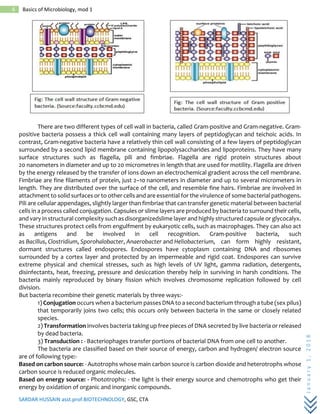
There are two different types of cell wall in bacteria, called Gram-positive and Gram-negative. Gram-
positive bacteria possess a thick cell wall containing many layers of peptidoglycan and teichoic acids. In
contrast, Gram-negative bacteria have a relatively thin cell wall consisting of a few layers of peptidoglycan
surrounded by a second lipid membrane containing lipopolysaccharides and lipoproteins. They have many
surface structures such as flagella, pili and fimbriae. Flagella are rigid protein structures about
20 nanometers in diameter and up to 20 micrometres in length that are used for motility. Flagella are driven
by the energy released by the transfer of ions down an electrochemical gradient across the cell membrane.
Fimbriae are fine filaments of protein, just 2–10 nanometers in diameter and up to several micrometers in
length. They are distributed over the surface of the cell, and resemble fine hairs. Fimbriae are involved in
attachment to solid surfaces or to other cells and are essential for the virulence of some bacterial pathogens.
Pili are cellular appendages, slightly larger than fimbriae that can transfer genetic material between bacterial
cells in a process called conjugation. Capsules or slime layers are produced by bacteria to surround their cells,
and vary in structural complexity such as disorganizedslime layer and highly structured capsule or glycocalyx.
These structures protect cells from engulfment by eukaryotic cells, such as macrophages. They can also act
as antigens and be involved in cell recognition. Gram-positive bacteria, such
as Bacillus, Clostridium, Sporohalobacter, Anaerobacter and Heliobacterium, can form highly resistant,
dormant structures called endospores. Endospores have cytoplasm containing DNA and ribosomes
surrounded by a cortex layer and protected by an impermeable and rigid coat. Endospores can survive
extreme physical and chemical stresses, such as high levels of UV light, gamma radiation, detergents,
disinfectants, heat, freezing, pressure and desiccation thereby help in surviving in harsh conditions. The
bacteria mainly reproduced by binary fission which involves chromosome replication followed by cell
division.
But bacteria recombine their genetic materials by three ways:-
1) Conjugation occurs when a bacterium passes DNA to a second bacterium through a tube (sex pilus)
that temporarily joins two cells; this occurs only between bacteria in the same or closely related
species.
2) Transformation involves bacteria taking up free pieces of DNA secreted by live bacteria or released
by dead bacteria.
3) Transduction : - Bacteriophages transfer portions of bacterial DNA from one cell to another.
The bacteria are classified based on their source of energy, carbon and hydrogen/ electron source
are of following type:-
Based on carbon source: - Autotrophs whose main carbon source is carbon dioxide and heterotrophs whose
carbon source is reduced organic molecules.
Based on energy source: - Phototrophs: - the light is their energy source and chemotrophs who get their
energy by oxidation of organic and inorganic compounds.
SARDAR HUSSAIN asst.prof.BIOTECHNOLOGY, GSC, CTA
January1,2018
7 Basics of Microbiology, mod 1
Based on hydrogen and electron source: - Lithotrophs: - the electron source is reduced inorganic molecule
and organotrophs whose electron source is organic molecules.
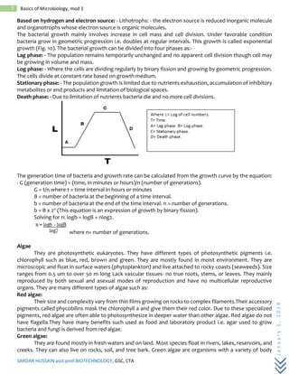
The bacterial growth mainly involves increase in cell mass and cell division. Under favorable condition
bacteria grow in geometric progression i.e. doubles at regular intervals. This growth is called exponential
growth (Fig. 10). The bacterial growth can be divided into four phases as:-
Lag phase: - The population remains temporarily unchanged and no apparent cell division though cell may
be growing in volume and mass.
Log phase: - Where the cells are dividing regularly by binary fission and growing by geometric progression.
The cells divide at constant rate based on growth medium.
Stationary phase: - The population growth is limited due to nutrients exhaustion, accumulation of inhibitory
metabolites or end products and limitation of biological spaces.
Death phase: - Due to limitation of nutrients bacteria die and no more cell divisions.
The generation time of bacteria and growth rate can be calculated from the growth curve by the equation:
- G (generation time) = (time, in minutes or hours)/n (number of generations).
G = t/n where t = time interval in hours or minutes
B = number of bacteria at the beginning of a time interval.
b = number of bacteria at the end of the time interval. n = number of generations.
b = B x 2n (This equation is an expression of growth by binary fission).
Solving for n: logb = logB + nlog2.
where n= number of generations.
Algae
They are photosynthetic eukaryotes. They have different types of photosynthetic pigments i.e.
chlorophyll such as blue, red, brown and green. They are mostly found in moist environment. They are
microscopic and float in surface waters (phytoplankton) and live attached to rocky coasts (seaweeds). Size
ranges from 0.5 um to over 50 m long Lack vascular tissues- no true roots, stems, or leaves. They mainly
reproduced by both sexual and asexual modes of reproduction and have no multicellular reproductive
organs. They are many different types of algae such as:
Red algae:
Their size and complexity vary from thin films growing on rocks to complex filaments.Their accessory
pigments called phycobilins mask the chlorophyll a and give them their red color. Due to these specialized
pigments, red algae are often able to photosynthesize in deeper water than other algae. Red algae do not
have flagella.They have many benefits such used as food and laboratory product i.e. agar used to grow
bacteria and fungi is derived from red algae.
Green algae:
They are found mostly in fresh waters and on land. Most species float in rivers, lakes, reservoirs, and
creeks. They can also live on rocks, soil, and tree bark. Green algae are organisms with a variety of body
SARDAR HUSSAIN asst.prof.BIOTECHNOLOGY, GSC, CTA
January1,2018
8 Basics of Microbiology, mod 1
forms including single cells, filaments, colonies, and thalli. They possess the same photosynthetic pigments
(chlorophyll a and b) and some green algae have stiff cell walls composed of cellulose, as do plants.
Dinoflagellates
Found in warm, Tropical Ocean. They are mainly unicellular. Green and colorless forms, phagotrophic
and parasitic. They are biflagellate. Theirnucleus is unusual. Some are bioluminescent forms- light up when
water is disturbed and they always reproduced by asexually.
Brown algae :
They are known as cold water algae and found in rocky coast in temperate zone or open sea. Most
brown algae contain the pigment fucoxanthin, which is responsible for the distinctive greenish-brown color.
They are multicellular and reproduced by flagellated spores.
Diatoms:
They are most common types of phytoplankton and also known as golden-brown algae. They are
mostly unicellular and can exist as colonies in the shape of filaments or ribbons. Their cell wall made of silica
called a frustule. They are commonly used in studies of water quality. Some diatoms are capable of
movement via flagellation. They reproduced by asexual for several generations, then sexual.
Fungi
They are eukaryotic organisms that include yeasts, molds and mushrooms. They are non-
photosynthetic and contain no chlorophyll pigments. Most of them are multicellular and some are unicellular
e.g. yeast. They are non motile and lack true leaves, roots and stems. Fungi needwarm, moist places to grow.
They are found mainly in moist foods, damp tree barks, and wet bathroom tiles etc. Fungi are heterotrophs
that feed by absorption. They absorb small organic molecules from the surrounding medium. The enzymes
and hydrolytic enzymes secreted by the fungus break down food outside its body into simpler compounds
that the fungus can absorb and use. The absorptive mode of nutrition is associated with the ecological roles
of fungi as decomposers, parasites, and mutualistic symbionts. Saprobic fungi absorb nutrients from
nonliving organisms. Parasitic fungi absorb nutrients from the cells of living hosts. The fungal cells contain
membrane-bound nuclei with chromosomes that contain DNA with noncoding regions called introns and
coding regions called exons. They also possess membrane-bound cytoplasmic organelles such as
mitochondria, sterol -containing membranes, and ribosomes of the 80S type. They have soluble
carbohydrates and storage compounds, including sugar alcohols, disaccharides, and polysaccharides. Fungi
lack chloroplasts and are heterotrophic organisms, requiring preformed organic compounds as energy
sources. Fungi possess a cell wall and vacuoles. They reproduce by both sexual and asexual means and
produce spores. They have haploid nuclei. The cells of most fungi grow as tubular, elongated, and thread-
like structures are called hyphae which may contain multiple nuclei. Some species grow as single-celled
yeasts that reproduce by budding or binary fission. The fungal cell wall is composed of glucans and chitin.
Most fungi grow as hyphae which are cylindrical, thread-like structures 2–10 µm in diameter and up to
several centimeters in length. Hyphae grow at their tips; new hyphae are typically formed by a process
called branching, or growing hyphal tips bifurcate giving rise to two parallel-growing hyphae. Hyphae can be
either septate or coenocytic: septate hyphae are divided into compartments separated by cross walls, with
each compartment containing one or more nuclei; coenocytic hyphae are not compartmentalized. Fungal
reproduction is complex. They reproduced by both sexually and asexually. Asexual reproduction via
vegetative spores (conidia) or through mycelial fragmentation. Sexual reproduction involves joining of
hyphae is called conjugation, two mating strains with differentnuclei form continuous membrane is known
as plasmogamy and sometimes thenuclei are fused is called karyogamy.
Protozoa
Protozoa are parasitic and animal-like protists because of their motility. Their sizes range from 10 to
52 micrometers. They moved by flagella, hair-like structures called cilia and foot-like structures called
pseudopodia (Fig. 13). Protozoa absorb food by their cell membranes e.g., amoebas, surround food and
engulf it. All protozoa digest their food in stomach-like compartments called vacuoles. Protozoa can
reproduce by binary fission or multiple fission. Some protozoa reproduce sexually, some asexually, while
SARDAR HUSSAIN asst.prof.BIOTECHNOLOGY, GSC, CTA
January1,2018
9 Basics of Microbiology, mod 1
some use a combination. They cause many diseases in human such as malaria, amoebiasis and leishmaniasis
etc.
The scope and relevance of microbiology
Microbes influence human society in countless ways. Sometimes, the influence of microorganisms
on human life is beneficial, whereas at other times, it is detrimental. For example, microorganisms are
required for the production of bread, cheese, yogurt, alcohol, wine, beer, antibiotics (e.g., penicillin,
streptomycin, and chloramphenicol), vaccines, vitamins and enzymes. Many products of microbes
contribute to public health as aids to nutrition, other products are used to interrupt the spread of disease,
and still others hold promise for improving the quality of life in the years ahead. Microbes are also an
important and essential component of an ecosystem. Molds and bacteria play key roles in the cycling of
important nutrients in plant nutrition particularly those of carbon, nitrogen and sulphur. Bacteria referred
to as nitrogen fixers live in the soil where they convert vast quantities of nitrogen in air into a form that
plants can use. Microorganisms also play major roles in energy production. Natural gas (methane) is a
product of bacterial activity, arising from the metabolism of methanogenic bacteria. Microorganisms are
also being used to clean up pollution caused by human activities, a process called bioremediation (the
introduction of microbes to restore stability to disturbed or polluted environments). Bacteria and fungi have
been used to consume spilled oil, solvents, pesticides and other environmentally toxic substances.
Agricultural microbiology – try to combat plant diseases that attack important food crops, work on
methods to increase soil fertility and crop yields etc. Currently there is a great interest in using bacterial or
viral insect pathogens as substitute for chemical pesticides.
Microbial ecology – biogeochemical cycles – bioremediation to reduce pollution effects
Food and dairy microbiology – try to prevent microbial spoilage of food and transmission of food borne
diseases such as botulism and salmonellolis. Use microorganisms to make foods such as cheese, yogurt,
pickles and beers.
Industrial microbiology – used to make products such as antibiotics, vaccines, steroids, alcohols and other
solvents, vitamins, amino acids and enzymes.
Microbial physiology and Biochemistry – study the synthesis of antibiotics and toxins, microbial energy
production, microbial nitrogen fixation, effects of chemical and physical agents on microbial growth and
survival etc.
Microbial genetics and Molecular biology – nature of genetic information and how it regulated the
development and function of cells and organisms. Development of new microbial strains that are more
efficient in synthesizing useful products.
Genetic engineering – arisen from work of microbial genetics and molecular biology. Engineered
microorganisms are used to make hormones, antibiotics, vaccines and other products. New genes can be
inserted into plants and animals.
Areas impacted by microbes include:
Medicine : Microbes produce valuable chemicals such as antibiotics. Many antibiotics are produced by
common soil bacteria called Streptomyces and actinomycetes. The ability of Streptomyces cultures to inhibit
the growth of other bacteria leads to discovery of many antibiotics. Streptomycin is an antibiotic and was
the first used for tuberculosis. It is produced by actinobacterium Streptomyces griseus. Many antibiotics are
produced by microbes such as Rifampicin produced by Amycolatopsis rifamycinica, Chloramphenicol by
bacterium Streptomyces venezuelae and Actinomycin D produced by genus Streptomyces etc.
Industry: - The microorganisms are used for the production of food, either human or animal. Yogurt, cheese,
chocolate, and silage (animal food) are all produced by industrial microbiology processes. Lactic acid
bacteria and Bifidobacteria are amongst the most important groups of microorganisms used in the food
industry. The microorganisms used in industrial processes may be natural isolates; laboratory selected
mutants or genetically engineered organisms.
Ecology: - Microbial life plays a primary role in regulating biogeochemical systems in all environment such as
frozen environments and acidic lakes, to hydrothermal vents at the bottom of deepest oceans, and human
SARDAR HUSSAIN asst.prof.BIOTECHNOLOGY, GSC, CTA
January1,2018
10 Basics of Microbiology, mod 1
small intestine. Microbes, often engage in symbiotic relationships (either positive or negative) with other
organisms, and these relationships affect the ecosystem. They are the backbone of all ecosystems. Other
microbes are decomposers, with the ability to recycle nutrients from other organisms' waste products.
These microbes play a vital role in biogeochemical cycles. The nitrogen cycle , the phosphorus cycle and the
carbon cycle all depend on microorganisms in one way or another. Presently, microbiologists facing many
challenges to solve many of society's problems including combating disease, reducing environmental
pollution, and maintaining improving the world's food supply.
Future of microbiology:
• Future challenges such as finding new ways to combat disease, reduce pollution and feed the
world's population.
• AIDS, hemorrhagic fevers and other infectious diseases
• Create new drugs, vaccines. Use the techniques in molecular biology and rDNA to solve the
problems
• Host-pathogen relationships
• Study the role of microorganisms as
• Sources of high-quality food and other practical products such as enzymes for industrial
application
• Degrade pollutants and toxic wastes
• Used as vectors to treat diseases and enhance agricultural productivity
Microbial diversity – less than 1% of the earth's microbial population has been cultured. Develop isolation
techniques and work needs to be done on microorganisms living in extreme environments. Discovery of new
organisms may lead to further advances in industrial processes and enhanced environmental control
• Microbe – microbe interactions.
• Analysis of genome – advances in the field of bioinformatics
• Symbiotic relationships – knowledge can help improve our appreciation of the living world, and
improvements in the health of plant, livestock's and humans.
Microbial taxonomy
Introduction
Living organisms are fascinating by its diversity whether it is plants, animals or microbes. A handful
of soil is populated with more than the human population on earth. They play important essential roles in
nature. So if we arrange these microbes in order or hierarchy by based on its similarity or differences in any
characteristics, we can easily get to know and get easy access to all the microbes. So it is desirable to
determine the classification. Greek Philosopher Aristotle who is the one classified the living things as plants
and animals around 2000 years ago. So in this lecture, we will learn about taxonomy, how is it classifified?
What methods are available to classify them? And then brief description about microbial evolution and
diversity and its phylogeny.
Taxonomy
Taxonomy [Greek taxis, arrangement, and nomos, law, or nemein, to distribute] is defined as the science of
biological classification. In simple term, taxonomy is orderly arranging organisms under study into groups
of larger units. It consists of three interrelated parts namely
1. Classification is the arrangement of organisms into groups or taxa (s., taxon) based on mutual
similarity or evolutionary relatedness.
2. Nomenclature is concerned with the assignment of names to taxonomic groups in agreement with
published rules.
3. Identification is the practical side of taxonomy, the process of determining that a particular
isolate belongs to a recognized taxon. (So in short Identify-Naming them and classify them)
Classification
SARDAR HUSSAIN asst.prof.BIOTECHNOLOGY, GSC, CTA
January1,2018
11 Basics of Microbiology, mod 1
It is bringing order to the diverse variety of organisms present in nature. So there are two general ways
the classification can be constructed. First one is based on the morphological characters (phenetic
classification) and second is based on evolutionary relationship (phylogenetic classification)
 Phenetic classification - Grouping organisms together based on the mutual similarity of their phenotypic
characteristics. It does not provide information about phylogenetic relations.
Phylogenetic classification- These are systems based on evolutionary relationships rather than external
appearance (the term phylogeny [Greek phylon, tribe or race, and genesis, generation or origin] refers to the
evolutionary development of a species). It is based on the direct comparison of genetic materials and/or
gene product.
Nomenclature (Binomial system)
Biologists in the middle ages used to follow polynomial system, i.e naming organisms with many names
(poly -many, nomo - name). For example name for the European honeybee, was Apis pubescens, thorace
subgriseo, abdomine fusco, pedibus posticis glabris utrinque margine ciliatis (just for example no need to be
memorized). Later Binomial systems were developed by Swedish biologist Carolus Linnaeus (1707–1778)
based on the anatomical characteristics of plants and animals. Nomenclature in microbiology is developed
based on the principals established for the plant and Animal kingdom by Linnaeus. The first word in the
binomial is the genus name and is always capitalized. The second word is species name and never capitalized.
For example honeybee, Apis mellifera
Taxonomic ranks:
In prokaryotic taxonomy the most commonly used levels or ranks (in ascending order) are species, genera,
families, orders, classes, phyla, kingdom or domain. In order to remember the seven categories of the
taxonomic hierarchy in their proper order, it may be useful to memorize a phrase such as
“ k indly p ay c ash o r f urnish g ood s ecurity”
(k ingdom– p hylum– c lass– o rder– f amily– g enus– s pecies). The basic taxonomic group in microbial
taxonomy is the species.
A species is a collection of strains that have a similar G+C composition and 70% or greater similarity as judged
by DNA hybridization. Ideally a species also should be phenotypically distinguishable from other similar
species. An example of hierarchy in taxonomy is given below.
A strain is a population of organisms that is distinguishable from at least some other populations within a
particular taxonomic category. It is considered to have descended from a single organism or pure culture
isolate. Strains within a species may differ slightly from one another in many ways. Biovars are variant
prokaryotic strains characterized by biochemical or physiological
differences, morphovars differ morphologically, and serovars have distinctive antigenic properties . One
strain of a species is designated as the type strain. It is usually one of the first strains studied and often
is more fully characterized than other strains; however, it does not have to be the most representative
member but this strain can be considered as reference strain and can be compared with other strains. Each
species is assigned to a genus, the next rank in the taxonomic hierarchy. A genus is a well-defined group of
one or more species that is clearly separate from other genera.
SARDAR HUSSAIN asst.prof.BIOTECHNOLOGY, GSC, CTA
January1,2018
12 Basics of Microbiology, mod 1
Techniques for identifying or determining taxonomical characters
In order to identify and classify microorganisms, we need to know about their characteristics. There
are two ways to determine the taxonomical characters; classical and molecular characters
Classical characteristics:- This approach uses morphological, biochemical, physiological, ecological and
genetic characteristics. It is mainly used in microbial taxonomy.
1. Morphology:- Morphology is the one which can be easily studied and analyzed. Structural features (cell
shape, size, colony morphology, appendages, and etc.) depend on the expression of many genes, are usually
genetically stable.
2. Physiology and metabolism:- Organisms are classified based on the requirements for growth characters
like carbon and nitrogen sources, cell wall constituents, general nutritional type, energy sources, optimum
growth temperature, Motility.
3. Ecology:- These are taxonomically valuable because even very closely related microorganisms can differ
considerably with respect to ecological characteristics. The ability to cause disease in a particular host;
and habitat preferences such as requirements for temperature, pH, oxygen, and osmotic concentration are
examples of ecological characteristics.
4. Genetic analysis:- The study of chromosomal gene exchange between species through transformation and
conjugation (in Enteric bacteria) is sometimes useful in their classification. Most bacteria are harboring
plasmids, classification based on plasmid is also an important part of classification.
Molecular characteristics:-This is the most powerful approaches to study taxonomy by analyzing proteins
and nucleic acids. Because these are either direct gene products or the genes themselves, comparisons of
proteins and nucleic acids yield considerable information about true relatedness.
1. Comparing amino acid sequences:- Comparison of amino acid sequences of proteins from different
organisms reveals its taxonomic relations. The most direct approach is to determine the amino acid
sequence of proteins with the same function. If the sequences of proteins with the same function are similar,
the organisms possessing them are probably closely related . The electrophoretic mobility of proteins is useful
in studying relationships at the species and subspecies levels. Antibodies can discriminate between very
similar proteins, and immunologic techniques are used to compare proteins from different microorganisms.
2. Nucleic acid composition:- By direct comparison of microbial genomes and based on the G+C content of
different organisms (Escherichia coli 48-52 %). And genomic fingerprinting (RFLP, AFLP) reveals its
relatedness with others.
3. Nucleic acid hybridization: - It uses the property of complementarities in double stranded DNA. More
distantly related organism can be identified based on DNA-RNA hybridization
4. Nucleic acid sequencing: - Techniques are now available to sequence both DNA and RNA. 5S and 16S RNA
(prokaryotes), 18S (fungi) analysis of microorganisms can reveal their relatedness because of its functional
role is same in all ribosomes and slow structural changes with time.
Microbial evolution and Diversity
It has been estimated that our planet is about 4.6 billion years old. Around 3.5 to 3.8 billion years old
fossilized remains of prokaryotic cells have been discovered in sedimentary rocks. Thus earlier prokaryotes
were anaerobic and arose shortly after the earth cooled. Cyanobacteria and oxygen-producing
photosynthesis probably developed 2.5 to 3.0 billion or more years ago.
It appears likely that modern eukaryotic cells arose from prokaryotes about 1.4 billion years ago.
Two hypotheses for the evolution of eukaryotic cells
1. Organelles arose within prokaryotes from the invagination of the plasma membrane
2. Endosymbiotic hypothesis
Fusion of ancient true bacteria and archaea to form a nucleus. They proposed that the eukaryotic line
diverged from the Archaea and then the nucleus formed, possibly from the Golgi apparatus
SARDAR HUSSAIN asst.prof.BIOTECHNOLOGY, GSC, CTA
January1,2018
13 Basics of Microbiology, mod 1
Mitochondria and chloroplasts develop later from a permanent symbiotic relationship with other bacteria,
e.g., cyanelle (cyanobacterium) living inside the protist Cyanophora paradoxa
Cyanobacteria have been considered the most likely ancestors of chloroplasts. More recently Prochloron has
become the favorite candidate. The existence of this bacterium suggests that chloroplasts arose from a
common ancestor of prochlorophytes and cyanobacteria. Mitochondria arose from an endosymbiotic
relationship between the free-living primitive eukaryotic and bacteria with aerobic respiration (possibly an
ancestor of three modern groups: Agrobacterium, Rhizobium, and Rickettsia).
Divisions of Life
Kingdom systems of classification
- Five-kingdom system (Whittaker, 1960s) - based upon cell type, organization, and the means of nutrient
acquisition (Monera, Protista, Fungi, Plantae, Animalia)
- Six-kingdom system - differs from five-kingdom system by dividing prokaryotes into bacteria and archaea
(Bacteria, Archaea, Protista, Fungi, Plantae, Animalia)
- Eight-kingdom system (Cavalier-Smith) - further division of the protists using rRNA data and grouping
organisms into two empires (Eucaryota and Bacteria) containing a total of eight kingdoms [(Bacteria,
Archaea), (Archezoa, Protista, Plantae, Chromista, Fungi, Animalia)
Domains
Advances in genomic DNA sequencing of the microorganisms, biologists are increasingly adapting the
classification of living organisms that recognizes three domains, a taxonomic level higher than kingdom.
Archaebacteria are in one domain, eubacteria in a second, and eukaryotes in the third. Domain Eukarya is
subdivided into four kingdoms plants, animals, fungi, protists.
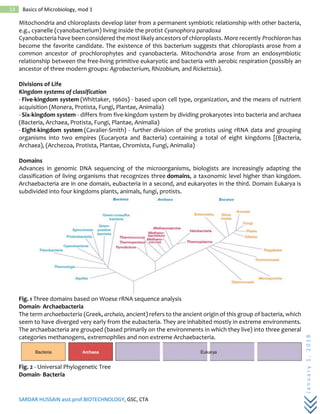
Fig. 1 Three domains based on Woese rRNA sequence analysis
Domain- Archaebacteria
The term archaebacteria (Greek, archaio, ancient) refers to the ancient origin of this group of bacteria, which
seem to have diverged very early from the eubacteria. They are inhabited mostly in extreme environments.
The archaebacteria are grouped (based primarily on the environments in which they live) into three general
categories methanogens, extremophiles and non extreme Archaebacteria.
Fig. 2 - Universal Phylogenetic Tree
Domain- Bacteria
SARDAR HUSSAIN asst.prof.BIOTECHNOLOGY, GSC, CTA
January1,2018
14 Basics of Microbiology, mod 1
The Eubacteria are the most abundant organisms on earth. It plays critical roles like cycling carbon and sulfur.
Much of the world's photosynthesis is carried out by eubacteria. However, certain groups of eubacteria are
also responsible for many forms of disease.
Domain- Eukarya
It consists of four kingdoms. The first of which is protista, mostly unicellular organism like amoeba. The other
three kingdoms are plants, fungi, animals. Multicellularity and sexuality are the two unique characters that
differentiate from prokaryote and eukaryotes.
Fig. 3 . Phylogenetic tree. a) unrooted tree, b) rooted tree.
Molecular chronometers
This concept, first suggested by Zuckerkandl and Pauling (1965), which is based on thought that the
sequences of many rRNAs and proteins gradually change over time without destroying or severely altering
their functions. Changes increases with time linearly. If sequences of similar molecules from two organisms
differs, it means that they diverged very long time ago.
Phylogenetic tree
Phylogenetic relationships are illustrated in the form of branched diagrams or trees (denrograms).
A phylogenetic tree is a graph made of branches that connect nodes. The nodes represent taxonomic units
such as species or genes; the external nodes, those at the end of the branches, represent living organisms.
The tree may have a time scale, or the length of the branches may represent the number of molecular
changes that have taken place between the two nodes. Finally, a tree may be unrooted or rooted.
An unrooted tree simply represents phylogenetic relationships but does not provide an evolutionary path.
Figure 3. a. shows that A is more closely related to C than it is to either B or D, but does not specify the
common ancestor for the four species or the direction of change. In contrast, the rooted tree Figure 3. b. does
give a node that serves as the common ancestor and shows the development of the four species from this
root.
Parsimony analysis
Phylogenetic relationships also can be estimated by techniques such as parsimony analysis. In this approach,
relationships are determined by estimating the minimum number of sequence changes required to give the
final sequences being compared. It is presumed that evolutionary change occurs along the shortest pathway
with the fewest changes or steps from an ancestor to the organism in question.
Oligonucleotide signature sequences
The 16S rRNA of most major phylogenetic groups has one or more characteristic nucleotide sequences called
oligonucleotide signatures. Oligonucleotide signature sequences are specific oligonucleotide sequences
that occur in most or all members of a particular phylogenetic group. They are rarely or never present in
SARDAR HUSSAIN asst.prof.BIOTECHNOLOGY, GSC, CTA
January1,2018
15 Basics of Microbiology, mod 1
other groups, even closely related ones. Thus signature sequences can be used to place microorganisms in
the proper group.
Polyphasic taxonomy
Studying phylogeny based on both genotypic and phenotypic information ranging from molecular
characteristics to ecological characters.
Numerical Taxonomy
Computer based approaches of grouping organisms is called Numerical taxonomy which is based on
presence or absence of selected characters in the group of organisms. It is method of estimating percent
similarity (ratio between the number of characters same and total number of characters organisms having).
This method has great practical usefulness as well as being relatively unbiased in its approach. It has high
degree of stability and predictability.
Bacteria
Bacteria are prokaryotes, evolved first on living earth. They does not contain membrane bound nucleus and
organelles. Almost all the bacteria are having circular genome and extrachromasomal DNA which helps in
survive different environments. Bacteria reproduce by prokaryotic fission,resulting in two genetically
identical daughter cells. Most of the bacteria are unicellular in nature but sometimes they form aggregates.
The most common shapes of bacteria are spheres (cocci), rods (bacilli), spirals.
Bacterial cell walls are made of peptidoglycan contains sugar moieties (N-acetylgluscosamine, and N-
acetylmuramic acid cross linked with pentapeptide (D-aminoacids). Gram stain is a valuable tool to identify
the bacteria based on the cell wall constituents. Gram-positive bacteria have simple cell walls with large
amounts of peptidoglycan and which retains crystal violet. Gram-negative bacteria have more complex cell
walls with less peptidoglycan and which retains saffron, the counter strain. Presence of lipid layer is a unique
characteristic of Gram negative bacteria. Most of the gram negative bacteria are causative agent of many
human diseases than Gram positive bacteria. The lipopolysaccharides on the walls of gram-negative bacteria
are often toxic, and the outermembrane protects the pathogens from the defenses of their hosts.
Capsules are the slimy layer produced by most of the bacteria which helps them to adhere together and
form colonies. Two kinds of filamentous structures may be attached to the cell wall: The bacterial flagellum
rotates like a propeller to pull the cell along while movement. Pili help bacteria attach to one another in
conjugation, and fimbriae help them attach to surfaces. Many prokaryotes are capable of taxis, movement
toward nutrients or oxygen (positive chemotaxis) away from a toxic substance (negative chemotaxis).
Some bacteria form resistant cells called endospores when an essential nutrient is lacking in the
environment and it may remain dormant but viable for centuries or longer.
Classification based on nutrition and metabolism
Organisms can be categorized by their nutrition, based on how they obtain energy and carbon to build the
organic molecules that make up their cells. Organisms that obtain energy from light are phototrophs.
Organisms that obtain energy from chemicals in their environment are chemotrophs. Organisms that need
only an inorganic compound such as CO2 as a carbon source are autotrophs. Organisms that require at least
one organic nutrient—such as glucose—as a carbon source are heterotrophs. Based on requirement of
oxygen they are classified as obligate aerobes (requires O2for respiration), facultative anaerobes (can grow
both aerobically and anerobically), and obligate anaerobes (does not require O2).
Bacterial taxonomy
Until the late 20th century, biologists based prokaryotic taxonomy on criteria such as shape, motility,
nutritional mode, and Gram staining. Although these criteria may be valuable in culturing and identifying
pathogenic bacteria, they may not reflect evolutionary relationships. Applying molecular data to the
investigation of prokaryotic phylogeny has been very fruitful. Microbiologists began comparing sequences
of prokaryotic genes in the 1970s. Carl Woese and his colleagues used ribosomal RNA (rRNA) as a marker for
evolutionary relationships.
SARDAR HUSSAIN asst.prof.BIOTECHNOLOGY, GSC, CTA
January1,2018
16 Basics of Microbiology, mod 1
In 1923, David Bergey and colleagues set out to publish a definitive book on the identification and
classification of bacteria. A Survey of Bacterial Phylogeny and Diversity - based on the 2nd edition of Bergey's
olume 1: The Archaea, Cyanobacteria, Phototrophs and Deeply Branching Genera
Archaea - divided into two kingdoms
a. Crenarchaeota - diverse kingdom that contains thermophilic and hyperthermophilic
b. Euryarchaeota - contains primarily mathanogenic and halophilic bacteria and also
Eubacteria - complex with several small groups of phototrophs, cyanobacteria, and deeply branching
eubacteria
Based on Bergeys' manual Domain Bacteria contains six phyla in volume 1
• Phylum Aquificiae- earliest branch of bacteria that contain autotrophs which utilize hydrogen for
energy production
• Phylum Thermotogae - anaerobic, thermophilic, and fermentative Gram negative bacteria
• Phylum “ Deinococcus Thermus ” - radiation resistant bacteria
• Phylum Chloroflexi - green non-sulfur bacteria that carries out anoxygenic photosynthesis
• Phylum Cyanobacteria - oxygenic photosynthetic bacteria
• Phylum Chlorobi - green sulfur bacteria that carry out anoxygenic photosynthesis
Volume 2 - Gram negative proteobacteria (purple bacteria)
Based on the nutritional type and rRNA data Gram negative- Proteobacteria have been classified into five
classes.
• Alphaproteobacteria -oligotrophic forms including the purple nonsulfur photosynthesizers
• Betaproteobacteria - metabolically similar to alphaproteobacteria
• Gammaproteobacteria - diverse methods of energy metabolism
• Deltaproteobacteria - includes predators and the fruiting myxobacteria
• Epsilonproteobacteria - contains pathogens
Volume 3 - Gram positive bacteria with low G + C content (< 50%)
Three classes of the phylum Firmicutes
• Clostridia - tend to be anaerobic and endospore formers
• Mollicutes - mycoplasmas (no cell walls)
• Bacilli - Gram-positive aerobes or facultative anaerobes, rods or cocci, some endospore formers
Volume 4 - Gram positive bacteria with high G + C content (> 50-55%)
• All belong to the phylum Actinobacteria
• Some are filamentous
Volume 5 - Gram negative with various morphologies
Nine phyla of which four are presented below
• Phylum Planctomycetes - some have a membrane-bound nucleus
• Phylum Chlamydiae - obligate intracellular parasites important in disease
• Phylum Spirochaetes - helical-shaped, Gram-negative motile bacteria (axial filaments)
• Phylum Bacteroidetes - ecologically significant species are found in this phylum
THE PROKARYOTIC CELL
The members of the prokaryotic world make up a vast heterogeneous group of very small unicellular
organisms. This group includes eubacteria, or true bacteria, and archaebacteria. Bacteria are one of the most
important groups of the microbial world.
The chief distinguishing characteristics of prokaryotic cells are:
• Their genetic material (DNA) is not enclosed within a membrane.
• They lack other membrane-bounded organelles
• Their DNA is not associated with histone proteins (special chromosomal proteins found in
eukaryotes).
SARDAR HUSSAIN asst.prof.BIOTECHNOLOGY, GSC, CTA
January1,2018
17 Basics of Microbiology, mod 1
• Their cell walls most of the time contains the complex polysaccharide peptidoglycan.
• They usually divide by binary fission. During this process, the DNA is copied and the cell splits into
two cells.
Size, shape and arrangement of bacterial cells
Size:
Prokaryotes are among the smallest of all organisms (0.5 to 2.0 m m). Because of their small size,
bacteria have a large surface-to-volume ratio. The smallest member of the genus is about 0.3µm in diameter.
Even smaller cells have been reported like the nanobacteria or ultramicrobacteria appear to range from
around 0.2µm to not less than 0.05µm. E. coli , a bacillusof about average size is 1.1 to 1.5 µm wide by 2.0 to
6.0 µm long. Spirochaetes occasionally reach 500 µm in length and the cyanobacterium Oscillatoria is about
7 µm in diameter. The bacterium, Epulosiscium fishelsoni , can be seen with the naked eye (600 m m long by
80 m m in diameter). Thus a few bacteria are much larger than the average eukaryotic cell (typical plant and
animal cells are around 10 to 50 µm in diameter).
Shape and arrangement:
Typically bacteria have three basic shapes – spherical, rod like and spiral.
Spherical bacteria :
Coccus (pluralcocci ) in pairs– diplococcic; in chains– Streptococci (Streptococcus); in cube like groups of
eight– Sarcinae (Sarcina); in grape like structures– Staphylococci (Staphylococcus) (Fig.1)
Fig. 1. Arrangement of spherical cells
We shall be dealing with the structures internal to the cell wall of a bacterial cell.
SARDAR HUSSAIN asst.prof.BIOTECHNOLOGY, GSC, CTA
January1,2018
18 Basics of Microbiology, mod 1
They include plasma membrane, organelles in the cytoplasm like nuclear area, ribosomes, inclusion bodies
and endospores.
Plasma (cytoplasmic) membrane
Membranes are absolute requirement of all living organisms. It is the chief point of contact with the
cell's environment and thus is responsible for much of its relationship with the outside world. Plasma
membrane – encloses the cytoplasm and consists of phospholipids and proteins (fluid mosaic model).Most
membrane-associated lipids are structurally asymmetric with polar and nonpolar ends. The polar ends
interact with water and are hydrophilic and the nonpolar hydrophobic ends are insoluble in water. The lipid
composition of bacterial membranes varies with environmental temperature in such a way that the
membrane remains fluid during growth. Bacterial membranes usually differ from eukaryotic membranes in
lacking sterols such as cholesterol and they contain pentacyclic sterol-like molecules called hopanoids and
these are said to stabilize the bacterial membranes. Cell membranes are very thin structures about 5 to 10
nm thick and can be seen only with electron microscope. Plasma membranes have a complex internal
structure; the small globular particles seen in these membranes are thought to be membrane proteins that
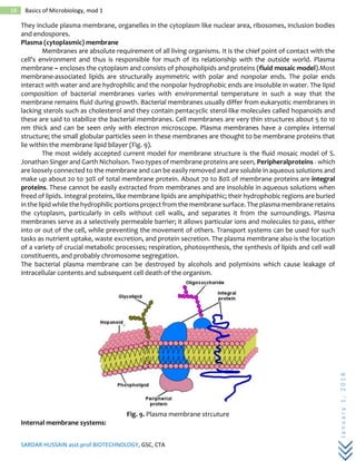
lie within the membrane lipid bilayer (Fig. 9).
The most widely accepted current model for membrane structure is the fluid mosaic model of S.
Jonathan Singer and Garth Nicholson. Two types of membrane proteins are seen, Peripheralproteins - which
are loosely connected to the membrane and can be easily removed and are soluble in aqueous solutions and
make up about 20 to 30% of total membrane protein. About 70 to 80% of membrane proteins are integral
proteins. These cannot be easily extracted from membranes and are insoluble in aqueous solutions when
freed of lipids. Integral proteins, like membrane lipids are amphipathic; their hydrophobic regions are buried
in the lipid while the hydrophilic portions project from the membrane surface. The plasma membrane retains
the cytoplasm, particularly in cells without cell walls, and separates it from the surroundings. Plasma
membranes serve as a selectively permeable barrier; it allows particular ions and molecules to pass, either
into or out of the cell, while preventing the movement of others. Transport systems can be used for such
tasks as nutrient uptake, waste excretion, and protein secretion. The plasma membrane also is the location
of a variety of crucial metabolic processes; respiration, photosynthesis, the synthesis of lipids and cell wall
constituents, and probably chromosome segregation.
The bacterial plasma membrane can be destroyed by alcohols and polymixins which cause leakage of
intracellular contents and subsequent cell death of the organism.
Fig. 9. Plasma membrane strcuture
Internal membrane systems:
SARDAR HUSSAIN asst.prof.BIOTECHNOLOGY, GSC, CTA
January1,2018
19 Basics of Microbiology, mod 1
Prokaryotes do not contain complex membrane systems as present in eukaryotes like chloroplast
and mitochondria. They contain membranous structures like the one observed most is mesosome.
Mesosomes – irregular infoldings or invaginations of the plasma membrane in the shape of vesicles, tubules,
or lamellae. They can be seen in both gram positive and gram-negative bacteria. These are often found next
to the septa or cross-walls in dividing bacteria and sometimes seems attached to the bacterial chromosome.
Thus they seem to be involved in cell wall formation during division or play a role in chromosome replication
and distribution to daughter cells.
Some bacteria have internal membrane systems quite different from the mesosomes. The infoldings of the
plasma membrane can become extensive and complex in photosynthetic bacteria such as the cyanobacteria
and purple bacteria or in bacteria with very high respiratory activity like the nitrifying bacteria. They may be
aggregates of spherical vesicles, flattened vesicles, or tubular membranes. Their function may be to provide
a larger membrane surface for greater metabolic activity.
Cytoplasm
Is the fluid component inside the plasma membrane. Cytoplasm is about 80% water and contains
primarily proteins (enzymes), carbohydrates, lipids, inorganic ions and many low MW compounds. Major
structures in the cytoplasm are DNA, ribosomes and inclusions.
Nuclear area
The striking difference between prokaryotic and eukaryotic systems is the way in which their genetic
material is packaged. Prokaryotes lack a membrane-delimited nucleus. Contains a single long circular
molecule of double-stranded DNA bacterial chromosomes do not include histones and are not surrounded
by a nuclear envelope and located in an irregularly shaped region called nucleiod . The nuclear area can be
spherical, elongated, or dumb-bell shaped. It is attached to the plasma membrane and proteins of plasma
membrane are believed to be responsible for replication of the DNA. It has been discovered recently
that Vibrio cholera has more than one chromosome. Electron microscope studies have shown the nucleiod
in contact with either the mesosome or the plasma membrane and hence evidence that the membranes may
be involved in the separation of DNA into daughter cells during division. Chemical analysis reveals that they
nucleoids are composed to about 60% DNA, 30% RNA and 10% protein. E. coli, which is about 2 to 6um long,
the closed DNA circle measures approximately 1400um. Hence, it is evident that the DNA is efficiently
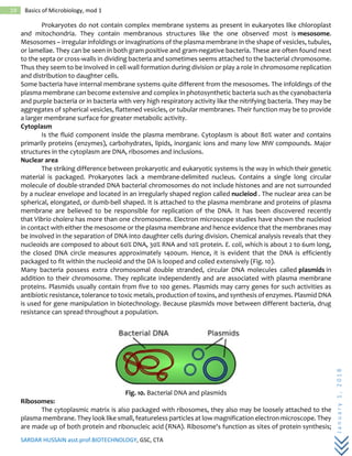
packaged to fit within the nucleoid and the DA is looped and coiled extensively (Fig. 10).
Many bacteria possess extra chromosomal double stranded, circular DNA molecules called plasmids in
addition to their chromosome. They replicate independently and are associated with plasma membrane
proteins. Plasmids usually contain from five to 100 genes. Plasmids may carry genes for such activities as
antibiotic resistance, tolerance to toxic metals, production of toxins, and synthesis of enzymes. Plasmid DNA
is used for gene manipulation in biotechnology. Because plasmids move between different bacteria, drug
resistance can spread throughout a population.
Fig. 10. Bacterial DNA and plasmids
Ribosomes:
The cytoplasmic matrix is also packaged with ribosomes, they also may be loosely attached to the
plasma membrane. They look like small, featureless particles at low magnification electron microscope. They
are made up of both protein and ribonucleic acid (RNA). Ribosome's function as sites of protein synthesis;
SARDAR HUSSAIN asst.prof.BIOTECHNOLOGY, GSC, CTA
January1,2018
20 Basics of Microbiology, mod 1
matrix ribosomes synthesize proteins destined to remain within the cell, whereas the plasma membrane
ribosomes make proteins for transport to the outside.The shape of each protein is determined by its amino
acids sequence and the special proteins called molecular chaperones or chaperones aid the polypeptide in
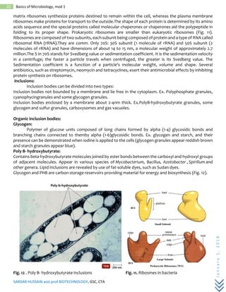
folding to its proper shape. Prokaryotic ribosomes are smaller than eukaryotic ribosomes (Fig. 11).
Ribosomes are composed of two subunits, each subunit being composed of protein and a type of RNA called
ribosomal RNA (rRNA).They are comm. Only 70S: 30S subunit (1 molecule of rRNA) and 50S subunit (2
molecules of rRNA) and have dimensions of about 14 to 15 nm, a molecular weight of approximately 2.7
million.The S in 70S stands for Svedberg value or sedimentation coefficient. It is the sedimentation velocity
in a centrifuge; the faster a particle travels when centrifuged, the greater is its Svedberg value. The
Sedimentation coefficient is a function of a particle's molecular weight, volume and shape. Several
antibiotics, such as streptomycin, neomycin and tetracyclines, exert their antimicrobial effects by inhibiting
protein synthesis on ribosomes.
Inclusions:
Inclusion bodies can be divided into two types:
Inclusion bodies not bounded by a membrane and lie free in the cytoplasm. Ex. Polyphosphate granules,
cyanophycingranules and some glycogen granules.
Inclusion bodies enclosed by a membrane about 2-4nm thick. Ex.PolyB-hydroxybutyrate granules, some
glycogen and sulfur granules, carboxysomes and gas vacuoles.
Organic inclusion bodies:
Glycogen:
Polymer of glucose units composed of long chains formed by alpha (1-4) glycosidic bonds and
branching chains connected to themby alpha (1-6)glycosidic bonds. Ex. glycogen and starch, and their
presence can be demonstrated when iodine is applied to the cells (glycogen granules appear reddish brown
and starch granules appear blue).
Poly B- hydroxybutyrate:
Contains beta-hydroxybutyrate molecules joined by ester bonds between the carboxyl and hydroxyl groups
of adjacent molecules. Appear in various species of Mycobacterium, Bacillus, Azotobacter , Spirillum and
other genera. Lipid inclusions are revealed by use of fat-soluble dyes, such as Sudan dyes.
Glycogen and PHB are carbon storage reservoirs providing material for energy and biosynthesis (Fig. 12).
Fig. 12 . Poly B- hydroxybutyrate inclusions Fig. 11. Ribosmes in bacteria
SARDAR HUSSAIN asst.prof.BIOTECHNOLOGY, GSC, CTA
January1,2018
21 Basics of Microbiology, mod 1
Cyanophycin granules:
Cyanobacteria are composed of large amino acids containing approximately equal amounts of amino
acids arginine and aspartic acid. These are used to store extra nitrogen for the bacteria.
Carboxysomes:
These are polyhedral and hexagonal inclusions that contain the enzyme ribulose 1,5-diphosphate
carboxylase. ·Bacteria that use carbon dioxide as their sole source of carbon require this enzyme for carbon
dioxide fixation during photosynthesis (Ex.nitrifying bacteria, cyanobacteria, and Thiobacilli ).
Gas vacuoles:
These are hollow cavities found in many aquatic prokaryotes, including cyanobacteria, anoxygenic
photosynthetic bacteria and halobacteria. Each vacuole consists of rows of several individual gas vesicles,
which are hollow cylinders covered by protein. Their function is to maintain buoyancy so that the cells can
remain at the depth in the water appropriate for them to receive sufficient amounts of oxygen, light and
nutrients. They are impermeable to water and permeable to atmospheric gases.
Inorganic inclusion bodies:
Polyphosphate granules or Metachromatic granules:
Linear polymer of organo phosphates joined by ester bonds. Reservoirs for phosphate, an important
component of cell nucleic acids and also energy reserves. Represents a reserve of inorganic phosphate
(polyphosphate) that can be used in the synthesis of ATP. Stain red with certain blue dyes, such as methylene
blue, and are collectively known as volutin. Found in algae, fungi and protozoans, as well as bacteria. These
granules are quite large and are characteristic of Corynebacterium diphtheriae, the causative agent of
diphtheria, thus they have diagnostic significance.
Sulphur granules:
Sulphur bacteria, which belong to the genus Thiobacillus, derive energy by oxidizing sulfur and sulfur
containing compounds. These bacteria may deposit sulfur granules in the cell, where they serve as an energy
reserve. Purple photosynthetic bacteria use H2S as electron donor and accumulate resulting sulfurin either
the periplasmic space or in special cytoplasmic globules.
Magnetosomes:
Not for storage, but these are used by some bacteria to orient in the earth's magnetic field. These
inclusion bodies contain iron in the form of magnetite (greigite or pyrite) (Fig. 13). Ex. Aquaspirillum
magnetotacticum. Also present in heads of birds, dolphins, and turtles etc which aid in navigation.
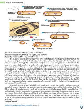
Endospores “An escape pod for DNA”
Endospores are a survival mechanism:they are triggered to form during adverse environmental conditions.
They are NOT reproductive structures as only one cell gives rise to one spore and endospores can be
identified with special stains and differentiated from the vegetative cell (Fig. 14). Endospores are resistant
to: heat: withstand boiling for over one hour; desiccation, UV radiation and chemical disinfectants. The
resistance of these spores has serious consequence and some very pathogenic bacteria have the ability of
produce such spores
SARDAR HUSSAIN asst.prof.BIOTECHNOLOGY, GSC, CTA
January1,2018
22 Basics of Microbiology, mod 1
Fig. 14. Endospore formation
The structures external to the cell wall of bacterial cells.
This includes glycocalyx, fimbriae, pili, flagella, axial filaments
Glycocalyx (Capsules, Slime layers and S-layers)
It is a viscous (sticky), gelatinous polymer composed of polysaccharide, polypeptide or both. If the
substance is organized and is firmly attached to the cell wall, the glycocalyx is described as
a capsule (negative staining). If the substance is unorganized and only loosely attached to the cell wall, the
glycocalyx is described as a slime layer. Capsules protect pathogenic bacteria from phagocytosis (process
by which certain white blood cells engulf and destroy microbes) and contribute to virulence.
Unencapsulated Streptomyces pneumoniae and Bacillus anthracis does not cause disease because the cells
are readily phagocytosized. This allows the bacteria to attach to various surfaces, such as rocks in fast-
moving streams, plant roots, human tooth and tissues and even other bacteria. Capsules also contain water
which prevents them from desiccation. Other examples are Streptococcus mutans (dental caries), Klebsiella
pneumoniae (respiratory tract). These can protect a cell against dehydration. Capsules and slime layers
usually are made up of polysaccharides, but they may be constructed of othermaterial, like Bacillus
anthracis has a capsule of poly D-glutamic acid. Capsules are clearly visible in the light microscope by using
stains or special capsule stains.
A regularly structured layer called S-layer is usually seen in many gram positive and gram negative
bacteria. It consists of proteins or glycoproteins and resembles a pattern something similar to floor tiles. The
S-layer adheres directly to the outer membrane in case of gram negative bacteria and with the peptidoglycan
surface in gram positive bacteria. These protect the bacteria against ion and pH fluctuations, osmotic stress,
enzymes, or the predacious bacterium Bdellovibrio . The S layer also helps maintain the shape and envelope
rigidity of at least bacterial cells and also promotes cell adhesion to surfaces. Sometimes, the layer also
seems to protect some pathogens against complement attack and phagocytosis, thus contributing to their
virulence.
Fimbriae and Pili:
SARDAR HUSSAIN asst.prof.BIOTECHNOLOGY, GSC, CTA
January1,2018
23 Basics of Microbiology, mod 1
Many gram negative bacteria have hairlike appendages that are shorter, straighter and thinner than
flagella and are used for attachment rather than for motility. They are usually called fimbriae. These
structures contain a protein called pilin. Fimbriae - occur at the poles of the bacterial cell, or they can be
evenly distributed over the entire surface of the cell. Fimbriae of Neisseria gonorrhoeae the causative agent
of gonorrhea help the microbe to colonize mucous membranes to cause the disease. At least some types of
fimbriae attach bacteria to solid surfaces such as rocks in streams and host tissues.
Pilior sex pili or pilus - usually longer than fimbriae and number only one to ten per cell. Pili function to join
bacterial cells prior to the transfer to DNA from one cell to another (sometimes called sex pili). They are
genetically determined by sex factors or conjugative plasmids and are required for bacterial mating. Some
bacterial viruses attach specifically to receptors on sex pili at the start of their reproductive cycle.
Flagella:
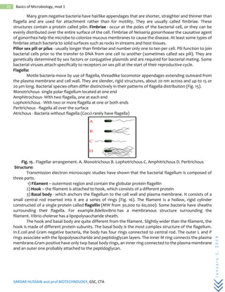
Motile bacteria move by use of flagella, threadlike locomotor appendages extending outward from
the plasma membrane and cell wall. They are slender, rigid structures, about 20 nm across and up to 15 or
20 µm long. Bacterial species often differ distinctively in their patterns of flagella distribution (Fig. 15).
Monotrichous- single polar flagellum located at one end
Amphitrochous- With two flagella, one at each end
Lophotrichous - With two or more flagella at one or both ends
Peritrichous - flagella all over the surface
Atrichous - Bacteria without flagella (Cocci rarely have flagella)
Fig. 15 . Flagellar arrangement. A. Monotrichous B. Lophotrichous C. Amphitrichous D. Peritrichous
Structure:
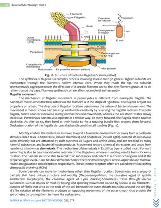
Transmission electron microscopic studies have shown that the bacterial flagellum is composed of
three parts.
1) Filament – outermost region and contain the globular protein flagellin
2) Hook – the filament is attached to hook, which consists of a different protein
3) Basal body - which anchors the flagellum to the cell wall and plasma membrane. It consists of a
small central rod inserted into it are a series of rings (Fig. 16). The filament is a hollow, rigid cylinder
constructed of a single protein called flagellin (MW from 30,000 to 60,000). Some bacteria have sheaths
surrounding their flagella. For example Bdellovibrio has a membranous structure surrounding the
filament. Vibrio cholerae has a lipopolysaccharide sheath.
The hook and basal body are quite different from the filament. Slightly wider than the filament, the
hook is made of different protein subunits. The basal body is the most complex structure of the flagellum.
In E.coli and Gram negative bacteria, the body has four rings connected to central rod. The outer L and P
rings associate with the lipopolysaccharide and peptidoglycan layers. The inner M ring connects the plasma
membrane.Gram positive have only twp basal body rings, an inner ring connected to the plasma membrane
and an outer one probably attached to the peptidoglycan.
SARDAR HUSSAIN asst.prof.BIOTECHNOLOGY, GSC, CTA
January1,2018
24 Basics of Microbiology, mod 1
Fig. 16. Structure of bacterial flagella (Gram negative)
The synthesis of flagella is a complex process involving atleast 20 to 30 genes. Flagellin subunits are
transported through the filament's hollow internal core. When they reach the tip, the subunits
spontaneously aggregate under the direction of a special filament cap so that the filament grows at its tip
rather than at the base. Filament synthesis is an excellent example of self-assembly.
Flagellar movement:
The mechanism of flagellar movement in prokaryotes is different from eukaryotic flagella. The
bacterium moves when the helix rotates as the filament is in the shape of rigid helix. The flagella act just like
propellers on a boat. The direction of flagellar rotation determines the nature of bacterial movement. The
movement in monotrichous bacteria stop and tumble randomly by reversing the flagellar rotation. The polar
flagella, rotate counter clockwise during normal forward movement, whereas the cell itself rotates slowly
clockwise. Peritrichous bacteria also operate in a similar way. To move forward, the flagella rotate counter
clockwise. As they do so, they bend at their hooks to for a rotating bundle that propels them forward.
Clockwise rotation of the flagella disrupts the bundle and the cell tumbles (Fig. 17).
Motility enables the bacterium to move toward a favorable environment or away from a particular
stimulus called taxis . Chemotaxis (include chemicals) and phototaxis (include light). Bacteria do not always
swim aimlessly but are attracted by such nutrients as sugars and amino acids, and are repelled by many
harmful substances and bacterial waste products. Movement toward chemical attractants and away from
repellents is known as chemotaxis. The mechanism ofchemotaxis in E.coli has been studied most. Forward
swimming is due to counterclockwise rotation of the flagellum, whereas tumbling results from clockwise
rotation. The bacteria must be able to avoid toxic substances and collect in nutrient-rich regions and at the
proper oxygen levels. E.coli has four different chemoreceptors that recognize serine, aspartate and maltose,
ribose and galactose and dipeptides respectively. These chemoreceptors often are called methyl-accepting
chemotaxis proteins (MCPs)
Some bacteria can move by mechanisms other than flagellar rotation. Spirochetes are a group of
bacteria that have unique structure and motility (Treponemapallidum, the causative agent of syphilis
and Borrelia burgdorgeri , the causative agent of Lyme disease).Spirochetes travel through viscous
substances such as mucus or mud by flexing and spinning movement caused by special a xial filaments -
bundles of fibrils that arise at the ends of the cell beneath the outer sheath and spiral around the cell (fig.
18).The rotation of the filaments produces an opposing movement of the outer sheath that propels the
spirochetes by causing them to move like corkscrews.
SARDAR HUSSAIN asst.prof.BIOTECHNOLOGY, GSC, CTA
January1,2018
25 Basics of Microbiology, mod 1
Fig. 18 . Axial filaments seen in spricohetes
A very different type of motility, gliding motility, is employed by many bacteria; cyanobacteria,
myxobacteria and cytophagas and some mycoplasmas.
Fungi
Basic features:
It belongs to the domain Eukarya. They are unicellular (yeast) as well as multicellular organism
(filamentous fungi). Multicellular fungi are composed of filaments called hyphae (singular: hypha). Hyphae
may contain internal cross walls, called septa, which divide the hyphae into separate cells. The hyphae may
be branched. A mass of hyphae that is not a reproductive structure is called a mycelium. Fungi are
saprophytic; absorb nutrients after degrading the organic matter and heterotrophs; require organic
compounds. They have cell walls composed of chitin. The hyphae of some symbiotic fungi become
specialized for penetrating the cells of the host. These hyphae are called haustoria. Reproduce both sexually
and asexually, typically through the production of spores. Sexually produced spores are resting spores. In
general, the life cycle involves the fusion of hyphae from two individuals, forming a mycelium that contains
haploid nuclei of both individuals. The fusion of hyphae is called plasmogamy. The fused hyphae containing
haploid nuclei from two individuals are heterokaryotic. In some cases, plasmogamy results in cells with one
nucleus from each individual. This condition is called dikaryotic. It has been classified based on the mode of
reprodouction
• Lower fungi -
Fungi belongs to this family are having nonseptate walls and spores contained in small sporangia. These
classes of fungi include three groups: Chytridiomycota, Oomycota, Myxomycota
• Higher fungi - true fungi (Kingdom Fungi)
Fungi belongs to this family are having septate cross walls spores contained in complex structures
Include three groups: Zygomycota, Ascomycota, Basidiomycota
Asexual Spores
Produced by mitosis and cell division
1. Sporangiospore
• Spores form sac called sporangium
• Sporangium forms at end of aerial hyphae called a sporangiophore. Hundreds of sporangiospores
in a single sporangium
2. Conidiospore
• Spores produced at the end of an aerial hyphae is called as conidiophore
• Conidia: chains of conidiospores on conidiophores
Sexual Spores
Sexual spores formed by fusion of two haploid nuclei into single diploid zygote. zygote then
undergoes meiosis to generate haploid spores (usually multiples of four)
1. Zygospores
One thick spore between two parent hyphae
SARDAR HUSSAIN asst.prof.BIOTECHNOLOGY, GSC, CTA
January1,2018
26 Basics of Microbiology, mod 1
2. Ascospores
Four spores in a sac called an ascus, at the end of one hyphae
3. Basidiospores
Four spores on the end of a basidium
Fungi organized into three Phyla based on the type of sexual spore:
Phylum Chytridiomycota (chytrids or water molds)
Basic features:
Aquatic , unicellular and coenocytic (multinucleate), hyphae are typically haploid (but some diploid) -
typically composed of a microscopic sphere, cell walls are made of chitin. They have Rhizoids to penetrate
food source. Many are parasitic on plants and other fungi. Some are saprophytics.
Reproduction:
Asexual reproduction - Sporangium with single nucleus that splits off to produce a flagellate zoospore with
one flagellum. Sexual production through the formation of sporophyte.
Phylum Oomycota (water molds & mildews)
Basic features:
Aquatic, extensive nonseptate mycelium (unicellular, coenocytic), hyphae are diploid, cell walls made of
cellulose , they are heterotrophic: parasites on fish, plant pathogen, also initial decomposers of dead insects.
Asexual Reproduction:
Hyphae grow, terminal portions of a mycelium pinch off to produce Zoospores , each with two flagella.
Sexual Reproduction
Reproductive cells produced by meiosis several eggs per Oogonium , Antheridium long and skinny (clavate).
Phylum Myxomycota (plasmodial slime molds)
Basic features:
They are terrestrial; contain no cell walls, vegetative structure are called as plasmodium which is diploid
(coenocytic), Amoeboid -feed by phagocytosis. With adverse environmental conditions (ex. drought)-
formation of a hardened Sclerotium (multicellular resting structure)
Sexual Reproduction
Sporangium development -- meiosis to form 4 haploid spores, 3 of the 4 spores disintegrate before release.
Germination into amoeboid cells, some cells become flagellate, fusion to form a zygote which later develops
into a new plasmodium
Phylum Zygomycota (pin and bread molds)
Basic features:
Terrestrial, hyphae haploid, septate, cell walls made of chitin
Saprophytic Produced complex reproductive structure; zygosporangium with one zygospore
AsexualReproduction:
Nonmotile spores on aerial sporangia
Spores are air dispersed
Sexual Reproduction
When two hyphae come in contact, they produce Gametangia by the initiation of process of conjugation
(fusion), and two haploid nuclei into a common cell and fertilize with a thickened wall; Zygosporangium
Phylum Chytridiomycota (chytrids or water molds)
Basic features:
Aquatic, unicellular and coenocytic (multinucleate), hyphae are typically haploid (but some diploid) - typically
composed of a microscopic sphere, cell walls are made of chitin. They have Rhizoids to penetrate food
source. Many are parasitic on plants and other fungi. Some are saprophytics.
Reproduction:
Asexual reproduction - Sporangium with single nucleus that splits off to produce a flagellate zoospore with
one flagellum. Sexual production through the formation of sporophyte.
Phylum Oomycota (water molds & mildews)
SARDAR HUSSAIN asst.prof.BIOTECHNOLOGY, GSC, CTA
January1,2018
27 Basics of Microbiology, mod 1
Basic features:
Aquatic, extensive nonseptate mycelium (unicellular, coenocytic), hyphae are diploid, cell walls made of
cellulose, they are heterotrophic: parasites on fish, plant pathogen, also initial decomposers of dead insects.
Asexual Reproduction:
Hyphae grow, terminal portions of a mycelium pinch off to produce Zoospores, each with two flagella.
Sexual Reproduction
Reproductive cells produced by meiosis several eggs per Oogonium, Antheridium long and skinny (clavate).
Phylum Myxomycota (plasmodial slime molds)
Basic features:
They are terrestrial; contain no cell walls, vegetative structure are called as plasmodium which is diploid
(coenocytic), Amoeboid -feed by phagocytosis.
With adverse environmental conditions (ex. drought)-formation of a hardened Sclerotium (multicellular
resting structure)
Sexual Reproduction
Sporangium development -- meiosis to form 4 haploid spores, 3 of the 4 spores disintegrate before release.
Germination into amoeboid cells, some cells become flagellate, fusion to form a zygote which later develops
into a new plasmodium
Phylum Zygomycota (pin and bread molds)
Basic features:
Terrestrial, hyphae haploid, septate, cell walls made of chitin
Saprophytic
Produced complex reproductive structure; zygosporangium with one zygospore
Asexual Reproduction:
Nonmotile spores on aerial sporangia
Spores are air dispersed
Sexual Reproduction
When two hyphae come in contact, they produce Gametangia by the initiation of process of conjugation
(fusion), and two haploid nuclei into a common cell and fertilize with a thickened wall; Zygosporangium
Phylum Ascomycota (cup or sac fungi)
Include: morels, truffles, yeasts, dutch elm disease, corn blight
Basic features:
Hyphae are septate and monokaryotic (having one nucleus per compartment)
Produce complete reproductive structure called ascocarp with 8 ascospores
Asexual Reproduction:
Produce a sporangium-like conidium , within it called as conidiospores
Some species - no sexual reproduction seen, produce only conidia:
a) Penicillium - flavoring in cheese (blue, Roquefort, Camembert)
b) Aspergillus - aid in fermentation of soybean to produce Tofu
Sexual Reproduction
Hypal fusion leads to production of dikaryotic cells which in turn form into ascocarp. and through meiosis it
produces 8 ascospores.
Three types of sporocarps:
1) Apothecium - most common, cup fungi, morels
2) Perithecium - small flask-shape with small opening
3) Cleistothecium - no opening, release by decomposition
Yeasts: most common Saccharomyces
Yeast are single celled having diploid nucleus, mostly reproduce asexually by budding
Sexual reproduction through meiosis to from 4 ascospores.
Phylum Basidiomycota (club fungi)
SARDAR HUSSAIN asst.prof.BIOTECHNOLOGY, GSC, CTA
January1,2018
28 Basics of Microbiology, mod 1
Include: mushrooms, coral fungi, rusts, smuts
Basic features:
 Mainly terrestrial
Hyphae are septate and monokaryotic (having one nucleus per compartment)
Produce complete reproductive structure called basidiocarp with 4 basidiospores
Sexual Reproduction
 Sexual process similar to that of ascomycetes
Fusion to get dikaryotic mycelia which develop into sporocarp- Basidiocarp
Immediate meiosis to form 4 Basidiospores on a Basidium (club-like structure)
Symbiosis Involving Fungi
Two types involving fungi:
1) mutualism - both species benefit
2) parasitism - one benefits, one harmed
Mutualism
Two examples: mycorrhizae & lichens
Mycorrhizae are the result of fungi in the roots of vascular plants
Fungus benefits: obtains photosynthates (esp.: sugar)
Plant benefits: obtains minerals (esp.: N, P)
Two types of mycorrhizae based on type of infection
1) Ectomycorhhizae (sheathing)
Grow between root cells of epidermis & cortex, not into cells, not beyond endodermis
results in short, stubby roots
most common in: conifers, oaks, willows which are infected with basidiomycetes.
2) Endomycorhhizae (internal)
Fungal hyphae grow into root cells
Within cell walls, NOT cell membrane
Mainly in epidermal & cortex cells
Most common in angiosperms (ex.: tulip tree) which are infected by zygomycetes
Lichens
are the result of a fungus and an algae living together
a) The mycobiont - a fungus
Mostly ascomycetes, but some basidiomycetes
Provides a suitable environment & minerals to algae
b) The photobiont - an algae
An algae (green) or cyanobacterium
Provide carbohydrate & nitrogen compounds to fungus
symbiosis allows for them to live in very harsh environments: rock surfaces, tree trunks
Ability to survive related to ability to dehydrate quickly
Fungal surface blocks UV light
Algae
Algae are photoautotrophic, unicellular (colony) as well as multicellular (filamentous). Cell walls are made
of cellulose or pectin, and require high moisture for their growth. Reproduction is through sexual as well as
asexual.
Five phyla
1. Brown algae
They are dark pigment producing, non motile multicellular organism, contains chlorophyll a and
b. Example. Sea weed.
2. Red alage
SARDAR HUSSAIN asst.prof.BIOTECHNOLOGY, GSC, CTA
January1,2018
29 Basics of Microbiology, mod 1
They are red pigment producing, non motile multicellular organism, contains chlorophyll a and d. Example -
Sea weed.
3. Green Algae
They are filamentous unicellular as well as multicellular organisms, contains chlorophyll a and b.
Example- Pond scum
4. Diatoms
They are light brown pigment producing unicellular organisms. Cell wall is made of pectin and silicon oxide.
5.Dinoflagellates
They are unicellular, flagellated organisms. Their cellulose walls are interlocked. Example- Plankton, red tide
Protozoa
Most of the protozoa are unicellular, aerobic, and chemoheterotrophic in nature. Reproduction is
through sexual as well as asexually. They also require high moisture for their growth as algae. They have
specialized structures to take food. Protozoa usually covered with pellicle. Digestion occurs in vacuoles.
Their life cycle switch between two forms; one is trophozoite (vegetative and growing stage), and another
one is cyst stage. This is the survival stage for protozoa where they will move from one host to another,
resisting to the different environmental conditions. Cyst will turn back to their vegetative stage when it finds
favorable conditions.
Based on gene sequencing and motility it has been grouped into five major phyla
1. Archaeoa
Archaeoa are spindle shaped, lack mitochondria. They are having flagella at the front end and common
symbionts in animal. Example- Giardia
2. Apicomplexa
Organisms present in this groups are obligate intracellular parasites, and non motile in mature form. Usually
transmitted by insects, and having complex life cycle with different stages in different hosts.
Example- Plasmodioum (malaria)
3. Amoebozoa
This group contains causative agent of dysentery, and they move with the help of pseudopods.
Example- Entamoeba
4. Ciliophora
Only one pathogen in group called Balantium coli, which is also a causative agent of dysentery. They move
with the help of cilia present on the surface of cell. Example- Paramacium.
5. Euglenozoa
Asexual mode of reproduction and movement with the help of flagella called zooflagellates. It contains two
groups; a) Euglenoids: they are photoautotroph as well as chemoautotrophs, has chlorophyll a, movement
via flagella, b) Hemoflagellates: they are long slender cells with undulating membrane and flagellum.
Transmitted through insects and live in host blood as the name implies. Example- Trypansoma
Slime Molds
They are having both the properties of fungi and amoeba, and mostly related to amoebazoa. They are the
parasites of bacteria and fungi and produce spores in unfavorable conditions. It has been divided into two
phyla; cellular slime molds and plasmodial slime molds.
1. Cellular slime molds
In favorable conditions, they exist as unicellular amoeba and in unfavorable conditions; they form as
aggregate of multicellular mushroom like structure to generate spores. When return to favorable
conditions, spores germinate into unicellular amoeba.
2. Plasmodial slime molds
In favorable conditions, they exist as plasmodium containing multinucleated mass of protoplasm. They used
to adapt amoeba like movement. In unfavorable conditions, they form into mycelium, which in turn
SARDAR HUSSAIN asst.prof.BIOTECHNOLOGY, GSC, CTA
January1,2018
30 Basics of Microbiology, mod 1
produces spores on aerial hyphae. When it returns to the favorable conditions, spores germinate and
undergo rapid cell division to form new plasmodium
Viruses
• Infectious acellular agents i.e. devoid of cell components like nucleus, organelles, cytoplasm, or plasma
membrane
• Replicate or multiply only inside living host cell
• Hence, also called obligate intracellular parasites
• Possesses only one type of nucleic acids- either DNA or RNA but never both (exception is
cytomegalovirus)
Fig. 1. Virions of mimivirus, one of the largest viruses and a parvovirus (arrowed), one of the smallest viruses.
History of Virology
What are Viruses?
• Infectious acellular agents i.e. devoid of cell components like nucleus, organelles, cytoplasm, or plasma
membrane
• Replicate or multiply only inside living host cell
• Hence, also called obligate intracellular parasites
• Possesses only one type of nucleic acids- either DNA or RNA but never both (exception is
cytomegalovirus)
SARDAR HUSSAIN asst.prof.BIOTECHNOLOGY, GSC, CTA
January1,2018
31 Basics of Microbiology, mod 1
Fig. 1. Virions of mimivirus, one of the largest viruses and a parvovirus (arrowed), one of the smallest viruses.
General Characteristics of Viruses
• Viral structure: Typical viral components are shown in Fig. 2. These components are a nucleic acid core and
a surrounding protein coat called a capsid. In addition some viruses have a surrounding lipid bilayer
membrane called an envelope.
Fig. 2. The components of helical virus
A. Nucleic acid
• Viral genomes are either DNA or RNA (not both)
• Nucleic acid may be single- or double-stranded
B. Capsid
• protein coat
• Protection of Nucleic Acid
• Provides Specificity for Attachment
• Capsomeres are subunits of the capsid
Fig. 4. Capsid structure
SARDAR HUSSAIN asst.prof.BIOTECHNOLOGY, GSC, CTA
January1,2018
32 Basics of Microbiology, mod 1
C. Envelope
• Outer covering of some viruses
• Envelope is derived from the host cell plasma membrane when the virus buds out
• Some enveloped viruses have spikes, which are viral glycoproteins that project from the envelope
• Naked (non-enveloped) viruses are protected by their capsid alone
Fig. 5. Enveloped helical virus
2. Size of viruses:
• Determined by electron microscopy
• Ranges from 20 to 14000 nm in length
Fig. 6. Size of different viruses
3. Shape of viruses:
Four basic morphologies
 • Icosahedral - efficient means to conserve and enclose space; form capsomers (planar faces formed by
association of proteins)
 • Helical - capsid is shaped like a hollow protein tube
 • Enveloped - outer covering derived from the host cell's nuclear or plasma membrane and often
possessing spikes or peplomer projections involved in attachment and entry into a host cell sometimes via
their enzymatic activity
SARDAR HUSSAIN asst.prof.BIOTECHNOLOGY, GSC, CTA
January1,2018
33 Basics of Microbiology, mod 1
 • Complex symmetry - viruses that fit neither of the above categories or which may employ portions in
combination, e.g., bacteriophage
Fig. 7. Types of viral symmetry
4. Host Range: The specific types of cells a virus can infect in its host species represent the host range of
the virus.
 • Animal virus
• Plant virus
• Bacterial virus (bacteriophage)
Host range is determined by attachment sites (receptors)
Important points to remember:
 • VIRION – a complete single viral particle
• Obligatory intracellular parasites
• Contain DNA or RNA
• Do not undergo binary fission
• Sensitive to interferon
• Contain a protein coat
• Some are enclosed by an envelope
• Some viruses have spikes
• Most viruses infect only specific types of cells in one host
• Host range is determined by specific host attachment sites and cellular factors (receptors)
• Viruses replicate through replication of their nucleic acid and synthesis of the viral protein.
• Viruses do not multiply in chemically defined media
• All ss-RNA viruses with negative polarity have the enzyme transcriptase (RNA dependent RNA
polymerase) inside virions.
• Retroviruses and hepatitis B virus contain the enzyme reverse transcriptase.
What are Bacteriophages?
Bacteriophages are obligate intracellular parasite on bacteria that uses bacterial machinery system for its
own multiplication anddevelopment. These are commonly referred as “phage”. Bacteriophages were jointly
discovered by Frederick Twort (1915) in England and by Felix d'Herelle (1917) at the Pasteur Institute in
France. “Bacteriophage” term was coined by Felix d'Herelle. Some of the examples of bacteriophages are,
Spherical phages such as φX174 (ssDNA), Filamentous phages such as M13(ssDNA), T-even phages such as
T2, T4 and T6 that infect E.coli, Temperate phages such as λ and μ.
SARDAR HUSSAIN asst.prof.BIOTECHNOLOGY, GSC, CTA
January1,2018
34 Basics of Microbiology, mod 1
Fig. 8. Basic structure of Bacteriophages
Composition:
All bacteriophages contain nucleic acid as genetic material and protein. Depending upon the phage, the
genetic material may be either DNA or RNA. Certain unusual modified bases are present in the genetic
material of phages which protect the phage genetic material from nucleases during infection. Protein
surrounds the genetic materials and protects to the surrounding environment.
Structure:
The basic structural features of T4 bacteriophages are illustrated in Figure 2. It is approximately 200 nm long
and 80-100 nm wide. Size of other phages is of 20 – 200nm in length. All bacteriophages contain head and
tail part. Head part is also termed ad capsid which composed of one or different types of proteins. Genetic
materials are present inside and protected by capsid. Tails are attached to the capsid in most of the phages.
These are hollow tube like structure through which viruses inject their genetic material inside the host during
infection. Tail part is more complex structure in phages. In T4 phage, tail part is surrounded by a contractile
sheath and basal plate like structure present at the end of tail from which certain tail fibres are attached. Tail
fibres help in attachment phages to bacteria and contractile sheath helps in contraction during infection.
Some of the phagesdo not contain tail fibres at the end. Certain other structures are involved in these phages
for binding to the bacterium during infection.
Fig. 9. Structure of T4 Bacteriophage
Infection of Host Cells:
The first step in the infections is binding of phage to bacterium which is mediated by tail fibres are some
other structures on those phages that lack tail fibres. Binding of phage tail fibre to bacterium is through
adsorption process and it is reversible. There are specific receptors are present on bacterial cell surface
through which phages bind on it by its tail fibre. These receptors are proteins, lipopolysaccharides, pili and
Microbial world
Microbial world

More Related Content

What's hot

Isolation of fungi from soil sample
Isolation of fungi from soil sampleIsolation of fungi from soil sample
Isolation of fungi from soil sampleMicrobiology
 
Microbes isolation from different environments
Microbes isolation from different environmentsMicrobes isolation from different environments
Microbes isolation from different environmentsMicrobiology
 
Secondary screening of industrial important microbes
Secondary screening of industrial important microbes   Secondary screening of industrial important microbes
Secondary screening of industrial important microbes DhruviSuvagiya
 
Archeabacteria presentation
Archeabacteria presentationArcheabacteria presentation
Archeabacteria presentationHina Zamir Noori
 
History & Scope of Microbiology SMG
History &  Scope of Microbiology   SMGHistory &  Scope of Microbiology   SMG
History & Scope of Microbiology SMGsajigeorge64
 
Sources of microorganisms in air
Sources of microorganisms in airSources of microorganisms in air
Sources of microorganisms in airMeera C R
 
Microbial associations or microbial interactions
Microbial associations or microbial interactionsMicrobial associations or microbial interactions
Microbial associations or microbial interactionsHARINATHA REDDY ASWARTHA
 
Air sampling devices
Air sampling devicesAir sampling devices
Air sampling devicessoniaangeline
 
Plant Microbe Interaction
Plant Microbe InteractionPlant Microbe Interaction
Plant Microbe InteractionManisha Thakur
 
Measurement of microbial growth
Measurement of microbial growthMeasurement of microbial growth
Measurement of microbial growthNOOR ARSHIA
 
General Characteristics of Fungi
General Characteristics of FungiGeneral Characteristics of Fungi
General Characteristics of FungiSaajida Sultaana
 
Interaction involving microorganisms(competitions, amensalism, commensalism, ...
Interaction involving microorganisms(competitions, amensalism, commensalism, ...Interaction involving microorganisms(competitions, amensalism, commensalism, ...
Interaction involving microorganisms(competitions, amensalism, commensalism, ...Tahir Ali,Punjab University Lahore
 
Classification of algae
Classification of algaeClassification of algae
Classification of algaesaran96
 

What's hot (20)

Isolation of fungi from soil sample
Isolation of fungi from soil sampleIsolation of fungi from soil sample
Isolation of fungi from soil sample
 
Microbes isolation from different environments
Microbes isolation from different environmentsMicrobes isolation from different environments
Microbes isolation from different environments
 
Production of mushroom
Production of mushroomProduction of mushroom
Production of mushroom
 
Secondary screening of industrial important microbes
Secondary screening of industrial important microbes   Secondary screening of industrial important microbes
Secondary screening of industrial important microbes
 
Antonie Van Leeuwenhoek
Antonie Van LeeuwenhoekAntonie Van Leeuwenhoek
Antonie Van Leeuwenhoek
 
Archeabacteria presentation
Archeabacteria presentationArcheabacteria presentation
Archeabacteria presentation
 
History & Scope of Microbiology SMG
History &  Scope of Microbiology   SMGHistory &  Scope of Microbiology   SMG
History & Scope of Microbiology SMG
 
Virus structure
Virus structure Virus structure
Virus structure
 
Sources of microorganisms in air
Sources of microorganisms in airSources of microorganisms in air
Sources of microorganisms in air
 
Microbial associations or microbial interactions
Microbial associations or microbial interactionsMicrobial associations or microbial interactions
Microbial associations or microbial interactions
 
Microbial intraction
Microbial  intractionMicrobial  intraction
Microbial intraction
 
Air sampling devices
Air sampling devicesAir sampling devices
Air sampling devices
 
Plant Microbe Interaction
Plant Microbe InteractionPlant Microbe Interaction
Plant Microbe Interaction
 
Aeromicrobiology
AeromicrobiologyAeromicrobiology
Aeromicrobiology
 
Measurement of microbial growth
Measurement of microbial growthMeasurement of microbial growth
Measurement of microbial growth
 
Rhizobium ppt
Rhizobium pptRhizobium ppt
Rhizobium ppt
 
General Characteristics of Fungi
General Characteristics of FungiGeneral Characteristics of Fungi
General Characteristics of Fungi
 
Interaction involving microorganisms(competitions, amensalism, commensalism, ...
Interaction involving microorganisms(competitions, amensalism, commensalism, ...Interaction involving microorganisms(competitions, amensalism, commensalism, ...
Interaction involving microorganisms(competitions, amensalism, commensalism, ...
 
Classification of algae
Classification of algaeClassification of algae
Classification of algae
 
The Microbial World
The Microbial WorldThe Microbial World
The Microbial World
 

Similar to Microbial world

1 intro and history (bio 211)vo
1  intro and history (bio 211)vo1  intro and history (bio 211)vo
1 intro and history (bio 211)voSam Smith
 
MICROBIOLOGY QUICK LEARN Food Microbiology Introduction and Development
MICROBIOLOGY QUICK LEARNFood MicrobiologyIntroduction and DevelopmentMICROBIOLOGY QUICK LEARNFood MicrobiologyIntroduction and Development
MICROBIOLOGY QUICK LEARN Food Microbiology Introduction and DevelopmentSaajida Sultaana
 
lecture 1 microbiology.pptx
lecture 1 microbiology.pptxlecture 1 microbiology.pptx
lecture 1 microbiology.pptxOsmanAli92
 
History of microbiology
History of microbiologyHistory of microbiology
History of microbiologyRinaldo John
 
1. INTRODUCTION TO MEDICAL MICROBIOLOGY-1 - Copy.ppt
1. INTRODUCTION TO MEDICAL MICROBIOLOGY-1 - Copy.ppt1. INTRODUCTION TO MEDICAL MICROBIOLOGY-1 - Copy.ppt
1. INTRODUCTION TO MEDICAL MICROBIOLOGY-1 - Copy.pptstevemash5
 
MLSC 417 HISTORY OF MICROBIOLOGY.ppt
MLSC 417 HISTORY OF MICROBIOLOGY.pptMLSC 417 HISTORY OF MICROBIOLOGY.ppt
MLSC 417 HISTORY OF MICROBIOLOGY.pptBiswadipMukherjee1
 
MLSC 417 HISTORY OF MICROBIOLOGY.ppt
MLSC 417 HISTORY OF MICROBIOLOGY.pptMLSC 417 HISTORY OF MICROBIOLOGY.ppt
MLSC 417 HISTORY OF MICROBIOLOGY.pptGounderKirthika2
 
Introduction to microbiology UNIT 1
Introduction to microbiology UNIT 1Introduction to microbiology UNIT 1
Introduction to microbiology UNIT 1RUBINAAKBAR1
 
History of microbiology
History of microbiologyHistory of microbiology
History of microbiologyVIGNESH. O
 
Introduction to Microbiology.pptx
Introduction to Microbiology.pptxIntroduction to Microbiology.pptx
Introduction to Microbiology.pptxssuser504dda
 
1. INTRODUCTION TO MICROBIOLOGY.pdf hpp@
1. INTRODUCTION TO MICROBIOLOGY.pdf hpp@1. INTRODUCTION TO MICROBIOLOGY.pdf hpp@
1. INTRODUCTION TO MICROBIOLOGY.pdf hpp@davidndonji83
 
History and Scope of Microbiology
History and Scope of MicrobiologyHistory and Scope of Microbiology
History and Scope of MicrobiologyPharmacy Universe
 

Similar to Microbial world (20)

History of microbiology
History of microbiologyHistory of microbiology
History of microbiology
 
Presentation1
Presentation1Presentation1
Presentation1
 
1 intro and history (bio 211)vo
1  intro and history (bio 211)vo1  intro and history (bio 211)vo
1 intro and history (bio 211)vo
 
I_Introduction.pptx
I_Introduction.pptxI_Introduction.pptx
I_Introduction.pptx
 
MICROBIOLOGY QUICK LEARN Food Microbiology Introduction and Development
MICROBIOLOGY QUICK LEARNFood MicrobiologyIntroduction and DevelopmentMICROBIOLOGY QUICK LEARNFood MicrobiologyIntroduction and Development
MICROBIOLOGY QUICK LEARN Food Microbiology Introduction and Development
 
lecture 1 microbiology.pptx
lecture 1 microbiology.pptxlecture 1 microbiology.pptx
lecture 1 microbiology.pptx
 
The history and scope of microbiology
The history and scope of microbiologyThe history and scope of microbiology
The history and scope of microbiology
 
history of microbiology .ppt
history of microbiology .ppthistory of microbiology .ppt
history of microbiology .ppt
 
Microsoft word bs5 bp303-t_pmb_unit_i
Microsoft word   bs5 bp303-t_pmb_unit_iMicrosoft word   bs5 bp303-t_pmb_unit_i
Microsoft word bs5 bp303-t_pmb_unit_i
 
History of microbiology
History of microbiologyHistory of microbiology
History of microbiology
 
Introduction to microbiology
Introduction to microbiologyIntroduction to microbiology
Introduction to microbiology
 
1. INTRODUCTION TO MEDICAL MICROBIOLOGY-1 - Copy.ppt
1. INTRODUCTION TO MEDICAL MICROBIOLOGY-1 - Copy.ppt1. INTRODUCTION TO MEDICAL MICROBIOLOGY-1 - Copy.ppt
1. INTRODUCTION TO MEDICAL MICROBIOLOGY-1 - Copy.ppt
 
MLSC 417 HISTORY OF MICROBIOLOGY.ppt
MLSC 417 HISTORY OF MICROBIOLOGY.pptMLSC 417 HISTORY OF MICROBIOLOGY.ppt
MLSC 417 HISTORY OF MICROBIOLOGY.ppt
 
MLSC 417 HISTORY OF MICROBIOLOGY.ppt
MLSC 417 HISTORY OF MICROBIOLOGY.pptMLSC 417 HISTORY OF MICROBIOLOGY.ppt
MLSC 417 HISTORY OF MICROBIOLOGY.ppt
 
Introduction to microbiology UNIT 1
Introduction to microbiology UNIT 1Introduction to microbiology UNIT 1
Introduction to microbiology UNIT 1
 
History of microbiology
History of microbiologyHistory of microbiology
History of microbiology
 
Introduction to Microbiology.pptx
Introduction to Microbiology.pptxIntroduction to Microbiology.pptx
Introduction to Microbiology.pptx
 
History of Microbiology
History of MicrobiologyHistory of Microbiology
History of Microbiology
 
1. INTRODUCTION TO MICROBIOLOGY.pdf hpp@
1. INTRODUCTION TO MICROBIOLOGY.pdf hpp@1. INTRODUCTION TO MICROBIOLOGY.pdf hpp@
1. INTRODUCTION TO MICROBIOLOGY.pdf hpp@
 
History and Scope of Microbiology
History and Scope of MicrobiologyHistory and Scope of Microbiology
History and Scope of Microbiology
 

More from MSCW Mysore

Mod 4 regulation of gene expression -notes SH.pdf
Mod 4 regulation of gene expression -notes SH.pdfMod 4 regulation of gene expression -notes SH.pdf
Mod 4 regulation of gene expression -notes SH.pdfMSCW Mysore
 
Regulation of Gene Expression-SH.pdf
Regulation of Gene Expression-SH.pdfRegulation of Gene Expression-SH.pdf
Regulation of Gene Expression-SH.pdfMSCW Mysore
 
unit 1.Ribosomes.pdf sh.pdf
unit 1.Ribosomes.pdf sh.pdfunit 1.Ribosomes.pdf sh.pdf
unit 1.Ribosomes.pdf sh.pdfMSCW Mysore
 
unit 1 cell wall , vacuole.pdf
unit 1 cell wall , vacuole.pdfunit 1 cell wall , vacuole.pdf
unit 1 cell wall , vacuole.pdfMSCW Mysore
 
unit 1 SCOPE OF BIOTECHNOLOGY .pdf
unit 1 SCOPE OF BIOTECHNOLOGY .pdfunit 1 SCOPE OF BIOTECHNOLOGY .pdf
unit 1 SCOPE OF BIOTECHNOLOGY .pdfMSCW Mysore
 
unit 1 cytoskeletal structures ECM docx.pdf sh.pdf
unit 1 cytoskeletal structures ECM docx.pdf sh.pdfunit 1 cytoskeletal structures ECM docx.pdf sh.pdf
unit 1 cytoskeletal structures ECM docx.pdf sh.pdfMSCW Mysore
 
Biotechnology III sem Practical manual
Biotechnology III sem Practical manual Biotechnology III sem Practical manual
Biotechnology III sem Practical manual MSCW Mysore
 
Vitamins -Biochemistry /Biotechnology
Vitamins -Biochemistry /BiotechnologyVitamins -Biochemistry /Biotechnology
Vitamins -Biochemistry /BiotechnologyMSCW Mysore
 
Waste water treatment technology SH/pdf
Waste water treatment technology SH/pdfWaste water treatment technology SH/pdf
Waste water treatment technology SH/pdfMSCW Mysore
 
Biomolecules lecture notes
Biomolecules  lecture notesBiomolecules  lecture notes
Biomolecules lecture notesMSCW Mysore
 
AVENUES AND Careers IN BIOTECHNOLOGY
AVENUES AND Careers IN BIOTECHNOLOGYAVENUES AND Careers IN BIOTECHNOLOGY
AVENUES AND Careers IN BIOTECHNOLOGYMSCW Mysore
 
Immunology and cell culture techniques
Immunology and cell culture techniquesImmunology and cell culture techniques
Immunology and cell culture techniquesMSCW Mysore
 
Cell membrane permeability and functions
Cell membrane permeability and functionsCell membrane permeability and functions
Cell membrane permeability and functionsMSCW Mysore
 
DNA REPLICATION DAMAGE AND REPAIR
DNA REPLICATION DAMAGE AND REPAIRDNA REPLICATION DAMAGE AND REPAIR
DNA REPLICATION DAMAGE AND REPAIRMSCW Mysore
 
Role of genetically engineered microorganisms in biodegradation
Role of genetically engineered microorganisms in biodegradationRole of genetically engineered microorganisms in biodegradation
Role of genetically engineered microorganisms in biodegradationMSCW Mysore
 
structural biology-Protein structure function relationship
structural biology-Protein structure function relationshipstructural biology-Protein structure function relationship
structural biology-Protein structure function relationshipMSCW Mysore
 
BIOCHEMISTRY LAB MANUAL
BIOCHEMISTRY LAB MANUALBIOCHEMISTRY LAB MANUAL
BIOCHEMISTRY LAB MANUALMSCW Mysore
 
Protein: structure, classification,function and assay methods
Protein: structure, classification,function and assay methodsProtein: structure, classification,function and assay methods
Protein: structure, classification,function and assay methodsMSCW Mysore
 
practical manual on molecular biology and genetic engineering,recombinant DNA...
practical manual on molecular biology and genetic engineering,recombinant DNA...practical manual on molecular biology and genetic engineering,recombinant DNA...
practical manual on molecular biology and genetic engineering,recombinant DNA...MSCW Mysore
 
lab manual on biophysics, bioinformatics and biostaistics for under graduates...
lab manual on biophysics, bioinformatics and biostaistics for under graduates...lab manual on biophysics, bioinformatics and biostaistics for under graduates...
lab manual on biophysics, bioinformatics and biostaistics for under graduates...MSCW Mysore
 

More from MSCW Mysore (20)

Mod 4 regulation of gene expression -notes SH.pdf
Mod 4 regulation of gene expression -notes SH.pdfMod 4 regulation of gene expression -notes SH.pdf
Mod 4 regulation of gene expression -notes SH.pdf
 
Regulation of Gene Expression-SH.pdf
Regulation of Gene Expression-SH.pdfRegulation of Gene Expression-SH.pdf
Regulation of Gene Expression-SH.pdf
 
unit 1.Ribosomes.pdf sh.pdf
unit 1.Ribosomes.pdf sh.pdfunit 1.Ribosomes.pdf sh.pdf
unit 1.Ribosomes.pdf sh.pdf
 
unit 1 cell wall , vacuole.pdf
unit 1 cell wall , vacuole.pdfunit 1 cell wall , vacuole.pdf
unit 1 cell wall , vacuole.pdf
 
unit 1 SCOPE OF BIOTECHNOLOGY .pdf
unit 1 SCOPE OF BIOTECHNOLOGY .pdfunit 1 SCOPE OF BIOTECHNOLOGY .pdf
unit 1 SCOPE OF BIOTECHNOLOGY .pdf
 
unit 1 cytoskeletal structures ECM docx.pdf sh.pdf
unit 1 cytoskeletal structures ECM docx.pdf sh.pdfunit 1 cytoskeletal structures ECM docx.pdf sh.pdf
unit 1 cytoskeletal structures ECM docx.pdf sh.pdf
 
Biotechnology III sem Practical manual
Biotechnology III sem Practical manual Biotechnology III sem Practical manual
Biotechnology III sem Practical manual
 
Vitamins -Biochemistry /Biotechnology
Vitamins -Biochemistry /BiotechnologyVitamins -Biochemistry /Biotechnology
Vitamins -Biochemistry /Biotechnology
 
Waste water treatment technology SH/pdf
Waste water treatment technology SH/pdfWaste water treatment technology SH/pdf
Waste water treatment technology SH/pdf
 
Biomolecules lecture notes
Biomolecules  lecture notesBiomolecules  lecture notes
Biomolecules lecture notes
 
AVENUES AND Careers IN BIOTECHNOLOGY
AVENUES AND Careers IN BIOTECHNOLOGYAVENUES AND Careers IN BIOTECHNOLOGY
AVENUES AND Careers IN BIOTECHNOLOGY
 
Immunology and cell culture techniques
Immunology and cell culture techniquesImmunology and cell culture techniques
Immunology and cell culture techniques
 
Cell membrane permeability and functions
Cell membrane permeability and functionsCell membrane permeability and functions
Cell membrane permeability and functions
 
DNA REPLICATION DAMAGE AND REPAIR
DNA REPLICATION DAMAGE AND REPAIRDNA REPLICATION DAMAGE AND REPAIR
DNA REPLICATION DAMAGE AND REPAIR
 
Role of genetically engineered microorganisms in biodegradation
Role of genetically engineered microorganisms in biodegradationRole of genetically engineered microorganisms in biodegradation
Role of genetically engineered microorganisms in biodegradation
 
structural biology-Protein structure function relationship
structural biology-Protein structure function relationshipstructural biology-Protein structure function relationship
structural biology-Protein structure function relationship
 
BIOCHEMISTRY LAB MANUAL
BIOCHEMISTRY LAB MANUALBIOCHEMISTRY LAB MANUAL
BIOCHEMISTRY LAB MANUAL
 
Protein: structure, classification,function and assay methods
Protein: structure, classification,function and assay methodsProtein: structure, classification,function and assay methods
Protein: structure, classification,function and assay methods
 
practical manual on molecular biology and genetic engineering,recombinant DNA...
practical manual on molecular biology and genetic engineering,recombinant DNA...practical manual on molecular biology and genetic engineering,recombinant DNA...
practical manual on molecular biology and genetic engineering,recombinant DNA...
 
lab manual on biophysics, bioinformatics and biostaistics for under graduates...
lab manual on biophysics, bioinformatics and biostaistics for under graduates...lab manual on biophysics, bioinformatics and biostaistics for under graduates...
lab manual on biophysics, bioinformatics and biostaistics for under graduates...
 

Recently uploaded

Bentham & Hooker's Classification. along with the merits and demerits of the ...
Bentham & Hooker's Classification. along with the merits and demerits of the ...Bentham & Hooker's Classification. along with the merits and demerits of the ...
Bentham & Hooker's Classification. along with the merits and demerits of the ...Nistarini College, Purulia (W.B) India
 
Unlocking the Potential: Deep dive into ocean of Ceramic Magnets.pptx
Unlocking  the Potential: Deep dive into ocean of Ceramic Magnets.pptxUnlocking  the Potential: Deep dive into ocean of Ceramic Magnets.pptx
Unlocking the Potential: Deep dive into ocean of Ceramic Magnets.pptxanandsmhk
 
Call Us ≽ 9953322196 ≼ Call Girls In Mukherjee Nagar(Delhi) |
Call Us ≽ 9953322196 ≼ Call Girls In Mukherjee Nagar(Delhi) |Call Us ≽ 9953322196 ≼ Call Girls In Mukherjee Nagar(Delhi) |
Call Us ≽ 9953322196 ≼ Call Girls In Mukherjee Nagar(Delhi) |aasikanpl
 
Disentangling the origin of chemical differences using GHOST
Disentangling the origin of chemical differences using GHOSTDisentangling the origin of chemical differences using GHOST
Disentangling the origin of chemical differences using GHOSTSérgio Sacani
 
Boyles law module in the grade 10 science
Boyles law module in the grade 10 scienceBoyles law module in the grade 10 science
Boyles law module in the grade 10 sciencefloriejanemacaya1
 
Nanoparticles synthesis and characterization​ ​
Nanoparticles synthesis and characterization​  ​Nanoparticles synthesis and characterization​  ​
Nanoparticles synthesis and characterization​ ​kaibalyasahoo82800
 
Cultivation of KODO MILLET . made by Ghanshyam pptx
Cultivation of KODO MILLET . made by Ghanshyam pptxCultivation of KODO MILLET . made by Ghanshyam pptx
Cultivation of KODO MILLET . made by Ghanshyam pptxpradhanghanshyam7136
 
zoogeography of pakistan.pptx fauna of Pakistan
zoogeography of pakistan.pptx fauna of Pakistanzoogeography of pakistan.pptx fauna of Pakistan
zoogeography of pakistan.pptx fauna of Pakistanzohaibmir069
 
Lucknow 💋 Russian Call Girls Lucknow Finest Escorts Service 8923113531 Availa...
Lucknow 💋 Russian Call Girls Lucknow Finest Escorts Service 8923113531 Availa...Lucknow 💋 Russian Call Girls Lucknow Finest Escorts Service 8923113531 Availa...
Lucknow 💋 Russian Call Girls Lucknow Finest Escorts Service 8923113531 Availa...anilsa9823
 
Nightside clouds and disequilibrium chemistry on the hot Jupiter WASP-43b
Nightside clouds and disequilibrium chemistry on the hot Jupiter WASP-43bNightside clouds and disequilibrium chemistry on the hot Jupiter WASP-43b
Nightside clouds and disequilibrium chemistry on the hot Jupiter WASP-43bSérgio Sacani
 
PossibleEoarcheanRecordsoftheGeomagneticFieldPreservedintheIsuaSupracrustalBe...
PossibleEoarcheanRecordsoftheGeomagneticFieldPreservedintheIsuaSupracrustalBe...PossibleEoarcheanRecordsoftheGeomagneticFieldPreservedintheIsuaSupracrustalBe...
PossibleEoarcheanRecordsoftheGeomagneticFieldPreservedintheIsuaSupracrustalBe...Sérgio Sacani
 
Recombinant DNA technology (Immunological screening)
Recombinant DNA technology (Immunological screening)Recombinant DNA technology (Immunological screening)
Recombinant DNA technology (Immunological screening)PraveenaKalaiselvan1
 
GFP in rDNA Technology (Biotechnology).pptx
GFP in rDNA Technology (Biotechnology).pptxGFP in rDNA Technology (Biotechnology).pptx
GFP in rDNA Technology (Biotechnology).pptxAleenaTreesaSaji
 
Animal Communication- Auditory and Visual.pptx
Animal Communication- Auditory and Visual.pptxAnimal Communication- Auditory and Visual.pptx
Animal Communication- Auditory and Visual.pptxUmerFayaz5
 
Physiochemical properties of nanomaterials and its nanotoxicity.pptx
Physiochemical properties of nanomaterials and its nanotoxicity.pptxPhysiochemical properties of nanomaterials and its nanotoxicity.pptx
Physiochemical properties of nanomaterials and its nanotoxicity.pptxAArockiyaNisha
 
Stunning ➥8448380779▻ Call Girls In Panchshil Enclave Delhi NCR
Stunning ➥8448380779▻ Call Girls In Panchshil Enclave Delhi NCRStunning ➥8448380779▻ Call Girls In Panchshil Enclave Delhi NCR
Stunning ➥8448380779▻ Call Girls In Panchshil Enclave Delhi NCRDelhi Call girls
 
Isotopic evidence of long-lived volcanism on Io
Isotopic evidence of long-lived volcanism on IoIsotopic evidence of long-lived volcanism on Io
Isotopic evidence of long-lived volcanism on IoSérgio Sacani
 
SOLUBLE PATTERN RECOGNITION RECEPTORS.pptx
SOLUBLE PATTERN RECOGNITION RECEPTORS.pptxSOLUBLE PATTERN RECOGNITION RECEPTORS.pptx
SOLUBLE PATTERN RECOGNITION RECEPTORS.pptxkessiyaTpeter
 
STERILITY TESTING OF PHARMACEUTICALS ppt by DR.C.P.PRINCE
STERILITY TESTING OF PHARMACEUTICALS ppt by DR.C.P.PRINCESTERILITY TESTING OF PHARMACEUTICALS ppt by DR.C.P.PRINCE
STERILITY TESTING OF PHARMACEUTICALS ppt by DR.C.P.PRINCEPRINCE C P
 

Recently uploaded (20)

Bentham & Hooker's Classification. along with the merits and demerits of the ...
Bentham & Hooker's Classification. along with the merits and demerits of the ...Bentham & Hooker's Classification. along with the merits and demerits of the ...
Bentham & Hooker's Classification. along with the merits and demerits of the ...
 
Unlocking the Potential: Deep dive into ocean of Ceramic Magnets.pptx
Unlocking  the Potential: Deep dive into ocean of Ceramic Magnets.pptxUnlocking  the Potential: Deep dive into ocean of Ceramic Magnets.pptx
Unlocking the Potential: Deep dive into ocean of Ceramic Magnets.pptx
 
Call Us ≽ 9953322196 ≼ Call Girls In Mukherjee Nagar(Delhi) |
Call Us ≽ 9953322196 ≼ Call Girls In Mukherjee Nagar(Delhi) |Call Us ≽ 9953322196 ≼ Call Girls In Mukherjee Nagar(Delhi) |
Call Us ≽ 9953322196 ≼ Call Girls In Mukherjee Nagar(Delhi) |
 
Disentangling the origin of chemical differences using GHOST
Disentangling the origin of chemical differences using GHOSTDisentangling the origin of chemical differences using GHOST
Disentangling the origin of chemical differences using GHOST
 
Boyles law module in the grade 10 science
Boyles law module in the grade 10 scienceBoyles law module in the grade 10 science
Boyles law module in the grade 10 science
 
Nanoparticles synthesis and characterization​ ​
Nanoparticles synthesis and characterization​  ​Nanoparticles synthesis and characterization​  ​
Nanoparticles synthesis and characterization​ ​
 
Cultivation of KODO MILLET . made by Ghanshyam pptx
Cultivation of KODO MILLET . made by Ghanshyam pptxCultivation of KODO MILLET . made by Ghanshyam pptx
Cultivation of KODO MILLET . made by Ghanshyam pptx
 
zoogeography of pakistan.pptx fauna of Pakistan
zoogeography of pakistan.pptx fauna of Pakistanzoogeography of pakistan.pptx fauna of Pakistan
zoogeography of pakistan.pptx fauna of Pakistan
 
Lucknow 💋 Russian Call Girls Lucknow Finest Escorts Service 8923113531 Availa...
Lucknow 💋 Russian Call Girls Lucknow Finest Escorts Service 8923113531 Availa...Lucknow 💋 Russian Call Girls Lucknow Finest Escorts Service 8923113531 Availa...
Lucknow 💋 Russian Call Girls Lucknow Finest Escorts Service 8923113531 Availa...
 
9953056974 Young Call Girls In Mahavir enclave Indian Quality Escort service
9953056974 Young Call Girls In Mahavir enclave Indian Quality Escort service9953056974 Young Call Girls In Mahavir enclave Indian Quality Escort service
9953056974 Young Call Girls In Mahavir enclave Indian Quality Escort service
 
Nightside clouds and disequilibrium chemistry on the hot Jupiter WASP-43b
Nightside clouds and disequilibrium chemistry on the hot Jupiter WASP-43bNightside clouds and disequilibrium chemistry on the hot Jupiter WASP-43b
Nightside clouds and disequilibrium chemistry on the hot Jupiter WASP-43b
 
PossibleEoarcheanRecordsoftheGeomagneticFieldPreservedintheIsuaSupracrustalBe...
PossibleEoarcheanRecordsoftheGeomagneticFieldPreservedintheIsuaSupracrustalBe...PossibleEoarcheanRecordsoftheGeomagneticFieldPreservedintheIsuaSupracrustalBe...
PossibleEoarcheanRecordsoftheGeomagneticFieldPreservedintheIsuaSupracrustalBe...
 
Recombinant DNA technology (Immunological screening)
Recombinant DNA technology (Immunological screening)Recombinant DNA technology (Immunological screening)
Recombinant DNA technology (Immunological screening)
 
GFP in rDNA Technology (Biotechnology).pptx
GFP in rDNA Technology (Biotechnology).pptxGFP in rDNA Technology (Biotechnology).pptx
GFP in rDNA Technology (Biotechnology).pptx
 
Animal Communication- Auditory and Visual.pptx
Animal Communication- Auditory and Visual.pptxAnimal Communication- Auditory and Visual.pptx
Animal Communication- Auditory and Visual.pptx
 
Physiochemical properties of nanomaterials and its nanotoxicity.pptx
Physiochemical properties of nanomaterials and its nanotoxicity.pptxPhysiochemical properties of nanomaterials and its nanotoxicity.pptx
Physiochemical properties of nanomaterials and its nanotoxicity.pptx
 
Stunning ➥8448380779▻ Call Girls In Panchshil Enclave Delhi NCR
Stunning ➥8448380779▻ Call Girls In Panchshil Enclave Delhi NCRStunning ➥8448380779▻ Call Girls In Panchshil Enclave Delhi NCR
Stunning ➥8448380779▻ Call Girls In Panchshil Enclave Delhi NCR
 
Isotopic evidence of long-lived volcanism on Io
Isotopic evidence of long-lived volcanism on IoIsotopic evidence of long-lived volcanism on Io
Isotopic evidence of long-lived volcanism on Io
 
SOLUBLE PATTERN RECOGNITION RECEPTORS.pptx
SOLUBLE PATTERN RECOGNITION RECEPTORS.pptxSOLUBLE PATTERN RECOGNITION RECEPTORS.pptx
SOLUBLE PATTERN RECOGNITION RECEPTORS.pptx
 
STERILITY TESTING OF PHARMACEUTICALS ppt by DR.C.P.PRINCE
STERILITY TESTING OF PHARMACEUTICALS ppt by DR.C.P.PRINCESTERILITY TESTING OF PHARMACEUTICALS ppt by DR.C.P.PRINCE
STERILITY TESTING OF PHARMACEUTICALS ppt by DR.C.P.PRINCE
 

Microbial world

  • 1. SARDAR HUSSAIN asst.prof.BIOTECHNOLOGY, GSC, CTA January1,2018 0 Basics of Microbiology, mod 1 1/1/2018 Basics of Microbiology, mod 1 History and scope, Microbial world classification, Taxonomy, General features By SARDAR HUSSAIN GSC, CTA
  • 2. SARDAR HUSSAIN asst.prof.BIOTECHNOLOGY, GSC, CTA January1,2018 1 Basics of Microbiology, mod 1 History and scope of Microbiology: The environment: • Microbes are responsible for the cycling of carbon, nitrogen phosphorus (geochemical cycles) • Maintain ecological balance on earth • They are found in association with plants in symbiotic relationships, maintain soil fertility and may also be used to clean up the environment of toxic compounds (bio-remediation). • Some are devasting plant pathogens, but others act as biological control agents against these diseases. Medicine: • Disease causing ability of some microbes such as • Small Pox (Variola virus) • Cholera ( Vibrio cholera ) • Malaria ( Plasmodium , protozoa) etc. • They have also provided us with the means of their control in the form of antibiotics and other medically important drugs. Food: • Microorganisms have been used to produce food, from brewing and wine making, through cheese production and bread making, to manufacture of soy sauce. • Microbes are also responsible for food spoilage. Biotechnology: • Commercial applications include the synthesis of acetone, organic acids, enzymes, alcohols and many drugs. • Genetic engineering – bacteria can produce important therapeutic substances such as insulin, human growth hormone, and interferon. Research: • Because of their simple structure they are easier to study most life processes in simple unicellular organisms than in complex multicellular ones. • Millions of copies of the same single cell can be produced in large numbers very quickly and at low cost to give plenty of homogenous experimental material. • Because they reproduce very quickly, they are useful for studies involving the transfer of genetic information. Brief history of microbiology • Robert Hook (1665) – reported that life's smallest structural units were ‘little boxes' or ‘cells'. This marked the beginning of cell theory – that all living things are composed of cells. • Van Leuwenhoek (1673) – discovered the ‘invisible' world of microorganisms ‘animalcules'. • Until second half of nineteenth century many believed that some forms of life could arise spontaneously from non-living matter – spontaneous generation. • Francesco Redi (1668) – Strong opponent of spontaneous generation. He demonstrated that maggots appear on decaying meat only when flies are able to lay eggs on the meat. • John Needham (1745) – claimed that microorganisms could arise spontaneously from heated nutrient broth. • Lazzaro Spallanzani (1765) – repeated Needhams experiments and suggested that Needham's results were due to microorganisms in the air entering the broth. • Rudolf Virchow (1858) – concept of biogenesis – living cells can arise only from preexisting cells. • Louis Pasteur (1822-1895) – Pasteur's experiments on swan shaped necks resolved the controversy of spontaneous generation. His discoveries led to the development of aseptic techniques used in the laboratory and medical procedure to prevent contamination by microorganisms that are in the air. Golden age of microbiology:
  • 3. SARDAR HUSSAIN asst.prof.BIOTECHNOLOGY, GSC, CTA January1,2018 2 Basics of Microbiology, mod 1 • Rapid advances in the science of microbiology were made between 1857 and 1914. Fermentation and Pasteurization: • Pasteur found that yeast ferments sugars to alcohols and that bacterium can oxidize the alcohol to acetic acid. • Heating processes called pasteurization is used to kill bacteria in some alcoholic beverages and milk. The Germ theory of disease: • Agostino Bassi (1934) and Pasteur (1865) – showed a casual relationship between microorganisms and disease. • Joseph Lister (1860s) – introduced the use of disinfectant to clean surgical dressings in order to control infection in humans • Robert Koch (1876) – proved that microorganisms transmit disease – Koch's postulates which are used today to prove that a particular microorganism causes a particular disease. • Introduced pure cultures • Koch's postulates (Henle-Koch's Postulates) are, 1. A specific organism should be found constantly in association with the disease. 2. The organism should be isolated and grown in a pure culture in the laboratory. 3. The pure culture when inoculated into a healthy susceptible animal should produce symptoms/lesions of the same disease 4. From the inoculated animal, the microorganism should be isolated in pure culture. 5. An additional criterion introduced is that specific antibodies to the causative organism shouldbe demonstrable in patient's serum. • Angelina – American wife of Koch's assistant suggested solidifying broths with agar as an aid to obtaining pure cultures. • Koch also developed techniques for isolating organisms. Identified the bacillus that causes tuberculosis and anthrax, developed tuberculin and studied various diseases in Africa and Asia. His studies on Tuberculosis won him Nobel prize for philosophy and medicine in 1905. Vaccination: • Immunity is conferred by inoculation with a vaccine. • Edward Jenner(1798 ) – demonstrated that inoculations with cowpox material provides humans with immunity from small pox • Pasteur (1880) – discovered that avirulent bacteria could be used as a vaccine for chicken cholera; he coined the word vaccine • Modern vaccines are prepared from living avirulent microorganisms or killed pathogens, from isolated components of pathogens, and by recombinant DNA techniques. Emergence of special fields of Microbiology: Immunology: • Immunization was first used against small pox. Edward Jenner used fluid from cowpox blisters to immunize against it. • Pasteur developed techniques to weaken organisms so they would produce immunity without producing disease. • Elie Metchnikoff discovered that certain cells in the body would ingest microbes and named them as phagocytes. Industrial Microbiology and Microbial ecology: • Pasteur – fermentation technology and pasteurization. One of his most important discoveries was that some fermentative microorganisms were anaerobic and others were able to live either aerobically or anaerobically. Microbial ecology – Two pioneers –
  • 4. SARDAR HUSSAIN asst.prof.BIOTECHNOLOGY, GSC, CTA January1,2018 3 Basics of Microbiology, mod 1 • Sergei N. Winogradsky (1856-1953) – Soil microbiology – discovered that soil bacteria could oxidize iron, sulfur and ammonia to obtain energy and many bacteria incorporate CO2 into organic matter. He also isolated anaerobic nitrogen fixing soil bacteria and studied the decomposition of cellulose. • Martinus Beijerinck (1851-1931) – He isolated aerobic nitrogen fixing bacterium Azotobacte r, a root nodule bacterium also capable of fixing nitrogen (later renamed as Rhizobium ); and sulfate reducing bacteria. Both of them developed enrichment culture technique and use of selective media, which have been of great importance in microbiology. Virology: • Beijerinck characterized viruses as pathogenic molecules that could take over a host cells mechanisms for their own use • Wendell Stanley (1935) – crystallized TMV and crystals consisted of protein and RNA. • Viruses were first observed with an EM in 1939. • Alfred Hershey and Martha Chase (1952) – demonstrated that the genetic material of some viruses is DNA • James Watson and Francis Crick (1953) -determined the structure of DNA Chemotherapy: • There are two types of chemotherapeutic agents: synthetic drugs and antibiotics. • Elrlich (1910) introduced an arsenic containing chemical called Salvarsan to treat Syphilis. • Alexander Fleming (1928) – observed that the mold Penicillium inhibited the growth of bacteria and named the active ingredient as penicillin. Penicillin has been used clinically as an antibiotic since the 1940s. Domagk and others developed sulfa drugs. • Waksman and others developed Streptomycin and other antibiotics derived from soil organisms. • Researchers are tackling the problem of drug-resistant microbes. Genetics and Molecular Biology: • 1900 – Modern genetics began with the rediscovery of Gregor Mendel's principles of genetics. • Frederick Griffith (1928) - discovered that previously harmless bacteria could change their nature and become capable of causing disease • Avery, McCarty and MacLeod (1940's) – showed that this genetic change was due to DNA. After this finding came the crucial discovery of the structure of DNA by Watson and Crick • Edward Tatum and George Beadle – studied biochemical mutants of Neurospora to show how genetic information controls metabolism. • Barbara McClintock (1950) – discovered that some genes could move from one location to another on a chromosome. • Early 1960's witnessed a further explosion of discoveries relating to the way DNA controls protein synthesis. • Francois Jacob and Jacques Monod (1961) – discovered mRNA and later made the first major discoveries about regulation of gene function in bacteria. • Microorganisms can now be genetically engineered to manufacture large amounts of human hormones and other urgently needed medical substances. • Late 1960's Paul Berg showed that fragments of human or animal DNA that code for important proteins can be attached to bacterial DNA. The resulting hybrid was the first example of recombinant DNA. Tomorrow's history: Microbiology has been in the forefront of research in medicine and biology and continues to play a role in Genetic engineering and Gene therapy. Genetic engineering – scientists are attempting to redesign microorganisms for a variety of purposes (drugs, hormones, vaccines and a variety of biologically important compounds) rDNA technology – enabling us to produce improved varieties of plants and animals such as pest-resistant crops and may even enable us to correct genetic defects in human beings.
  • 5. SARDAR HUSSAIN asst.prof.BIOTECHNOLOGY, GSC, CTA January1,2018 4 Basics of Microbiology, mod 1 Human genome project: • Microbial genetic techniques have made possible a colossal scientific undertaking HGP. Begun in 1990 and supposed to complete by 2005 was completed in May 2000. • Humans have just over 30,000 genes instead of estimates that ranged up to 142,000 genes. 3 billion base pairs in the human genome do not all code for useful genes (75% of them code for ‘junk DNA') • Over 100 microbial genomes have been sequenced so far. • Approx. 113 genes have come to human genome directly from bacteria. • Venter has sequenced mouse genome and reports that humans have only 300 genes not found in the mouse. Scope of microbiology The microbiology has influence on genetics, agriculture, food science, ecology, immunology and various fields.  Genetics: Mainly involves engineered microbes to make hormones, vaccine, antibiotics and many other useful products for human being.  Agriculture: The influence of microbes on agriculture; the prevention of the diseases that mainly damage the useful crops.  Food science: It involves the prevention of spoilage of food and food borne diseases and the uses of microbes to produce cheese, yoghurt, pickles and beer.  Immunology: The study of immune system which protect the body from pathogens.  Medicine: deals with the identification of plans and measures to cure diseases of human and animals which are infectious to them.  Industry: it involves use of microbes to produce antibiotics, steroids, alcohol, vitamins and amino acids etc. Two fundamentally different types of cells exist, Prokaryotic cells having a simpler morphology and lack a true membrane de-limited nucleus. All bacteria are prokaryotic. • Eukaryotic cells have a membrane-bound nucleus; are more complex morphologically and larger than prokaryotes. Algae, fungi, protozoa, higher plants, and animals are eukaryotes • For many years biologists have divided organisms into five kingdoms; Monera, Protists, Fungi, Animalia and Plantae. • In the last few decades great progress in three areas has been made that affect microbial classification. • First – detailed structure of microbial cells has been studied using EM • Second – microbiologists have determined the biochemical and physiological characteristics of many different microorganisms • Third – sequences of nucleic acids and proteins from a wide variety of organisms have been compared. • It is now clear that there are two quite different groups of prokaryotic organisms; Bacteria and Archaea. • The differences between bacteria, archaea and eukaryotes seem so great that many microbiologists proposed that organisms should be divided among three domains; Bacteria (the true bacteria or eubacteria), Archaea and Eucarya (all eukaryotic organisms). Members of microbial world There are five major members of microorganisms, Archaea, Bacteria, Algae, Protozoa, and Fungi .The Archaea and Bacteria are prokaryotic cells. Unicellular algae and protozoa and fungi are eukaryotic cells. Archaea
  • 6. SARDAR HUSSAIN asst.prof.BIOTECHNOLOGY, GSC, CTA January1,2018 5 Basics of Microbiology, mod 1 The Archaea are a group of single-celled microorganisms. They have no cell nucleus or any other membrane-bound organelles within their cells. Archaea and bacteria are quite similar in size and shape, although a few archaea have very unusual shapes, such as the flat and square-shaped cells. Similarity to bacteria, archaea possess genes and several metabolic pathways that are more closely related to those of eukaryotes, notably the enzymes involved in transcription and translation. The archaea biochemistry are unique, such as presence of ether lipids in their cell membranes. Archaea use a much greater variety of sources of energy than eukaryotes: ranging from familiar organic compounds such as sugars, to ammonia, metal ions or even hydrogen gas. Archaea reproduce asexually by binary fission, fragmentation, or budding; unlike bacteria and eukaryotes, no known species form spores. Initially archaea were seen as extremophiles that lived in harsh environments, such as hot springs and salt lakes, but they are now found in a broad range of habitats, including soils, oceans, marshland. Archaea play roles in both the carbon cycle and the nitrogen cycle. No archaea pathogens or parasites are known, but they are often mutualists or commensals. Methanogens are used in biogas production and sewge treatmen, and enzymes from extremophile archaea that can endure high temperatures and organic solvents are exploited in biotechnology. Bacteria Bacteria are a large domain of prokaryotic microorganisms . Bacteria are present in most habitats on Earth , growing in soil, acidic hot springs , radioactive waste water, organic matter and live bodies of plants and animals. Bacteria have many shapes and sizes. Bacterial cells are about one tenth the size of eukaryotic cells and 0.5–5.0 micrometres in length. Most bacterial species are either spherical, called cocci or rod- shaped, called bacilli . Some rod-shaped bacteria are slightly curved called vibrio or comma-shaped. Many bacterial species exist as single cells and associate in characteristic patterns such as form pairs calleddiploids, form chains, and group together in clusters. Bacteria can also be elongated to form filaments. The bacterial cell is surrounded by cell membrane, which encloses the contents of the cell and acts as a barrier to hold nutrients, proteins and other essential components of the cytoplasm within the cell. They lack a true nucleus, mitochondria, chloroplasts, Golgi apparatus and endoplasmic reticulum. Most bacteria do not have a membrane-bound nucleus, and their genetic material is typically a single circular chromosome located in the cytoplasm in an irregularly shaped body called the nucleoid .The nucleoid contains the chromosome with associated proteins and RNA. The bacteria contain ribosomes for the production of proteinsbut different from those of eukaryotes and Archaea. Some bacteria produce intracellular nutrient storage granules, such as glycogen, polyphosphate, sulfur or polyhydroxyalkanoates.These granules enable bacteria to store compounds for later use. Certain bacterial species, such as the photosynthetic Cyanobacteria produce internal gas vesicles which they use to regulate their buoyancy – allowing them to move up or down into water layers with different light intensities and nutrient levels. The cell wall is present on the outside of the cytoplasmic membrane. A common bacterial cell wall material is peptidoglycan which is a polymer contains two sugar derivatives N-aetylglucosamine(NAG) and N-acetylmuramic acid (NAM) joined by glycosidic bond. A peptide chain of four alternating D- and L-amino acids called tetrapeptide is connected to the carboxyl group of the NAM. The amino acids present in the tetrapeptide include L-alanine, D-alanine, D-glutamic acid, and either lysine or diaminopimilic acid (DAP).The carboxyl group of terminal D- alanine is connected directly to amino group of DAD. The peptide interbridge connects the tetrapeptide chains.
  • 7. SARDAR HUSSAIN asst.prof.BIOTECHNOLOGY, GSC, CTA January1,2018 6 Basics of Microbiology, mod 1 There are two different types of cell wall in bacteria, called Gram-positive and Gram-negative. Gram- positive bacteria possess a thick cell wall containing many layers of peptidoglycan and teichoic acids. In contrast, Gram-negative bacteria have a relatively thin cell wall consisting of a few layers of peptidoglycan surrounded by a second lipid membrane containing lipopolysaccharides and lipoproteins. They have many surface structures such as flagella, pili and fimbriae. Flagella are rigid protein structures about 20 nanometers in diameter and up to 20 micrometres in length that are used for motility. Flagella are driven by the energy released by the transfer of ions down an electrochemical gradient across the cell membrane. Fimbriae are fine filaments of protein, just 2–10 nanometers in diameter and up to several micrometers in length. They are distributed over the surface of the cell, and resemble fine hairs. Fimbriae are involved in attachment to solid surfaces or to other cells and are essential for the virulence of some bacterial pathogens. Pili are cellular appendages, slightly larger than fimbriae that can transfer genetic material between bacterial cells in a process called conjugation. Capsules or slime layers are produced by bacteria to surround their cells, and vary in structural complexity such as disorganizedslime layer and highly structured capsule or glycocalyx. These structures protect cells from engulfment by eukaryotic cells, such as macrophages. They can also act as antigens and be involved in cell recognition. Gram-positive bacteria, such as Bacillus, Clostridium, Sporohalobacter, Anaerobacter and Heliobacterium, can form highly resistant, dormant structures called endospores. Endospores have cytoplasm containing DNA and ribosomes surrounded by a cortex layer and protected by an impermeable and rigid coat. Endospores can survive extreme physical and chemical stresses, such as high levels of UV light, gamma radiation, detergents, disinfectants, heat, freezing, pressure and desiccation thereby help in surviving in harsh conditions. The bacteria mainly reproduced by binary fission which involves chromosome replication followed by cell division. But bacteria recombine their genetic materials by three ways:- 1) Conjugation occurs when a bacterium passes DNA to a second bacterium through a tube (sex pilus) that temporarily joins two cells; this occurs only between bacteria in the same or closely related species. 2) Transformation involves bacteria taking up free pieces of DNA secreted by live bacteria or released by dead bacteria. 3) Transduction : - Bacteriophages transfer portions of bacterial DNA from one cell to another. The bacteria are classified based on their source of energy, carbon and hydrogen/ electron source are of following type:- Based on carbon source: - Autotrophs whose main carbon source is carbon dioxide and heterotrophs whose carbon source is reduced organic molecules. Based on energy source: - Phototrophs: - the light is their energy source and chemotrophs who get their energy by oxidation of organic and inorganic compounds.
  • 8. SARDAR HUSSAIN asst.prof.BIOTECHNOLOGY, GSC, CTA January1,2018 7 Basics of Microbiology, mod 1 Based on hydrogen and electron source: - Lithotrophs: - the electron source is reduced inorganic molecule and organotrophs whose electron source is organic molecules. The bacterial growth mainly involves increase in cell mass and cell division. Under favorable condition bacteria grow in geometric progression i.e. doubles at regular intervals. This growth is called exponential growth (Fig. 10). The bacterial growth can be divided into four phases as:- Lag phase: - The population remains temporarily unchanged and no apparent cell division though cell may be growing in volume and mass. Log phase: - Where the cells are dividing regularly by binary fission and growing by geometric progression. The cells divide at constant rate based on growth medium. Stationary phase: - The population growth is limited due to nutrients exhaustion, accumulation of inhibitory metabolites or end products and limitation of biological spaces. Death phase: - Due to limitation of nutrients bacteria die and no more cell divisions. The generation time of bacteria and growth rate can be calculated from the growth curve by the equation: - G (generation time) = (time, in minutes or hours)/n (number of generations). G = t/n where t = time interval in hours or minutes B = number of bacteria at the beginning of a time interval. b = number of bacteria at the end of the time interval. n = number of generations. b = B x 2n (This equation is an expression of growth by binary fission). Solving for n: logb = logB + nlog2. where n= number of generations. Algae They are photosynthetic eukaryotes. They have different types of photosynthetic pigments i.e. chlorophyll such as blue, red, brown and green. They are mostly found in moist environment. They are microscopic and float in surface waters (phytoplankton) and live attached to rocky coasts (seaweeds). Size ranges from 0.5 um to over 50 m long Lack vascular tissues- no true roots, stems, or leaves. They mainly reproduced by both sexual and asexual modes of reproduction and have no multicellular reproductive organs. They are many different types of algae such as: Red algae: Their size and complexity vary from thin films growing on rocks to complex filaments.Their accessory pigments called phycobilins mask the chlorophyll a and give them their red color. Due to these specialized pigments, red algae are often able to photosynthesize in deeper water than other algae. Red algae do not have flagella.They have many benefits such used as food and laboratory product i.e. agar used to grow bacteria and fungi is derived from red algae. Green algae: They are found mostly in fresh waters and on land. Most species float in rivers, lakes, reservoirs, and creeks. They can also live on rocks, soil, and tree bark. Green algae are organisms with a variety of body
  • 9. SARDAR HUSSAIN asst.prof.BIOTECHNOLOGY, GSC, CTA January1,2018 8 Basics of Microbiology, mod 1 forms including single cells, filaments, colonies, and thalli. They possess the same photosynthetic pigments (chlorophyll a and b) and some green algae have stiff cell walls composed of cellulose, as do plants. Dinoflagellates Found in warm, Tropical Ocean. They are mainly unicellular. Green and colorless forms, phagotrophic and parasitic. They are biflagellate. Theirnucleus is unusual. Some are bioluminescent forms- light up when water is disturbed and they always reproduced by asexually. Brown algae : They are known as cold water algae and found in rocky coast in temperate zone or open sea. Most brown algae contain the pigment fucoxanthin, which is responsible for the distinctive greenish-brown color. They are multicellular and reproduced by flagellated spores. Diatoms: They are most common types of phytoplankton and also known as golden-brown algae. They are mostly unicellular and can exist as colonies in the shape of filaments or ribbons. Their cell wall made of silica called a frustule. They are commonly used in studies of water quality. Some diatoms are capable of movement via flagellation. They reproduced by asexual for several generations, then sexual. Fungi They are eukaryotic organisms that include yeasts, molds and mushrooms. They are non- photosynthetic and contain no chlorophyll pigments. Most of them are multicellular and some are unicellular e.g. yeast. They are non motile and lack true leaves, roots and stems. Fungi needwarm, moist places to grow. They are found mainly in moist foods, damp tree barks, and wet bathroom tiles etc. Fungi are heterotrophs that feed by absorption. They absorb small organic molecules from the surrounding medium. The enzymes and hydrolytic enzymes secreted by the fungus break down food outside its body into simpler compounds that the fungus can absorb and use. The absorptive mode of nutrition is associated with the ecological roles of fungi as decomposers, parasites, and mutualistic symbionts. Saprobic fungi absorb nutrients from nonliving organisms. Parasitic fungi absorb nutrients from the cells of living hosts. The fungal cells contain membrane-bound nuclei with chromosomes that contain DNA with noncoding regions called introns and coding regions called exons. They also possess membrane-bound cytoplasmic organelles such as mitochondria, sterol -containing membranes, and ribosomes of the 80S type. They have soluble carbohydrates and storage compounds, including sugar alcohols, disaccharides, and polysaccharides. Fungi lack chloroplasts and are heterotrophic organisms, requiring preformed organic compounds as energy sources. Fungi possess a cell wall and vacuoles. They reproduce by both sexual and asexual means and produce spores. They have haploid nuclei. The cells of most fungi grow as tubular, elongated, and thread- like structures are called hyphae which may contain multiple nuclei. Some species grow as single-celled yeasts that reproduce by budding or binary fission. The fungal cell wall is composed of glucans and chitin. Most fungi grow as hyphae which are cylindrical, thread-like structures 2–10 µm in diameter and up to several centimeters in length. Hyphae grow at their tips; new hyphae are typically formed by a process called branching, or growing hyphal tips bifurcate giving rise to two parallel-growing hyphae. Hyphae can be either septate or coenocytic: septate hyphae are divided into compartments separated by cross walls, with each compartment containing one or more nuclei; coenocytic hyphae are not compartmentalized. Fungal reproduction is complex. They reproduced by both sexually and asexually. Asexual reproduction via vegetative spores (conidia) or through mycelial fragmentation. Sexual reproduction involves joining of hyphae is called conjugation, two mating strains with differentnuclei form continuous membrane is known as plasmogamy and sometimes thenuclei are fused is called karyogamy. Protozoa Protozoa are parasitic and animal-like protists because of their motility. Their sizes range from 10 to 52 micrometers. They moved by flagella, hair-like structures called cilia and foot-like structures called pseudopodia (Fig. 13). Protozoa absorb food by their cell membranes e.g., amoebas, surround food and engulf it. All protozoa digest their food in stomach-like compartments called vacuoles. Protozoa can reproduce by binary fission or multiple fission. Some protozoa reproduce sexually, some asexually, while
  • 10. SARDAR HUSSAIN asst.prof.BIOTECHNOLOGY, GSC, CTA January1,2018 9 Basics of Microbiology, mod 1 some use a combination. They cause many diseases in human such as malaria, amoebiasis and leishmaniasis etc. The scope and relevance of microbiology Microbes influence human society in countless ways. Sometimes, the influence of microorganisms on human life is beneficial, whereas at other times, it is detrimental. For example, microorganisms are required for the production of bread, cheese, yogurt, alcohol, wine, beer, antibiotics (e.g., penicillin, streptomycin, and chloramphenicol), vaccines, vitamins and enzymes. Many products of microbes contribute to public health as aids to nutrition, other products are used to interrupt the spread of disease, and still others hold promise for improving the quality of life in the years ahead. Microbes are also an important and essential component of an ecosystem. Molds and bacteria play key roles in the cycling of important nutrients in plant nutrition particularly those of carbon, nitrogen and sulphur. Bacteria referred to as nitrogen fixers live in the soil where they convert vast quantities of nitrogen in air into a form that plants can use. Microorganisms also play major roles in energy production. Natural gas (methane) is a product of bacterial activity, arising from the metabolism of methanogenic bacteria. Microorganisms are also being used to clean up pollution caused by human activities, a process called bioremediation (the introduction of microbes to restore stability to disturbed or polluted environments). Bacteria and fungi have been used to consume spilled oil, solvents, pesticides and other environmentally toxic substances. Agricultural microbiology – try to combat plant diseases that attack important food crops, work on methods to increase soil fertility and crop yields etc. Currently there is a great interest in using bacterial or viral insect pathogens as substitute for chemical pesticides. Microbial ecology – biogeochemical cycles – bioremediation to reduce pollution effects Food and dairy microbiology – try to prevent microbial spoilage of food and transmission of food borne diseases such as botulism and salmonellolis. Use microorganisms to make foods such as cheese, yogurt, pickles and beers. Industrial microbiology – used to make products such as antibiotics, vaccines, steroids, alcohols and other solvents, vitamins, amino acids and enzymes. Microbial physiology and Biochemistry – study the synthesis of antibiotics and toxins, microbial energy production, microbial nitrogen fixation, effects of chemical and physical agents on microbial growth and survival etc. Microbial genetics and Molecular biology – nature of genetic information and how it regulated the development and function of cells and organisms. Development of new microbial strains that are more efficient in synthesizing useful products. Genetic engineering – arisen from work of microbial genetics and molecular biology. Engineered microorganisms are used to make hormones, antibiotics, vaccines and other products. New genes can be inserted into plants and animals. Areas impacted by microbes include: Medicine : Microbes produce valuable chemicals such as antibiotics. Many antibiotics are produced by common soil bacteria called Streptomyces and actinomycetes. The ability of Streptomyces cultures to inhibit the growth of other bacteria leads to discovery of many antibiotics. Streptomycin is an antibiotic and was the first used for tuberculosis. It is produced by actinobacterium Streptomyces griseus. Many antibiotics are produced by microbes such as Rifampicin produced by Amycolatopsis rifamycinica, Chloramphenicol by bacterium Streptomyces venezuelae and Actinomycin D produced by genus Streptomyces etc. Industry: - The microorganisms are used for the production of food, either human or animal. Yogurt, cheese, chocolate, and silage (animal food) are all produced by industrial microbiology processes. Lactic acid bacteria and Bifidobacteria are amongst the most important groups of microorganisms used in the food industry. The microorganisms used in industrial processes may be natural isolates; laboratory selected mutants or genetically engineered organisms. Ecology: - Microbial life plays a primary role in regulating biogeochemical systems in all environment such as frozen environments and acidic lakes, to hydrothermal vents at the bottom of deepest oceans, and human
  • 11. SARDAR HUSSAIN asst.prof.BIOTECHNOLOGY, GSC, CTA January1,2018 10 Basics of Microbiology, mod 1 small intestine. Microbes, often engage in symbiotic relationships (either positive or negative) with other organisms, and these relationships affect the ecosystem. They are the backbone of all ecosystems. Other microbes are decomposers, with the ability to recycle nutrients from other organisms' waste products. These microbes play a vital role in biogeochemical cycles. The nitrogen cycle , the phosphorus cycle and the carbon cycle all depend on microorganisms in one way or another. Presently, microbiologists facing many challenges to solve many of society's problems including combating disease, reducing environmental pollution, and maintaining improving the world's food supply. Future of microbiology: • Future challenges such as finding new ways to combat disease, reduce pollution and feed the world's population. • AIDS, hemorrhagic fevers and other infectious diseases • Create new drugs, vaccines. Use the techniques in molecular biology and rDNA to solve the problems • Host-pathogen relationships • Study the role of microorganisms as • Sources of high-quality food and other practical products such as enzymes for industrial application • Degrade pollutants and toxic wastes • Used as vectors to treat diseases and enhance agricultural productivity Microbial diversity – less than 1% of the earth's microbial population has been cultured. Develop isolation techniques and work needs to be done on microorganisms living in extreme environments. Discovery of new organisms may lead to further advances in industrial processes and enhanced environmental control • Microbe – microbe interactions. • Analysis of genome – advances in the field of bioinformatics • Symbiotic relationships – knowledge can help improve our appreciation of the living world, and improvements in the health of plant, livestock's and humans. Microbial taxonomy Introduction Living organisms are fascinating by its diversity whether it is plants, animals or microbes. A handful of soil is populated with more than the human population on earth. They play important essential roles in nature. So if we arrange these microbes in order or hierarchy by based on its similarity or differences in any characteristics, we can easily get to know and get easy access to all the microbes. So it is desirable to determine the classification. Greek Philosopher Aristotle who is the one classified the living things as plants and animals around 2000 years ago. So in this lecture, we will learn about taxonomy, how is it classifified? What methods are available to classify them? And then brief description about microbial evolution and diversity and its phylogeny. Taxonomy Taxonomy [Greek taxis, arrangement, and nomos, law, or nemein, to distribute] is defined as the science of biological classification. In simple term, taxonomy is orderly arranging organisms under study into groups of larger units. It consists of three interrelated parts namely 1. Classification is the arrangement of organisms into groups or taxa (s., taxon) based on mutual similarity or evolutionary relatedness. 2. Nomenclature is concerned with the assignment of names to taxonomic groups in agreement with published rules. 3. Identification is the practical side of taxonomy, the process of determining that a particular isolate belongs to a recognized taxon. (So in short Identify-Naming them and classify them) Classification
  • 12. SARDAR HUSSAIN asst.prof.BIOTECHNOLOGY, GSC, CTA January1,2018 11 Basics of Microbiology, mod 1 It is bringing order to the diverse variety of organisms present in nature. So there are two general ways the classification can be constructed. First one is based on the morphological characters (phenetic classification) and second is based on evolutionary relationship (phylogenetic classification)  Phenetic classification - Grouping organisms together based on the mutual similarity of their phenotypic characteristics. It does not provide information about phylogenetic relations. Phylogenetic classification- These are systems based on evolutionary relationships rather than external appearance (the term phylogeny [Greek phylon, tribe or race, and genesis, generation or origin] refers to the evolutionary development of a species). It is based on the direct comparison of genetic materials and/or gene product. Nomenclature (Binomial system) Biologists in the middle ages used to follow polynomial system, i.e naming organisms with many names (poly -many, nomo - name). For example name for the European honeybee, was Apis pubescens, thorace subgriseo, abdomine fusco, pedibus posticis glabris utrinque margine ciliatis (just for example no need to be memorized). Later Binomial systems were developed by Swedish biologist Carolus Linnaeus (1707–1778) based on the anatomical characteristics of plants and animals. Nomenclature in microbiology is developed based on the principals established for the plant and Animal kingdom by Linnaeus. The first word in the binomial is the genus name and is always capitalized. The second word is species name and never capitalized. For example honeybee, Apis mellifera Taxonomic ranks: In prokaryotic taxonomy the most commonly used levels or ranks (in ascending order) are species, genera, families, orders, classes, phyla, kingdom or domain. In order to remember the seven categories of the taxonomic hierarchy in their proper order, it may be useful to memorize a phrase such as “ k indly p ay c ash o r f urnish g ood s ecurity” (k ingdom– p hylum– c lass– o rder– f amily– g enus– s pecies). The basic taxonomic group in microbial taxonomy is the species. A species is a collection of strains that have a similar G+C composition and 70% or greater similarity as judged by DNA hybridization. Ideally a species also should be phenotypically distinguishable from other similar species. An example of hierarchy in taxonomy is given below. A strain is a population of organisms that is distinguishable from at least some other populations within a particular taxonomic category. It is considered to have descended from a single organism or pure culture isolate. Strains within a species may differ slightly from one another in many ways. Biovars are variant prokaryotic strains characterized by biochemical or physiological differences, morphovars differ morphologically, and serovars have distinctive antigenic properties . One strain of a species is designated as the type strain. It is usually one of the first strains studied and often is more fully characterized than other strains; however, it does not have to be the most representative member but this strain can be considered as reference strain and can be compared with other strains. Each species is assigned to a genus, the next rank in the taxonomic hierarchy. A genus is a well-defined group of one or more species that is clearly separate from other genera.
  • 13. SARDAR HUSSAIN asst.prof.BIOTECHNOLOGY, GSC, CTA January1,2018 12 Basics of Microbiology, mod 1 Techniques for identifying or determining taxonomical characters In order to identify and classify microorganisms, we need to know about their characteristics. There are two ways to determine the taxonomical characters; classical and molecular characters Classical characteristics:- This approach uses morphological, biochemical, physiological, ecological and genetic characteristics. It is mainly used in microbial taxonomy. 1. Morphology:- Morphology is the one which can be easily studied and analyzed. Structural features (cell shape, size, colony morphology, appendages, and etc.) depend on the expression of many genes, are usually genetically stable. 2. Physiology and metabolism:- Organisms are classified based on the requirements for growth characters like carbon and nitrogen sources, cell wall constituents, general nutritional type, energy sources, optimum growth temperature, Motility. 3. Ecology:- These are taxonomically valuable because even very closely related microorganisms can differ considerably with respect to ecological characteristics. The ability to cause disease in a particular host; and habitat preferences such as requirements for temperature, pH, oxygen, and osmotic concentration are examples of ecological characteristics. 4. Genetic analysis:- The study of chromosomal gene exchange between species through transformation and conjugation (in Enteric bacteria) is sometimes useful in their classification. Most bacteria are harboring plasmids, classification based on plasmid is also an important part of classification. Molecular characteristics:-This is the most powerful approaches to study taxonomy by analyzing proteins and nucleic acids. Because these are either direct gene products or the genes themselves, comparisons of proteins and nucleic acids yield considerable information about true relatedness. 1. Comparing amino acid sequences:- Comparison of amino acid sequences of proteins from different organisms reveals its taxonomic relations. The most direct approach is to determine the amino acid sequence of proteins with the same function. If the sequences of proteins with the same function are similar, the organisms possessing them are probably closely related . The electrophoretic mobility of proteins is useful in studying relationships at the species and subspecies levels. Antibodies can discriminate between very similar proteins, and immunologic techniques are used to compare proteins from different microorganisms. 2. Nucleic acid composition:- By direct comparison of microbial genomes and based on the G+C content of different organisms (Escherichia coli 48-52 %). And genomic fingerprinting (RFLP, AFLP) reveals its relatedness with others. 3. Nucleic acid hybridization: - It uses the property of complementarities in double stranded DNA. More distantly related organism can be identified based on DNA-RNA hybridization 4. Nucleic acid sequencing: - Techniques are now available to sequence both DNA and RNA. 5S and 16S RNA (prokaryotes), 18S (fungi) analysis of microorganisms can reveal their relatedness because of its functional role is same in all ribosomes and slow structural changes with time. Microbial evolution and Diversity It has been estimated that our planet is about 4.6 billion years old. Around 3.5 to 3.8 billion years old fossilized remains of prokaryotic cells have been discovered in sedimentary rocks. Thus earlier prokaryotes were anaerobic and arose shortly after the earth cooled. Cyanobacteria and oxygen-producing photosynthesis probably developed 2.5 to 3.0 billion or more years ago. It appears likely that modern eukaryotic cells arose from prokaryotes about 1.4 billion years ago. Two hypotheses for the evolution of eukaryotic cells 1. Organelles arose within prokaryotes from the invagination of the plasma membrane 2. Endosymbiotic hypothesis Fusion of ancient true bacteria and archaea to form a nucleus. They proposed that the eukaryotic line diverged from the Archaea and then the nucleus formed, possibly from the Golgi apparatus
  • 14. SARDAR HUSSAIN asst.prof.BIOTECHNOLOGY, GSC, CTA January1,2018 13 Basics of Microbiology, mod 1 Mitochondria and chloroplasts develop later from a permanent symbiotic relationship with other bacteria, e.g., cyanelle (cyanobacterium) living inside the protist Cyanophora paradoxa Cyanobacteria have been considered the most likely ancestors of chloroplasts. More recently Prochloron has become the favorite candidate. The existence of this bacterium suggests that chloroplasts arose from a common ancestor of prochlorophytes and cyanobacteria. Mitochondria arose from an endosymbiotic relationship between the free-living primitive eukaryotic and bacteria with aerobic respiration (possibly an ancestor of three modern groups: Agrobacterium, Rhizobium, and Rickettsia). Divisions of Life Kingdom systems of classification - Five-kingdom system (Whittaker, 1960s) - based upon cell type, organization, and the means of nutrient acquisition (Monera, Protista, Fungi, Plantae, Animalia) - Six-kingdom system - differs from five-kingdom system by dividing prokaryotes into bacteria and archaea (Bacteria, Archaea, Protista, Fungi, Plantae, Animalia) - Eight-kingdom system (Cavalier-Smith) - further division of the protists using rRNA data and grouping organisms into two empires (Eucaryota and Bacteria) containing a total of eight kingdoms [(Bacteria, Archaea), (Archezoa, Protista, Plantae, Chromista, Fungi, Animalia) Domains Advances in genomic DNA sequencing of the microorganisms, biologists are increasingly adapting the classification of living organisms that recognizes three domains, a taxonomic level higher than kingdom. Archaebacteria are in one domain, eubacteria in a second, and eukaryotes in the third. Domain Eukarya is subdivided into four kingdoms plants, animals, fungi, protists. Fig. 1 Three domains based on Woese rRNA sequence analysis Domain- Archaebacteria The term archaebacteria (Greek, archaio, ancient) refers to the ancient origin of this group of bacteria, which seem to have diverged very early from the eubacteria. They are inhabited mostly in extreme environments. The archaebacteria are grouped (based primarily on the environments in which they live) into three general categories methanogens, extremophiles and non extreme Archaebacteria. Fig. 2 - Universal Phylogenetic Tree Domain- Bacteria
  • 15. SARDAR HUSSAIN asst.prof.BIOTECHNOLOGY, GSC, CTA January1,2018 14 Basics of Microbiology, mod 1 The Eubacteria are the most abundant organisms on earth. It plays critical roles like cycling carbon and sulfur. Much of the world's photosynthesis is carried out by eubacteria. However, certain groups of eubacteria are also responsible for many forms of disease. Domain- Eukarya It consists of four kingdoms. The first of which is protista, mostly unicellular organism like amoeba. The other three kingdoms are plants, fungi, animals. Multicellularity and sexuality are the two unique characters that differentiate from prokaryote and eukaryotes. Fig. 3 . Phylogenetic tree. a) unrooted tree, b) rooted tree. Molecular chronometers This concept, first suggested by Zuckerkandl and Pauling (1965), which is based on thought that the sequences of many rRNAs and proteins gradually change over time without destroying or severely altering their functions. Changes increases with time linearly. If sequences of similar molecules from two organisms differs, it means that they diverged very long time ago. Phylogenetic tree Phylogenetic relationships are illustrated in the form of branched diagrams or trees (denrograms). A phylogenetic tree is a graph made of branches that connect nodes. The nodes represent taxonomic units such as species or genes; the external nodes, those at the end of the branches, represent living organisms. The tree may have a time scale, or the length of the branches may represent the number of molecular changes that have taken place between the two nodes. Finally, a tree may be unrooted or rooted. An unrooted tree simply represents phylogenetic relationships but does not provide an evolutionary path. Figure 3. a. shows that A is more closely related to C than it is to either B or D, but does not specify the common ancestor for the four species or the direction of change. In contrast, the rooted tree Figure 3. b. does give a node that serves as the common ancestor and shows the development of the four species from this root. Parsimony analysis Phylogenetic relationships also can be estimated by techniques such as parsimony analysis. In this approach, relationships are determined by estimating the minimum number of sequence changes required to give the final sequences being compared. It is presumed that evolutionary change occurs along the shortest pathway with the fewest changes or steps from an ancestor to the organism in question. Oligonucleotide signature sequences The 16S rRNA of most major phylogenetic groups has one or more characteristic nucleotide sequences called oligonucleotide signatures. Oligonucleotide signature sequences are specific oligonucleotide sequences that occur in most or all members of a particular phylogenetic group. They are rarely or never present in
  • 16. SARDAR HUSSAIN asst.prof.BIOTECHNOLOGY, GSC, CTA January1,2018 15 Basics of Microbiology, mod 1 other groups, even closely related ones. Thus signature sequences can be used to place microorganisms in the proper group. Polyphasic taxonomy Studying phylogeny based on both genotypic and phenotypic information ranging from molecular characteristics to ecological characters. Numerical Taxonomy Computer based approaches of grouping organisms is called Numerical taxonomy which is based on presence or absence of selected characters in the group of organisms. It is method of estimating percent similarity (ratio between the number of characters same and total number of characters organisms having). This method has great practical usefulness as well as being relatively unbiased in its approach. It has high degree of stability and predictability. Bacteria Bacteria are prokaryotes, evolved first on living earth. They does not contain membrane bound nucleus and organelles. Almost all the bacteria are having circular genome and extrachromasomal DNA which helps in survive different environments. Bacteria reproduce by prokaryotic fission,resulting in two genetically identical daughter cells. Most of the bacteria are unicellular in nature but sometimes they form aggregates. The most common shapes of bacteria are spheres (cocci), rods (bacilli), spirals. Bacterial cell walls are made of peptidoglycan contains sugar moieties (N-acetylgluscosamine, and N- acetylmuramic acid cross linked with pentapeptide (D-aminoacids). Gram stain is a valuable tool to identify the bacteria based on the cell wall constituents. Gram-positive bacteria have simple cell walls with large amounts of peptidoglycan and which retains crystal violet. Gram-negative bacteria have more complex cell walls with less peptidoglycan and which retains saffron, the counter strain. Presence of lipid layer is a unique characteristic of Gram negative bacteria. Most of the gram negative bacteria are causative agent of many human diseases than Gram positive bacteria. The lipopolysaccharides on the walls of gram-negative bacteria are often toxic, and the outermembrane protects the pathogens from the defenses of their hosts. Capsules are the slimy layer produced by most of the bacteria which helps them to adhere together and form colonies. Two kinds of filamentous structures may be attached to the cell wall: The bacterial flagellum rotates like a propeller to pull the cell along while movement. Pili help bacteria attach to one another in conjugation, and fimbriae help them attach to surfaces. Many prokaryotes are capable of taxis, movement toward nutrients or oxygen (positive chemotaxis) away from a toxic substance (negative chemotaxis). Some bacteria form resistant cells called endospores when an essential nutrient is lacking in the environment and it may remain dormant but viable for centuries or longer. Classification based on nutrition and metabolism Organisms can be categorized by their nutrition, based on how they obtain energy and carbon to build the organic molecules that make up their cells. Organisms that obtain energy from light are phototrophs. Organisms that obtain energy from chemicals in their environment are chemotrophs. Organisms that need only an inorganic compound such as CO2 as a carbon source are autotrophs. Organisms that require at least one organic nutrient—such as glucose—as a carbon source are heterotrophs. Based on requirement of oxygen they are classified as obligate aerobes (requires O2for respiration), facultative anaerobes (can grow both aerobically and anerobically), and obligate anaerobes (does not require O2). Bacterial taxonomy Until the late 20th century, biologists based prokaryotic taxonomy on criteria such as shape, motility, nutritional mode, and Gram staining. Although these criteria may be valuable in culturing and identifying pathogenic bacteria, they may not reflect evolutionary relationships. Applying molecular data to the investigation of prokaryotic phylogeny has been very fruitful. Microbiologists began comparing sequences of prokaryotic genes in the 1970s. Carl Woese and his colleagues used ribosomal RNA (rRNA) as a marker for evolutionary relationships.
  • 17. SARDAR HUSSAIN asst.prof.BIOTECHNOLOGY, GSC, CTA January1,2018 16 Basics of Microbiology, mod 1 In 1923, David Bergey and colleagues set out to publish a definitive book on the identification and classification of bacteria. A Survey of Bacterial Phylogeny and Diversity - based on the 2nd edition of Bergey's olume 1: The Archaea, Cyanobacteria, Phototrophs and Deeply Branching Genera Archaea - divided into two kingdoms a. Crenarchaeota - diverse kingdom that contains thermophilic and hyperthermophilic b. Euryarchaeota - contains primarily mathanogenic and halophilic bacteria and also Eubacteria - complex with several small groups of phototrophs, cyanobacteria, and deeply branching eubacteria Based on Bergeys' manual Domain Bacteria contains six phyla in volume 1 • Phylum Aquificiae- earliest branch of bacteria that contain autotrophs which utilize hydrogen for energy production • Phylum Thermotogae - anaerobic, thermophilic, and fermentative Gram negative bacteria • Phylum “ Deinococcus Thermus ” - radiation resistant bacteria • Phylum Chloroflexi - green non-sulfur bacteria that carries out anoxygenic photosynthesis • Phylum Cyanobacteria - oxygenic photosynthetic bacteria • Phylum Chlorobi - green sulfur bacteria that carry out anoxygenic photosynthesis Volume 2 - Gram negative proteobacteria (purple bacteria) Based on the nutritional type and rRNA data Gram negative- Proteobacteria have been classified into five classes. • Alphaproteobacteria -oligotrophic forms including the purple nonsulfur photosynthesizers • Betaproteobacteria - metabolically similar to alphaproteobacteria • Gammaproteobacteria - diverse methods of energy metabolism • Deltaproteobacteria - includes predators and the fruiting myxobacteria • Epsilonproteobacteria - contains pathogens Volume 3 - Gram positive bacteria with low G + C content (< 50%) Three classes of the phylum Firmicutes • Clostridia - tend to be anaerobic and endospore formers • Mollicutes - mycoplasmas (no cell walls) • Bacilli - Gram-positive aerobes or facultative anaerobes, rods or cocci, some endospore formers Volume 4 - Gram positive bacteria with high G + C content (> 50-55%) • All belong to the phylum Actinobacteria • Some are filamentous Volume 5 - Gram negative with various morphologies Nine phyla of which four are presented below • Phylum Planctomycetes - some have a membrane-bound nucleus • Phylum Chlamydiae - obligate intracellular parasites important in disease • Phylum Spirochaetes - helical-shaped, Gram-negative motile bacteria (axial filaments) • Phylum Bacteroidetes - ecologically significant species are found in this phylum THE PROKARYOTIC CELL The members of the prokaryotic world make up a vast heterogeneous group of very small unicellular organisms. This group includes eubacteria, or true bacteria, and archaebacteria. Bacteria are one of the most important groups of the microbial world. The chief distinguishing characteristics of prokaryotic cells are: • Their genetic material (DNA) is not enclosed within a membrane. • They lack other membrane-bounded organelles • Their DNA is not associated with histone proteins (special chromosomal proteins found in eukaryotes).
  • 18. SARDAR HUSSAIN asst.prof.BIOTECHNOLOGY, GSC, CTA January1,2018 17 Basics of Microbiology, mod 1 • Their cell walls most of the time contains the complex polysaccharide peptidoglycan. • They usually divide by binary fission. During this process, the DNA is copied and the cell splits into two cells. Size, shape and arrangement of bacterial cells Size: Prokaryotes are among the smallest of all organisms (0.5 to 2.0 m m). Because of their small size, bacteria have a large surface-to-volume ratio. The smallest member of the genus is about 0.3µm in diameter. Even smaller cells have been reported like the nanobacteria or ultramicrobacteria appear to range from around 0.2µm to not less than 0.05µm. E. coli , a bacillusof about average size is 1.1 to 1.5 µm wide by 2.0 to 6.0 µm long. Spirochaetes occasionally reach 500 µm in length and the cyanobacterium Oscillatoria is about 7 µm in diameter. The bacterium, Epulosiscium fishelsoni , can be seen with the naked eye (600 m m long by 80 m m in diameter). Thus a few bacteria are much larger than the average eukaryotic cell (typical plant and animal cells are around 10 to 50 µm in diameter). Shape and arrangement: Typically bacteria have three basic shapes – spherical, rod like and spiral. Spherical bacteria : Coccus (pluralcocci ) in pairs– diplococcic; in chains– Streptococci (Streptococcus); in cube like groups of eight– Sarcinae (Sarcina); in grape like structures– Staphylococci (Staphylococcus) (Fig.1) Fig. 1. Arrangement of spherical cells We shall be dealing with the structures internal to the cell wall of a bacterial cell.
  • 19. SARDAR HUSSAIN asst.prof.BIOTECHNOLOGY, GSC, CTA January1,2018 18 Basics of Microbiology, mod 1 They include plasma membrane, organelles in the cytoplasm like nuclear area, ribosomes, inclusion bodies and endospores. Plasma (cytoplasmic) membrane Membranes are absolute requirement of all living organisms. It is the chief point of contact with the cell's environment and thus is responsible for much of its relationship with the outside world. Plasma membrane – encloses the cytoplasm and consists of phospholipids and proteins (fluid mosaic model).Most membrane-associated lipids are structurally asymmetric with polar and nonpolar ends. The polar ends interact with water and are hydrophilic and the nonpolar hydrophobic ends are insoluble in water. The lipid composition of bacterial membranes varies with environmental temperature in such a way that the membrane remains fluid during growth. Bacterial membranes usually differ from eukaryotic membranes in lacking sterols such as cholesterol and they contain pentacyclic sterol-like molecules called hopanoids and these are said to stabilize the bacterial membranes. Cell membranes are very thin structures about 5 to 10 nm thick and can be seen only with electron microscope. Plasma membranes have a complex internal structure; the small globular particles seen in these membranes are thought to be membrane proteins that lie within the membrane lipid bilayer (Fig. 9). The most widely accepted current model for membrane structure is the fluid mosaic model of S. Jonathan Singer and Garth Nicholson. Two types of membrane proteins are seen, Peripheralproteins - which are loosely connected to the membrane and can be easily removed and are soluble in aqueous solutions and make up about 20 to 30% of total membrane protein. About 70 to 80% of membrane proteins are integral proteins. These cannot be easily extracted from membranes and are insoluble in aqueous solutions when freed of lipids. Integral proteins, like membrane lipids are amphipathic; their hydrophobic regions are buried in the lipid while the hydrophilic portions project from the membrane surface. The plasma membrane retains the cytoplasm, particularly in cells without cell walls, and separates it from the surroundings. Plasma membranes serve as a selectively permeable barrier; it allows particular ions and molecules to pass, either into or out of the cell, while preventing the movement of others. Transport systems can be used for such tasks as nutrient uptake, waste excretion, and protein secretion. The plasma membrane also is the location of a variety of crucial metabolic processes; respiration, photosynthesis, the synthesis of lipids and cell wall constituents, and probably chromosome segregation. The bacterial plasma membrane can be destroyed by alcohols and polymixins which cause leakage of intracellular contents and subsequent cell death of the organism. Fig. 9. Plasma membrane strcuture Internal membrane systems:
  • 20. SARDAR HUSSAIN asst.prof.BIOTECHNOLOGY, GSC, CTA January1,2018 19 Basics of Microbiology, mod 1 Prokaryotes do not contain complex membrane systems as present in eukaryotes like chloroplast and mitochondria. They contain membranous structures like the one observed most is mesosome. Mesosomes – irregular infoldings or invaginations of the plasma membrane in the shape of vesicles, tubules, or lamellae. They can be seen in both gram positive and gram-negative bacteria. These are often found next to the septa or cross-walls in dividing bacteria and sometimes seems attached to the bacterial chromosome. Thus they seem to be involved in cell wall formation during division or play a role in chromosome replication and distribution to daughter cells. Some bacteria have internal membrane systems quite different from the mesosomes. The infoldings of the plasma membrane can become extensive and complex in photosynthetic bacteria such as the cyanobacteria and purple bacteria or in bacteria with very high respiratory activity like the nitrifying bacteria. They may be aggregates of spherical vesicles, flattened vesicles, or tubular membranes. Their function may be to provide a larger membrane surface for greater metabolic activity. Cytoplasm Is the fluid component inside the plasma membrane. Cytoplasm is about 80% water and contains primarily proteins (enzymes), carbohydrates, lipids, inorganic ions and many low MW compounds. Major structures in the cytoplasm are DNA, ribosomes and inclusions. Nuclear area The striking difference between prokaryotic and eukaryotic systems is the way in which their genetic material is packaged. Prokaryotes lack a membrane-delimited nucleus. Contains a single long circular molecule of double-stranded DNA bacterial chromosomes do not include histones and are not surrounded by a nuclear envelope and located in an irregularly shaped region called nucleiod . The nuclear area can be spherical, elongated, or dumb-bell shaped. It is attached to the plasma membrane and proteins of plasma membrane are believed to be responsible for replication of the DNA. It has been discovered recently that Vibrio cholera has more than one chromosome. Electron microscope studies have shown the nucleiod in contact with either the mesosome or the plasma membrane and hence evidence that the membranes may be involved in the separation of DNA into daughter cells during division. Chemical analysis reveals that they nucleoids are composed to about 60% DNA, 30% RNA and 10% protein. E. coli, which is about 2 to 6um long, the closed DNA circle measures approximately 1400um. Hence, it is evident that the DNA is efficiently packaged to fit within the nucleoid and the DA is looped and coiled extensively (Fig. 10). Many bacteria possess extra chromosomal double stranded, circular DNA molecules called plasmids in addition to their chromosome. They replicate independently and are associated with plasma membrane proteins. Plasmids usually contain from five to 100 genes. Plasmids may carry genes for such activities as antibiotic resistance, tolerance to toxic metals, production of toxins, and synthesis of enzymes. Plasmid DNA is used for gene manipulation in biotechnology. Because plasmids move between different bacteria, drug resistance can spread throughout a population. Fig. 10. Bacterial DNA and plasmids Ribosomes: The cytoplasmic matrix is also packaged with ribosomes, they also may be loosely attached to the plasma membrane. They look like small, featureless particles at low magnification electron microscope. They are made up of both protein and ribonucleic acid (RNA). Ribosome's function as sites of protein synthesis;
  • 21. SARDAR HUSSAIN asst.prof.BIOTECHNOLOGY, GSC, CTA January1,2018 20 Basics of Microbiology, mod 1 matrix ribosomes synthesize proteins destined to remain within the cell, whereas the plasma membrane ribosomes make proteins for transport to the outside.The shape of each protein is determined by its amino acids sequence and the special proteins called molecular chaperones or chaperones aid the polypeptide in folding to its proper shape. Prokaryotic ribosomes are smaller than eukaryotic ribosomes (Fig. 11). Ribosomes are composed of two subunits, each subunit being composed of protein and a type of RNA called ribosomal RNA (rRNA).They are comm. Only 70S: 30S subunit (1 molecule of rRNA) and 50S subunit (2 molecules of rRNA) and have dimensions of about 14 to 15 nm, a molecular weight of approximately 2.7 million.The S in 70S stands for Svedberg value or sedimentation coefficient. It is the sedimentation velocity in a centrifuge; the faster a particle travels when centrifuged, the greater is its Svedberg value. The Sedimentation coefficient is a function of a particle's molecular weight, volume and shape. Several antibiotics, such as streptomycin, neomycin and tetracyclines, exert their antimicrobial effects by inhibiting protein synthesis on ribosomes. Inclusions: Inclusion bodies can be divided into two types: Inclusion bodies not bounded by a membrane and lie free in the cytoplasm. Ex. Polyphosphate granules, cyanophycingranules and some glycogen granules. Inclusion bodies enclosed by a membrane about 2-4nm thick. Ex.PolyB-hydroxybutyrate granules, some glycogen and sulfur granules, carboxysomes and gas vacuoles. Organic inclusion bodies: Glycogen: Polymer of glucose units composed of long chains formed by alpha (1-4) glycosidic bonds and branching chains connected to themby alpha (1-6)glycosidic bonds. Ex. glycogen and starch, and their presence can be demonstrated when iodine is applied to the cells (glycogen granules appear reddish brown and starch granules appear blue). Poly B- hydroxybutyrate: Contains beta-hydroxybutyrate molecules joined by ester bonds between the carboxyl and hydroxyl groups of adjacent molecules. Appear in various species of Mycobacterium, Bacillus, Azotobacter , Spirillum and other genera. Lipid inclusions are revealed by use of fat-soluble dyes, such as Sudan dyes. Glycogen and PHB are carbon storage reservoirs providing material for energy and biosynthesis (Fig. 12). Fig. 12 . Poly B- hydroxybutyrate inclusions Fig. 11. Ribosmes in bacteria
  • 22. SARDAR HUSSAIN asst.prof.BIOTECHNOLOGY, GSC, CTA January1,2018 21 Basics of Microbiology, mod 1 Cyanophycin granules: Cyanobacteria are composed of large amino acids containing approximately equal amounts of amino acids arginine and aspartic acid. These are used to store extra nitrogen for the bacteria. Carboxysomes: These are polyhedral and hexagonal inclusions that contain the enzyme ribulose 1,5-diphosphate carboxylase. ·Bacteria that use carbon dioxide as their sole source of carbon require this enzyme for carbon dioxide fixation during photosynthesis (Ex.nitrifying bacteria, cyanobacteria, and Thiobacilli ). Gas vacuoles: These are hollow cavities found in many aquatic prokaryotes, including cyanobacteria, anoxygenic photosynthetic bacteria and halobacteria. Each vacuole consists of rows of several individual gas vesicles, which are hollow cylinders covered by protein. Their function is to maintain buoyancy so that the cells can remain at the depth in the water appropriate for them to receive sufficient amounts of oxygen, light and nutrients. They are impermeable to water and permeable to atmospheric gases. Inorganic inclusion bodies: Polyphosphate granules or Metachromatic granules: Linear polymer of organo phosphates joined by ester bonds. Reservoirs for phosphate, an important component of cell nucleic acids and also energy reserves. Represents a reserve of inorganic phosphate (polyphosphate) that can be used in the synthesis of ATP. Stain red with certain blue dyes, such as methylene blue, and are collectively known as volutin. Found in algae, fungi and protozoans, as well as bacteria. These granules are quite large and are characteristic of Corynebacterium diphtheriae, the causative agent of diphtheria, thus they have diagnostic significance. Sulphur granules: Sulphur bacteria, which belong to the genus Thiobacillus, derive energy by oxidizing sulfur and sulfur containing compounds. These bacteria may deposit sulfur granules in the cell, where they serve as an energy reserve. Purple photosynthetic bacteria use H2S as electron donor and accumulate resulting sulfurin either the periplasmic space or in special cytoplasmic globules. Magnetosomes: Not for storage, but these are used by some bacteria to orient in the earth's magnetic field. These inclusion bodies contain iron in the form of magnetite (greigite or pyrite) (Fig. 13). Ex. Aquaspirillum magnetotacticum. Also present in heads of birds, dolphins, and turtles etc which aid in navigation. Endospores “An escape pod for DNA” Endospores are a survival mechanism:they are triggered to form during adverse environmental conditions. They are NOT reproductive structures as only one cell gives rise to one spore and endospores can be identified with special stains and differentiated from the vegetative cell (Fig. 14). Endospores are resistant to: heat: withstand boiling for over one hour; desiccation, UV radiation and chemical disinfectants. The resistance of these spores has serious consequence and some very pathogenic bacteria have the ability of produce such spores
  • 23. SARDAR HUSSAIN asst.prof.BIOTECHNOLOGY, GSC, CTA January1,2018 22 Basics of Microbiology, mod 1 Fig. 14. Endospore formation The structures external to the cell wall of bacterial cells. This includes glycocalyx, fimbriae, pili, flagella, axial filaments Glycocalyx (Capsules, Slime layers and S-layers) It is a viscous (sticky), gelatinous polymer composed of polysaccharide, polypeptide or both. If the substance is organized and is firmly attached to the cell wall, the glycocalyx is described as a capsule (negative staining). If the substance is unorganized and only loosely attached to the cell wall, the glycocalyx is described as a slime layer. Capsules protect pathogenic bacteria from phagocytosis (process by which certain white blood cells engulf and destroy microbes) and contribute to virulence. Unencapsulated Streptomyces pneumoniae and Bacillus anthracis does not cause disease because the cells are readily phagocytosized. This allows the bacteria to attach to various surfaces, such as rocks in fast- moving streams, plant roots, human tooth and tissues and even other bacteria. Capsules also contain water which prevents them from desiccation. Other examples are Streptococcus mutans (dental caries), Klebsiella pneumoniae (respiratory tract). These can protect a cell against dehydration. Capsules and slime layers usually are made up of polysaccharides, but they may be constructed of othermaterial, like Bacillus anthracis has a capsule of poly D-glutamic acid. Capsules are clearly visible in the light microscope by using stains or special capsule stains. A regularly structured layer called S-layer is usually seen in many gram positive and gram negative bacteria. It consists of proteins or glycoproteins and resembles a pattern something similar to floor tiles. The S-layer adheres directly to the outer membrane in case of gram negative bacteria and with the peptidoglycan surface in gram positive bacteria. These protect the bacteria against ion and pH fluctuations, osmotic stress, enzymes, or the predacious bacterium Bdellovibrio . The S layer also helps maintain the shape and envelope rigidity of at least bacterial cells and also promotes cell adhesion to surfaces. Sometimes, the layer also seems to protect some pathogens against complement attack and phagocytosis, thus contributing to their virulence. Fimbriae and Pili:
  • 24. SARDAR HUSSAIN asst.prof.BIOTECHNOLOGY, GSC, CTA January1,2018 23 Basics of Microbiology, mod 1 Many gram negative bacteria have hairlike appendages that are shorter, straighter and thinner than flagella and are used for attachment rather than for motility. They are usually called fimbriae. These structures contain a protein called pilin. Fimbriae - occur at the poles of the bacterial cell, or they can be evenly distributed over the entire surface of the cell. Fimbriae of Neisseria gonorrhoeae the causative agent of gonorrhea help the microbe to colonize mucous membranes to cause the disease. At least some types of fimbriae attach bacteria to solid surfaces such as rocks in streams and host tissues. Pilior sex pili or pilus - usually longer than fimbriae and number only one to ten per cell. Pili function to join bacterial cells prior to the transfer to DNA from one cell to another (sometimes called sex pili). They are genetically determined by sex factors or conjugative plasmids and are required for bacterial mating. Some bacterial viruses attach specifically to receptors on sex pili at the start of their reproductive cycle. Flagella: Motile bacteria move by use of flagella, threadlike locomotor appendages extending outward from the plasma membrane and cell wall. They are slender, rigid structures, about 20 nm across and up to 15 or 20 µm long. Bacterial species often differ distinctively in their patterns of flagella distribution (Fig. 15). Monotrichous- single polar flagellum located at one end Amphitrochous- With two flagella, one at each end Lophotrichous - With two or more flagella at one or both ends Peritrichous - flagella all over the surface Atrichous - Bacteria without flagella (Cocci rarely have flagella) Fig. 15 . Flagellar arrangement. A. Monotrichous B. Lophotrichous C. Amphitrichous D. Peritrichous Structure: Transmission electron microscopic studies have shown that the bacterial flagellum is composed of three parts. 1) Filament – outermost region and contain the globular protein flagellin 2) Hook – the filament is attached to hook, which consists of a different protein 3) Basal body - which anchors the flagellum to the cell wall and plasma membrane. It consists of a small central rod inserted into it are a series of rings (Fig. 16). The filament is a hollow, rigid cylinder constructed of a single protein called flagellin (MW from 30,000 to 60,000). Some bacteria have sheaths surrounding their flagella. For example Bdellovibrio has a membranous structure surrounding the filament. Vibrio cholerae has a lipopolysaccharide sheath. The hook and basal body are quite different from the filament. Slightly wider than the filament, the hook is made of different protein subunits. The basal body is the most complex structure of the flagellum. In E.coli and Gram negative bacteria, the body has four rings connected to central rod. The outer L and P rings associate with the lipopolysaccharide and peptidoglycan layers. The inner M ring connects the plasma membrane.Gram positive have only twp basal body rings, an inner ring connected to the plasma membrane and an outer one probably attached to the peptidoglycan.
  • 25. SARDAR HUSSAIN asst.prof.BIOTECHNOLOGY, GSC, CTA January1,2018 24 Basics of Microbiology, mod 1 Fig. 16. Structure of bacterial flagella (Gram negative) The synthesis of flagella is a complex process involving atleast 20 to 30 genes. Flagellin subunits are transported through the filament's hollow internal core. When they reach the tip, the subunits spontaneously aggregate under the direction of a special filament cap so that the filament grows at its tip rather than at the base. Filament synthesis is an excellent example of self-assembly. Flagellar movement: The mechanism of flagellar movement in prokaryotes is different from eukaryotic flagella. The bacterium moves when the helix rotates as the filament is in the shape of rigid helix. The flagella act just like propellers on a boat. The direction of flagellar rotation determines the nature of bacterial movement. The movement in monotrichous bacteria stop and tumble randomly by reversing the flagellar rotation. The polar flagella, rotate counter clockwise during normal forward movement, whereas the cell itself rotates slowly clockwise. Peritrichous bacteria also operate in a similar way. To move forward, the flagella rotate counter clockwise. As they do so, they bend at their hooks to for a rotating bundle that propels them forward. Clockwise rotation of the flagella disrupts the bundle and the cell tumbles (Fig. 17). Motility enables the bacterium to move toward a favorable environment or away from a particular stimulus called taxis . Chemotaxis (include chemicals) and phototaxis (include light). Bacteria do not always swim aimlessly but are attracted by such nutrients as sugars and amino acids, and are repelled by many harmful substances and bacterial waste products. Movement toward chemical attractants and away from repellents is known as chemotaxis. The mechanism ofchemotaxis in E.coli has been studied most. Forward swimming is due to counterclockwise rotation of the flagellum, whereas tumbling results from clockwise rotation. The bacteria must be able to avoid toxic substances and collect in nutrient-rich regions and at the proper oxygen levels. E.coli has four different chemoreceptors that recognize serine, aspartate and maltose, ribose and galactose and dipeptides respectively. These chemoreceptors often are called methyl-accepting chemotaxis proteins (MCPs) Some bacteria can move by mechanisms other than flagellar rotation. Spirochetes are a group of bacteria that have unique structure and motility (Treponemapallidum, the causative agent of syphilis and Borrelia burgdorgeri , the causative agent of Lyme disease).Spirochetes travel through viscous substances such as mucus or mud by flexing and spinning movement caused by special a xial filaments - bundles of fibrils that arise at the ends of the cell beneath the outer sheath and spiral around the cell (fig. 18).The rotation of the filaments produces an opposing movement of the outer sheath that propels the spirochetes by causing them to move like corkscrews.
  • 26. SARDAR HUSSAIN asst.prof.BIOTECHNOLOGY, GSC, CTA January1,2018 25 Basics of Microbiology, mod 1 Fig. 18 . Axial filaments seen in spricohetes A very different type of motility, gliding motility, is employed by many bacteria; cyanobacteria, myxobacteria and cytophagas and some mycoplasmas. Fungi Basic features: It belongs to the domain Eukarya. They are unicellular (yeast) as well as multicellular organism (filamentous fungi). Multicellular fungi are composed of filaments called hyphae (singular: hypha). Hyphae may contain internal cross walls, called septa, which divide the hyphae into separate cells. The hyphae may be branched. A mass of hyphae that is not a reproductive structure is called a mycelium. Fungi are saprophytic; absorb nutrients after degrading the organic matter and heterotrophs; require organic compounds. They have cell walls composed of chitin. The hyphae of some symbiotic fungi become specialized for penetrating the cells of the host. These hyphae are called haustoria. Reproduce both sexually and asexually, typically through the production of spores. Sexually produced spores are resting spores. In general, the life cycle involves the fusion of hyphae from two individuals, forming a mycelium that contains haploid nuclei of both individuals. The fusion of hyphae is called plasmogamy. The fused hyphae containing haploid nuclei from two individuals are heterokaryotic. In some cases, plasmogamy results in cells with one nucleus from each individual. This condition is called dikaryotic. It has been classified based on the mode of reprodouction • Lower fungi - Fungi belongs to this family are having nonseptate walls and spores contained in small sporangia. These classes of fungi include three groups: Chytridiomycota, Oomycota, Myxomycota • Higher fungi - true fungi (Kingdom Fungi) Fungi belongs to this family are having septate cross walls spores contained in complex structures Include three groups: Zygomycota, Ascomycota, Basidiomycota Asexual Spores Produced by mitosis and cell division 1. Sporangiospore • Spores form sac called sporangium • Sporangium forms at end of aerial hyphae called a sporangiophore. Hundreds of sporangiospores in a single sporangium 2. Conidiospore • Spores produced at the end of an aerial hyphae is called as conidiophore • Conidia: chains of conidiospores on conidiophores Sexual Spores Sexual spores formed by fusion of two haploid nuclei into single diploid zygote. zygote then undergoes meiosis to generate haploid spores (usually multiples of four) 1. Zygospores One thick spore between two parent hyphae
  • 27. SARDAR HUSSAIN asst.prof.BIOTECHNOLOGY, GSC, CTA January1,2018 26 Basics of Microbiology, mod 1 2. Ascospores Four spores in a sac called an ascus, at the end of one hyphae 3. Basidiospores Four spores on the end of a basidium Fungi organized into three Phyla based on the type of sexual spore: Phylum Chytridiomycota (chytrids or water molds) Basic features: Aquatic , unicellular and coenocytic (multinucleate), hyphae are typically haploid (but some diploid) - typically composed of a microscopic sphere, cell walls are made of chitin. They have Rhizoids to penetrate food source. Many are parasitic on plants and other fungi. Some are saprophytics. Reproduction: Asexual reproduction - Sporangium with single nucleus that splits off to produce a flagellate zoospore with one flagellum. Sexual production through the formation of sporophyte. Phylum Oomycota (water molds & mildews) Basic features: Aquatic, extensive nonseptate mycelium (unicellular, coenocytic), hyphae are diploid, cell walls made of cellulose , they are heterotrophic: parasites on fish, plant pathogen, also initial decomposers of dead insects. Asexual Reproduction: Hyphae grow, terminal portions of a mycelium pinch off to produce Zoospores , each with two flagella. Sexual Reproduction Reproductive cells produced by meiosis several eggs per Oogonium , Antheridium long and skinny (clavate). Phylum Myxomycota (plasmodial slime molds) Basic features: They are terrestrial; contain no cell walls, vegetative structure are called as plasmodium which is diploid (coenocytic), Amoeboid -feed by phagocytosis. With adverse environmental conditions (ex. drought)- formation of a hardened Sclerotium (multicellular resting structure) Sexual Reproduction Sporangium development -- meiosis to form 4 haploid spores, 3 of the 4 spores disintegrate before release. Germination into amoeboid cells, some cells become flagellate, fusion to form a zygote which later develops into a new plasmodium Phylum Zygomycota (pin and bread molds) Basic features: Terrestrial, hyphae haploid, septate, cell walls made of chitin Saprophytic Produced complex reproductive structure; zygosporangium with one zygospore AsexualReproduction: Nonmotile spores on aerial sporangia Spores are air dispersed Sexual Reproduction When two hyphae come in contact, they produce Gametangia by the initiation of process of conjugation (fusion), and two haploid nuclei into a common cell and fertilize with a thickened wall; Zygosporangium Phylum Chytridiomycota (chytrids or water molds) Basic features: Aquatic, unicellular and coenocytic (multinucleate), hyphae are typically haploid (but some diploid) - typically composed of a microscopic sphere, cell walls are made of chitin. They have Rhizoids to penetrate food source. Many are parasitic on plants and other fungi. Some are saprophytics. Reproduction: Asexual reproduction - Sporangium with single nucleus that splits off to produce a flagellate zoospore with one flagellum. Sexual production through the formation of sporophyte. Phylum Oomycota (water molds & mildews)
  • 28. SARDAR HUSSAIN asst.prof.BIOTECHNOLOGY, GSC, CTA January1,2018 27 Basics of Microbiology, mod 1 Basic features: Aquatic, extensive nonseptate mycelium (unicellular, coenocytic), hyphae are diploid, cell walls made of cellulose, they are heterotrophic: parasites on fish, plant pathogen, also initial decomposers of dead insects. Asexual Reproduction: Hyphae grow, terminal portions of a mycelium pinch off to produce Zoospores, each with two flagella. Sexual Reproduction Reproductive cells produced by meiosis several eggs per Oogonium, Antheridium long and skinny (clavate). Phylum Myxomycota (plasmodial slime molds) Basic features: They are terrestrial; contain no cell walls, vegetative structure are called as plasmodium which is diploid (coenocytic), Amoeboid -feed by phagocytosis. With adverse environmental conditions (ex. drought)-formation of a hardened Sclerotium (multicellular resting structure) Sexual Reproduction Sporangium development -- meiosis to form 4 haploid spores, 3 of the 4 spores disintegrate before release. Germination into amoeboid cells, some cells become flagellate, fusion to form a zygote which later develops into a new plasmodium Phylum Zygomycota (pin and bread molds) Basic features: Terrestrial, hyphae haploid, septate, cell walls made of chitin Saprophytic Produced complex reproductive structure; zygosporangium with one zygospore Asexual Reproduction: Nonmotile spores on aerial sporangia Spores are air dispersed Sexual Reproduction When two hyphae come in contact, they produce Gametangia by the initiation of process of conjugation (fusion), and two haploid nuclei into a common cell and fertilize with a thickened wall; Zygosporangium Phylum Ascomycota (cup or sac fungi) Include: morels, truffles, yeasts, dutch elm disease, corn blight Basic features: Hyphae are septate and monokaryotic (having one nucleus per compartment) Produce complete reproductive structure called ascocarp with 8 ascospores Asexual Reproduction: Produce a sporangium-like conidium , within it called as conidiospores Some species - no sexual reproduction seen, produce only conidia: a) Penicillium - flavoring in cheese (blue, Roquefort, Camembert) b) Aspergillus - aid in fermentation of soybean to produce Tofu Sexual Reproduction Hypal fusion leads to production of dikaryotic cells which in turn form into ascocarp. and through meiosis it produces 8 ascospores. Three types of sporocarps: 1) Apothecium - most common, cup fungi, morels 2) Perithecium - small flask-shape with small opening 3) Cleistothecium - no opening, release by decomposition Yeasts: most common Saccharomyces Yeast are single celled having diploid nucleus, mostly reproduce asexually by budding Sexual reproduction through meiosis to from 4 ascospores. Phylum Basidiomycota (club fungi)
  • 29. SARDAR HUSSAIN asst.prof.BIOTECHNOLOGY, GSC, CTA January1,2018 28 Basics of Microbiology, mod 1 Include: mushrooms, coral fungi, rusts, smuts Basic features:  Mainly terrestrial Hyphae are septate and monokaryotic (having one nucleus per compartment) Produce complete reproductive structure called basidiocarp with 4 basidiospores Sexual Reproduction  Sexual process similar to that of ascomycetes Fusion to get dikaryotic mycelia which develop into sporocarp- Basidiocarp Immediate meiosis to form 4 Basidiospores on a Basidium (club-like structure) Symbiosis Involving Fungi Two types involving fungi: 1) mutualism - both species benefit 2) parasitism - one benefits, one harmed Mutualism Two examples: mycorrhizae & lichens Mycorrhizae are the result of fungi in the roots of vascular plants Fungus benefits: obtains photosynthates (esp.: sugar) Plant benefits: obtains minerals (esp.: N, P) Two types of mycorrhizae based on type of infection 1) Ectomycorhhizae (sheathing) Grow between root cells of epidermis & cortex, not into cells, not beyond endodermis results in short, stubby roots most common in: conifers, oaks, willows which are infected with basidiomycetes. 2) Endomycorhhizae (internal) Fungal hyphae grow into root cells Within cell walls, NOT cell membrane Mainly in epidermal & cortex cells Most common in angiosperms (ex.: tulip tree) which are infected by zygomycetes Lichens are the result of a fungus and an algae living together a) The mycobiont - a fungus Mostly ascomycetes, but some basidiomycetes Provides a suitable environment & minerals to algae b) The photobiont - an algae An algae (green) or cyanobacterium Provide carbohydrate & nitrogen compounds to fungus symbiosis allows for them to live in very harsh environments: rock surfaces, tree trunks Ability to survive related to ability to dehydrate quickly Fungal surface blocks UV light Algae Algae are photoautotrophic, unicellular (colony) as well as multicellular (filamentous). Cell walls are made of cellulose or pectin, and require high moisture for their growth. Reproduction is through sexual as well as asexual. Five phyla 1. Brown algae They are dark pigment producing, non motile multicellular organism, contains chlorophyll a and b. Example. Sea weed. 2. Red alage
  • 30. SARDAR HUSSAIN asst.prof.BIOTECHNOLOGY, GSC, CTA January1,2018 29 Basics of Microbiology, mod 1 They are red pigment producing, non motile multicellular organism, contains chlorophyll a and d. Example - Sea weed. 3. Green Algae They are filamentous unicellular as well as multicellular organisms, contains chlorophyll a and b. Example- Pond scum 4. Diatoms They are light brown pigment producing unicellular organisms. Cell wall is made of pectin and silicon oxide. 5.Dinoflagellates They are unicellular, flagellated organisms. Their cellulose walls are interlocked. Example- Plankton, red tide Protozoa Most of the protozoa are unicellular, aerobic, and chemoheterotrophic in nature. Reproduction is through sexual as well as asexually. They also require high moisture for their growth as algae. They have specialized structures to take food. Protozoa usually covered with pellicle. Digestion occurs in vacuoles. Their life cycle switch between two forms; one is trophozoite (vegetative and growing stage), and another one is cyst stage. This is the survival stage for protozoa where they will move from one host to another, resisting to the different environmental conditions. Cyst will turn back to their vegetative stage when it finds favorable conditions. Based on gene sequencing and motility it has been grouped into five major phyla 1. Archaeoa Archaeoa are spindle shaped, lack mitochondria. They are having flagella at the front end and common symbionts in animal. Example- Giardia 2. Apicomplexa Organisms present in this groups are obligate intracellular parasites, and non motile in mature form. Usually transmitted by insects, and having complex life cycle with different stages in different hosts. Example- Plasmodioum (malaria) 3. Amoebozoa This group contains causative agent of dysentery, and they move with the help of pseudopods. Example- Entamoeba 4. Ciliophora Only one pathogen in group called Balantium coli, which is also a causative agent of dysentery. They move with the help of cilia present on the surface of cell. Example- Paramacium. 5. Euglenozoa Asexual mode of reproduction and movement with the help of flagella called zooflagellates. It contains two groups; a) Euglenoids: they are photoautotroph as well as chemoautotrophs, has chlorophyll a, movement via flagella, b) Hemoflagellates: they are long slender cells with undulating membrane and flagellum. Transmitted through insects and live in host blood as the name implies. Example- Trypansoma Slime Molds They are having both the properties of fungi and amoeba, and mostly related to amoebazoa. They are the parasites of bacteria and fungi and produce spores in unfavorable conditions. It has been divided into two phyla; cellular slime molds and plasmodial slime molds. 1. Cellular slime molds In favorable conditions, they exist as unicellular amoeba and in unfavorable conditions; they form as aggregate of multicellular mushroom like structure to generate spores. When return to favorable conditions, spores germinate into unicellular amoeba. 2. Plasmodial slime molds In favorable conditions, they exist as plasmodium containing multinucleated mass of protoplasm. They used to adapt amoeba like movement. In unfavorable conditions, they form into mycelium, which in turn
  • 31. SARDAR HUSSAIN asst.prof.BIOTECHNOLOGY, GSC, CTA January1,2018 30 Basics of Microbiology, mod 1 produces spores on aerial hyphae. When it returns to the favorable conditions, spores germinate and undergo rapid cell division to form new plasmodium Viruses • Infectious acellular agents i.e. devoid of cell components like nucleus, organelles, cytoplasm, or plasma membrane • Replicate or multiply only inside living host cell • Hence, also called obligate intracellular parasites • Possesses only one type of nucleic acids- either DNA or RNA but never both (exception is cytomegalovirus) Fig. 1. Virions of mimivirus, one of the largest viruses and a parvovirus (arrowed), one of the smallest viruses. History of Virology What are Viruses? • Infectious acellular agents i.e. devoid of cell components like nucleus, organelles, cytoplasm, or plasma membrane • Replicate or multiply only inside living host cell • Hence, also called obligate intracellular parasites • Possesses only one type of nucleic acids- either DNA or RNA but never both (exception is cytomegalovirus)
  • 32. SARDAR HUSSAIN asst.prof.BIOTECHNOLOGY, GSC, CTA January1,2018 31 Basics of Microbiology, mod 1 Fig. 1. Virions of mimivirus, one of the largest viruses and a parvovirus (arrowed), one of the smallest viruses. General Characteristics of Viruses • Viral structure: Typical viral components are shown in Fig. 2. These components are a nucleic acid core and a surrounding protein coat called a capsid. In addition some viruses have a surrounding lipid bilayer membrane called an envelope. Fig. 2. The components of helical virus A. Nucleic acid • Viral genomes are either DNA or RNA (not both) • Nucleic acid may be single- or double-stranded B. Capsid • protein coat • Protection of Nucleic Acid • Provides Specificity for Attachment • Capsomeres are subunits of the capsid Fig. 4. Capsid structure
  • 33. SARDAR HUSSAIN asst.prof.BIOTECHNOLOGY, GSC, CTA January1,2018 32 Basics of Microbiology, mod 1 C. Envelope • Outer covering of some viruses • Envelope is derived from the host cell plasma membrane when the virus buds out • Some enveloped viruses have spikes, which are viral glycoproteins that project from the envelope • Naked (non-enveloped) viruses are protected by their capsid alone Fig. 5. Enveloped helical virus 2. Size of viruses: • Determined by electron microscopy • Ranges from 20 to 14000 nm in length Fig. 6. Size of different viruses 3. Shape of viruses: Four basic morphologies  • Icosahedral - efficient means to conserve and enclose space; form capsomers (planar faces formed by association of proteins)  • Helical - capsid is shaped like a hollow protein tube  • Enveloped - outer covering derived from the host cell's nuclear or plasma membrane and often possessing spikes or peplomer projections involved in attachment and entry into a host cell sometimes via their enzymatic activity
  • 34. SARDAR HUSSAIN asst.prof.BIOTECHNOLOGY, GSC, CTA January1,2018 33 Basics of Microbiology, mod 1  • Complex symmetry - viruses that fit neither of the above categories or which may employ portions in combination, e.g., bacteriophage Fig. 7. Types of viral symmetry 4. Host Range: The specific types of cells a virus can infect in its host species represent the host range of the virus.  • Animal virus • Plant virus • Bacterial virus (bacteriophage) Host range is determined by attachment sites (receptors) Important points to remember:  • VIRION – a complete single viral particle • Obligatory intracellular parasites • Contain DNA or RNA • Do not undergo binary fission • Sensitive to interferon • Contain a protein coat • Some are enclosed by an envelope • Some viruses have spikes • Most viruses infect only specific types of cells in one host • Host range is determined by specific host attachment sites and cellular factors (receptors) • Viruses replicate through replication of their nucleic acid and synthesis of the viral protein. • Viruses do not multiply in chemically defined media • All ss-RNA viruses with negative polarity have the enzyme transcriptase (RNA dependent RNA polymerase) inside virions. • Retroviruses and hepatitis B virus contain the enzyme reverse transcriptase. What are Bacteriophages? Bacteriophages are obligate intracellular parasite on bacteria that uses bacterial machinery system for its own multiplication anddevelopment. These are commonly referred as “phage”. Bacteriophages were jointly discovered by Frederick Twort (1915) in England and by Felix d'Herelle (1917) at the Pasteur Institute in France. “Bacteriophage” term was coined by Felix d'Herelle. Some of the examples of bacteriophages are, Spherical phages such as φX174 (ssDNA), Filamentous phages such as M13(ssDNA), T-even phages such as T2, T4 and T6 that infect E.coli, Temperate phages such as λ and μ.
  • 35. SARDAR HUSSAIN asst.prof.BIOTECHNOLOGY, GSC, CTA January1,2018 34 Basics of Microbiology, mod 1 Fig. 8. Basic structure of Bacteriophages Composition: All bacteriophages contain nucleic acid as genetic material and protein. Depending upon the phage, the genetic material may be either DNA or RNA. Certain unusual modified bases are present in the genetic material of phages which protect the phage genetic material from nucleases during infection. Protein surrounds the genetic materials and protects to the surrounding environment. Structure: The basic structural features of T4 bacteriophages are illustrated in Figure 2. It is approximately 200 nm long and 80-100 nm wide. Size of other phages is of 20 – 200nm in length. All bacteriophages contain head and tail part. Head part is also termed ad capsid which composed of one or different types of proteins. Genetic materials are present inside and protected by capsid. Tails are attached to the capsid in most of the phages. These are hollow tube like structure through which viruses inject their genetic material inside the host during infection. Tail part is more complex structure in phages. In T4 phage, tail part is surrounded by a contractile sheath and basal plate like structure present at the end of tail from which certain tail fibres are attached. Tail fibres help in attachment phages to bacteria and contractile sheath helps in contraction during infection. Some of the phagesdo not contain tail fibres at the end. Certain other structures are involved in these phages for binding to the bacterium during infection. Fig. 9. Structure of T4 Bacteriophage Infection of Host Cells: The first step in the infections is binding of phage to bacterium which is mediated by tail fibres are some other structures on those phages that lack tail fibres. Binding of phage tail fibre to bacterium is through adsorption process and it is reversible. There are specific receptors are present on bacterial cell surface through which phages bind on it by its tail fibre. These receptors are proteins, lipopolysaccharides, pili and