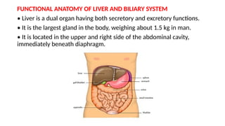
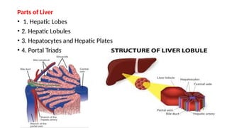
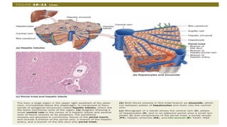
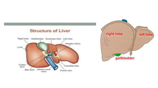
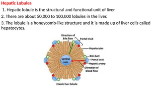
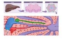
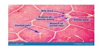
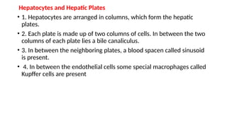
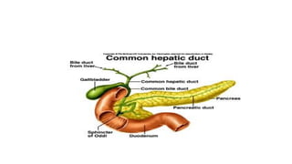
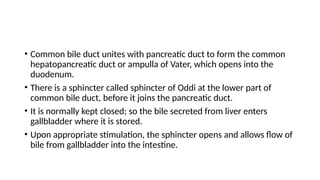
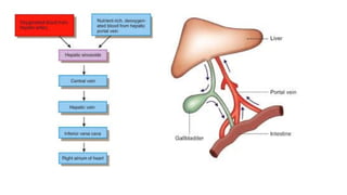
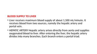
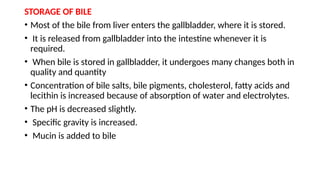
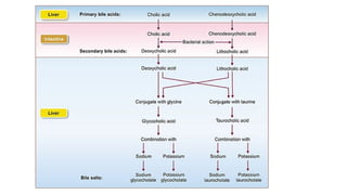
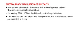
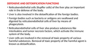
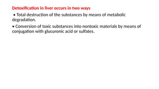
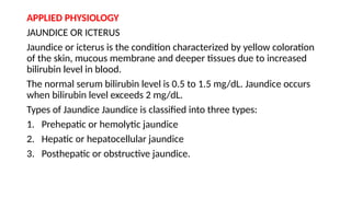
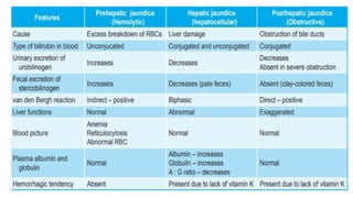
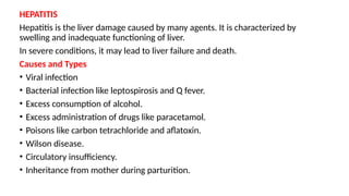
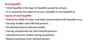
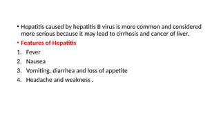
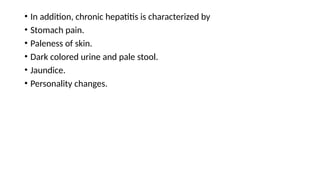
Liver
FUNCTIONAL ANATOMY OF LIVER AND BILIARY SYSTEM
• Liver is a dual organ having both secretory and excretory functions.
• It is the largest gland in the body, weighing about 1.5 kg in man.
• It is located in the upper and right side of the abdominal cavity,
immediately beneath diaphragm.
Parts of Liver
• 1. Hepatic Lobes
• 2. Hepatic Lobules
• 3. Hepatocytes and Hepatic Plates
• 4. Portal Triads
Hepatic Lobules
1. Hepatic lobule is the structural and functional unit of liver.
2. There are about 50,000 to 100,000 lobules in the liver.
3. The lobule is a honeycomb-like structure and it is made up of liver cells called
hepatocytes.
Hepatocytes and Hepatic Plates
• 1. Hepatocytes are arranged in columns, which form the hepatic
plates.
• 2. Each plate is made up of two columns of cells. In between the two
columns of each plate lies a bile canaliculus.
• 3. In between the neighboring plates, a blood spacen called sinusoid
is present.
• 4. In between the endothelial cells some special macrophages called
Kupffer cells are present
Portal Triads
Each lobule is surrounded by many portal triads.
Each portal triad consists of three vessels:
1. A branch of hepatic artery
2. A branch of portal vein
3. A tributary of bile duct
• Branches of hepatic artery and portal vein open into the sinusoid.
Sinusoid opens into the central vein.
• Central vein empties into hepatic vein.
• Bile is secreted by hepatic cells and emptied into bile canaliculus.
• From canaliculus, the bile enters the tributary of bile duct.
• Tributaries of bile duct from canaliculi of neighboring lobules unite to
form small bile ducts.
• These small bile ducts join together and finally form left and right
hepatic ducts, which emerge out of liver.
Functional anatomy of Biliary System
• 1. Biliary system or extrahepatic biliary apparatus is formed by gallbladder
and extrahepatic bile ducts (bile ducts outside the liver).
• 2. Right and left hepatic bile ducts which come out of liver join to form
common hepatic duct.
• 3. It unites with the cystic duct from gallbladder to form common bile duct.
• 4. All these ducts have similar structures.
•
• Common bile duct unites with pancreatic duct to form the common
hepatopancreatic duct or ampulla of Vater, which opens into the
duodenum.
• There is a sphincter called sphincter of Oddi at the lower part of
common bile duct, before it joins the pancreatic duct.
• It is normally kept closed; so the bile secreted from liver enters
gallbladder where it is stored.
• Upon appropriate stimulation, the sphincter opens and allows flow of
bile from gallbladder into the intestine.
BLOOD SUPPLY TO LIVER
• Liver receives maximum blood supply of about 1,500 mL/minute. It
receives blood from two sources, namely the hepatic artery and
portal vein.
• HEPATIC ARTERY Hepatic artery arises directly from aorta and supplies
oxygenated blood to liver. After entering the liver, the hepatic artery
divides into many branches. Each branch enters a portal triad.
PORTAL VEIN
• Portal vein is formed by superior mesenteric vein and splenic vein. It
brings deoxygenated blood from stomach, intestine, spleen and
pancreas.
• Portal blood is rich in monosaccharides and amino acids. It also
contains bile salts, bilirubin, urobilinogen and GI hormones.
• The oxygen content is less in portal blood. Flow of blood from
intestine to liver through portal vein is known as enterohepatic
circulation.
• The blood from hepatic artery mixes with blood from portal vein in
hepatic sinusoids. Hepatic cells obtain oxygen and nutrients from the
sinusoid
HEPATIC VEIN
• Substances synthesized by hepatic cells, waste products and carbon
dioxide are discharged into sinusoids.
• Sinusoids drain them into central vein of the lobule.
• Central veins from many lobules unite to form bigger veins, which
ultimately form hepatic veins (right and left) which open into inferior
vena cava.
PROPERTIES OF BILE
Volume : 800 to 1,200 mL/day
Reaction : Alkaline
pH : 8 to 8.6
Specific gravity : 1.010 to 1.011
Color : Golden yellow or green.
SECRETION OF BILE
• Bile is secreted by hepatocytes.
• The initial bile secreted by hepatocytes contains large quantity of bile
acids, bile pigments, cholesterol, lecithin and fatty acids.
• From hepatocytes, bile is released into canaliculi.
• From here, it passes through small ducts and hepatic ducts and reaches
the common hepatic duct.
• From common hepatic duct, bile is diverted either directly into the
intestine or into the gallbladder.
• Sodium, bicarbonate and water are added to bile when it passes
through the ducts.
• Addition of sodium, bicarbonate and water increases the total quantity
of bile.
STORAGE OF BILE
• Most of the bile from liver enters the gallbladder, where it is stored.
• It is released from gallbladder into the intestine whenever it is
required.
• When bile is stored in gallbladder, it undergoes many changes both in
quality and quantity
• Concentration of bile salts, bile pigments, cholesterol, fatty acids and
lecithin is increased because of absorption of water and electrolytes.
• The pH is decreased slightly.
• Specific gravity is increased.
• Mucin is added to bile
ENTEROHEPATIC CIRCULATION OF BILE SALTS
• 90% to 95% of bile salts from intestine are transported to liver
through enterohepatic circulation.
• Remaining 5% to 10% of the bile salts enter large intestine.
• The bile salts are converted into deoxycholate and lithocholate, which
are excreted in feces.
FUNCTIONS OF BILE SALTS
Bile salts are required for digestion and absorption of fats in the
intestine.
1. Emulsification of Fats
2. Absorption of Fats
3. Choleretic Action
4. Cholagogue Action
5. Laxative Action
6. Prevention of Gallstone Formation
BILE PIGMENTS
• Bile pigments are the excretory products in bile.
• Bilirubin and biliverdin are the two bile pigments and bilirubin is the
major bile pigment in human beings.
• Bile pigments are formed during the breakdown of hemoglobin,
which is released from the destroyed RBCs in the reticuloendothelial
system.
FORMATION AND EXCRETION OF BILE PIGMENTS
Stages of formation and circulation of bile pigments:
1. Senile erythrocytes are destroyed in reticuloendothelial system and hemoglobin is
released from them
2. Hemoglobin is broken into globin and heme
3. Heme is split into iron and the pigment biliverdin
4. Iron goes to iron pool and is reused
5. First formed pigment biliverdin is reduced to bilirubin.
6. Bilirubin is released into blood from the reticuloendothelial cells
7. In blood, the bilirubin is transported by the plasma protein, albumin. Bilirubin
circulating in the blood is called free bilirubin or unconjugated bilirubin
8. Within few hours after entering the circulation, the free bilirubin is taken up by the
liver cells
9. In the liver, it is conjugated with glucuronic acid to form conjugated bilirubin
10. Conjugated bilirubin is then excreted into intestine through bile
FATE OF CONJUGATED BILIRUBIN
Stages of excretion of conjugated bilirubin:
1. In intestine, 50% of the conjugated bilirubin is converted into
urobilinogen by intestinal bacteria. First the conjugated bilirubin is
deconjugated into free bilirubin, which is later reduced into
urobilinogen.
2. Remaining 50% of conjugated bilirubin from intestine is absorbed
into blood and enters the liver through portal vein (enterohepatic
circulation). From liver, it is re­
excreted in bile
3. Most of the urobilinogen from intestine enters liver via enterohepatic
circulation. Later, it is re­
excreted through bile
4. About 5% of urobilinogen is excreted by kidney through urine. In
urine, due to exposure to air, the urobilinogen is converted into urobilin
by oxidation
5. Some of the urobilinogen is excreted in feces as stercobilinogen. In
feces, stercobilinogen is oxidized to stercobilin.
NORMAL PLASMA LEVELS OF BILIRUBIN
• Normal bilirubin (Total bilirubin) content in plasma is 0.5 to 1.5
mg/dL.
• When it exceeds 1mg/dL, the condition is called hyperbilirubinemia.
• When it exceeds 2 mg/dL.
FUNCTIONS OF BILE
• DIGESTIVE FUNCTION
• ABSORPTIVE FUNCTIONS
• EXCRETORY FUNCTIONS
• LAXATIVE ACTION
• ANTISEPTIC ACTION
• CHOLERETIC ACTION
• MAINTENANCE OF pH IN GASTROINTESTINAL TRACT
• PREVENTION OF GALLSTONE FORMATION
• LUBRICATION FUNCTION
• CHOLAGOGUE ACTION
FUNCTIONS OF LIVER
1. METABOLIC FUNCTION
2. STORAGE FUNCTION
3. SYNTHETIC FUNCTION
4. SECRETION OF BILE
5. EXCRETORY FUNCTION
6. HEAT PRODUCTION
7. HEMOPOIETIC FUNCTION
8. HEMOLYTIC FUNCTION
9. INACTIVATION OF HORMONES AND DRUGS
10. DEFENSIVE AND DETOXIFICATION FUNCTIONS
METABOLIC FUNCTION
• Liver is the organ where maximum metabolic reactions such as
metabolism of carbohydrates, proteins, fats, vitamins and many
hormones are carried out.
STORAGE FUNCTION
• Many substances like glycogen, amino acids, iron, folic acid and vitamins
A, B12 and D are stored in liver.
SYNTHETIC FUNCTION
• Liver produces glucose by gluconeogenesis.
• It synthesizes all the plasma proteins and other proteins (except
immunoglobulins) such as clotting factors, complement factors and
hormone binding proteins.
• It also synthesizes steroids, somatomedin and heparin.
SECRETION OF BILE
• Liver secretes bile which contains bile salts, bile pigments,
cholesterol, fatty acids and lecithin.
• The functions of bile are mainly due to bile salts. Bile salts are
required for digestion an absorption of fats in the intestine.
• Bile helps to carry away waste products and breakdown fats, which
are excreted through feces or urine.
EXCRETORY FUNCTION
• Liver excretes cholesterol, bile pigments, heavy metals (like lead,
arsenic and bismuth), toxins, bacteria and virus (like that of yellow
fever) through bile.
HEAT PRODUCTION
• Enormous amount of heat is produced in the liver because of
metabolic reactions.
• Liver is the organ where maximum heat is produced.
HEMOPOIETIC FUNCTION
• In fetus (hepatic stage), liver produces the blood cells.
• It stores vitamin B12 necessary for erythropoiesis and iron necessary
for synthesis of hemoglobin.
• Liver produces thrombopoietin that promotes production of
thrombocytes.
HEMOLYTIC FUNCTION
• The senile RBCs after a lifespan of 120 days are destroyed by
reticuloendothelial cells (Kupffer cells) of liver.
INACTIVATION OF HORMONES AND DRUGS
• Liver catabolizes the hormones such as growth hormone,
parathormone, cortisol, insulin, glucagon and estrogen.
• It also inactivates the drugs, particularly the fat soluble drugs.
• The fatsoluble drugs are converted into water soluble substances,
which are excreted through bile or urine.
DEFENSIVE AND DETOXIFICATION FUNCTIONS
• Reticuloendothelial cells (Kupffer cells) of the liver play an important
role in the defense of the body.
• Liver is also involved in the detoxification of the foreign bodies.
• Foreign bodies such as bacteria or antigens are swallowed and
digested by reticuloendothelial cells of liver by means of
phagocytosis.
• Reticuloendothelial cells of liver also produce substances like
interleukins and tumor necrosis factors, which activate the immune
system of the body.
• Liver cells are involved in the removal of toxic property of various
harmful substances. Removal of toxic property of the harmful agent is
known as detoxification.
Detoxification in liver occurs in two ways
• Total destruction of the substances by means of metabolic
degradation.
• Conversion of toxic substances into nontoxic materials by means of
conjugation with glucuronic acid or sulfates.
APPLIED PHYSIOLOGY
JAUNDICE OR ICTERUS
Jaundice or icterus is the condition characterized by yellow coloration
of the skin, mucous membrane and deeper tissues due to increased
bilirubin level in blood.
The normal serum bilirubin level is 0.5 to 1.5 mg/dL. Jaundice occurs
when bilirubin level exceeds 2 mg/dL.
Types of Jaundice Jaundice is classified into three types:
1. Prehepatic or hemolytic jaundice
2. Hepatic or hepatocellular jaundice
3. Posthepatic or obstructive jaundice.
HEPATITIS
Hepatitis is the liver damage caused by many agents. It is characterized by
swelling and inadequate functioning of liver.
In severe conditions, it may lead to liver failure and death.
Causes and Types
• Viral infection
• Bacterial infection like leptospirosis and Q fever.
• Excess consumption of alcohol.
• Excess administration of drugs like paracetamol.
• Poisons like carbon tetrachloride and aflatoxin.
• Wilson disease.
• Circulatory insufficiency.
• Inheritance from mother during parturition.
Viral Hepatitis
• Viral hepatitis is the type of hepatitis caused by viruses.
• It is caused by two types of viruses, hepatitis A and hepatitis B.
Causes of viral hepatitis
• Mainly by intake of water and food contaminated with hepatitis virus
• Sharing needles with infected persons.
• Accidental prick by infected needle.
• Having unprotected sex with infected persons.
• Inheritance from mother during parturition.
• Blood transfusion from infected donors.
• Hepatitis caused by hepatitis B virus is more common and considered
more serious because it may lead to cirrhosis and cancer of liver.
• Features of Hepatitis
1. Fever
2. Nausea
3. Vomiting, diarrhea and loss of appetite
4. Headache and weakness .
• In addition, chronic hepatitis is characterized by
• Stomach pain.
• Paleness of skin.
• Dark­colored urine and pale stool.
• Jaundice.
• Personality changes.
CIRRHOSIS OF LIVER
Cirrhosis of liver refers to inflammation and damage of parenchyma of
liver. It results in degeneration of hepatic cells and dysfunction of liver.
Causes
1. Infection
2. Retention of bile in liver due to obstruction of ducts of biliary
system.
3. Enlargement of liver due to intoxication.
4. Inflammation around liver (perihepatitis).
5. Infiltration of fat in hepatic cells.
Features
1. Fever, nausea and vomiting
2. Jaundice
3. Increased heart rate and cardiac output
4. Portal hypertension
5. Muscular weakness and wasting of muscles
6. Drowsiness
7. Lack of concentration and confused state of mind
8. Coma in advanced stages.
GALLBLADDER
Bile secreted from liver is stored in gallbladder.
The capacity of gallbladder is approximately 50 mL.
Gallbladder is not essential for life and it is removed (cholecystectomy)
in patients suffering from gallbladder dysfunction.
FUNCTIONS OF GALLBLADDER
• Storage of Bile
• Concentration of Bile
• Alteration of pH of Bile
• Secretion of Mucin
• Maintenance of Pressure in Biliary System
FILLING AND EMPTYING OF GALLBLADDER
Usually, the sphincter of Oddi is closed during fasting and the pressure
in the biliary system is only 7 cm H2 O.
Because of this pressure, the bile from liver enters the gallbladder.
• While taking food or when chyme enters the intestine, gallbladder
contracts along with relaxation of sphincter of Oddi.
• The pressure increases to about 20 cm H2 O. Because of the increase
in pressure, the bile from gallbladder enters the intestine.
• Contraction of gallbladder is influenced by neural and hormonal
factors.
REGULATION OF BILE SECRETION
• Bile secretion is a continuous process though the amount is less
during fasting.
• It starts increasing after meals and continues for three hours.
• Secretion of bile from liver and release of bile from the gallbladder
are influenced by some chemical factors, which are categorized into
three groups:
• Choleretics
• Cholagogue
• Hydrocholeretic agents.
• Choleretics : Substances which increase the secretion of bile from
liver are known as choleretics.
• Effective choleretic agents are: Acetylcholine, Secretin,
Cholecystokinin, Acid chyme in intestine ,Bile salts.
• Cholagogues: Cholagogue is an agent which increases the release of
bile into the intestine by contracting gallbladder.
• Common cholagogues are:
• Bile salts
• Calcium
• Fatty acids
• Amino acids
• Inorganic acids
• All these substances stimulate the secretion of cholecystokinin, which
in turn causes contraction of gallbladder and flow of bile into
intestine.
• Hydrocholeretic Agents:
• Hydrocholeretic agent is a substance which causes the secretion of
bile from liver, with large amount of water and less amount of solids.
• Hydrochloric acid is a hydrocholeretic agent.
GALLSTONES:
• Gallstone is a solid crystal deposit that is formed by cholesterol, calcium
ions and bile pigments in the gallbladder or bile duct.
• Cholelithiasis is the presence of gallstones in gallbladder.
• Choledocholithiasis is the presence of gallstones in the bile ducts.
Causes for Gallstone Formation
1. Reduction in bile salts and/or lecithin
2. Excess of cholesterol
3. Disturbed cholesterol metabolism
4. Excess of calcium ions due to increased concentration of bile.
5. Damage or infection of gallbladder epithelium
6. Obstruction of bile flow from the gallbladder
Diagnosis of Gallstone
Ultrasound scanning and cholangiography.
Symptoms
Common feature of gallstone is the pain in stomach.
Nausea, vomiting, abdominal bloating and indigestion.
Treatment
Cholecystectomy.
Liver physiology . power point presentation

Liver physiology . power point presentation

  • 1.
  • 2.
    FUNCTIONAL ANATOMY OFLIVER AND BILIARY SYSTEM • Liver is a dual organ having both secretory and excretory functions. • It is the largest gland in the body, weighing about 1.5 kg in man. • It is located in the upper and right side of the abdominal cavity, immediately beneath diaphragm.
  • 3.
    Parts of Liver •1. Hepatic Lobes • 2. Hepatic Lobules • 3. Hepatocytes and Hepatic Plates • 4. Portal Triads
  • 6.
    Hepatic Lobules 1. Hepaticlobule is the structural and functional unit of liver. 2. There are about 50,000 to 100,000 lobules in the liver. 3. The lobule is a honeycomb-like structure and it is made up of liver cells called hepatocytes.
  • 9.
    Hepatocytes and HepaticPlates • 1. Hepatocytes are arranged in columns, which form the hepatic plates. • 2. Each plate is made up of two columns of cells. In between the two columns of each plate lies a bile canaliculus. • 3. In between the neighboring plates, a blood spacen called sinusoid is present. • 4. In between the endothelial cells some special macrophages called Kupffer cells are present
  • 11.
    Portal Triads Each lobuleis surrounded by many portal triads. Each portal triad consists of three vessels: 1. A branch of hepatic artery 2. A branch of portal vein 3. A tributary of bile duct
  • 12.
    • Branches ofhepatic artery and portal vein open into the sinusoid. Sinusoid opens into the central vein. • Central vein empties into hepatic vein. • Bile is secreted by hepatic cells and emptied into bile canaliculus. • From canaliculus, the bile enters the tributary of bile duct. • Tributaries of bile duct from canaliculi of neighboring lobules unite to form small bile ducts. • These small bile ducts join together and finally form left and right hepatic ducts, which emerge out of liver.
  • 13.
    Functional anatomy ofBiliary System • 1. Biliary system or extrahepatic biliary apparatus is formed by gallbladder and extrahepatic bile ducts (bile ducts outside the liver). • 2. Right and left hepatic bile ducts which come out of liver join to form common hepatic duct. • 3. It unites with the cystic duct from gallbladder to form common bile duct. • 4. All these ducts have similar structures.
  • 14.
  • 15.
    • Common bileduct unites with pancreatic duct to form the common hepatopancreatic duct or ampulla of Vater, which opens into the duodenum. • There is a sphincter called sphincter of Oddi at the lower part of common bile duct, before it joins the pancreatic duct. • It is normally kept closed; so the bile secreted from liver enters gallbladder where it is stored. • Upon appropriate stimulation, the sphincter opens and allows flow of bile from gallbladder into the intestine.
  • 17.
    BLOOD SUPPLY TOLIVER • Liver receives maximum blood supply of about 1,500 mL/minute. It receives blood from two sources, namely the hepatic artery and portal vein. • HEPATIC ARTERY Hepatic artery arises directly from aorta and supplies oxygenated blood to liver. After entering the liver, the hepatic artery divides into many branches. Each branch enters a portal triad.
  • 18.
    PORTAL VEIN • Portalvein is formed by superior mesenteric vein and splenic vein. It brings deoxygenated blood from stomach, intestine, spleen and pancreas. • Portal blood is rich in monosaccharides and amino acids. It also contains bile salts, bilirubin, urobilinogen and GI hormones. • The oxygen content is less in portal blood. Flow of blood from intestine to liver through portal vein is known as enterohepatic circulation. • The blood from hepatic artery mixes with blood from portal vein in hepatic sinusoids. Hepatic cells obtain oxygen and nutrients from the sinusoid
  • 19.
    HEPATIC VEIN • Substancessynthesized by hepatic cells, waste products and carbon dioxide are discharged into sinusoids. • Sinusoids drain them into central vein of the lobule. • Central veins from many lobules unite to form bigger veins, which ultimately form hepatic veins (right and left) which open into inferior vena cava. PROPERTIES OF BILE Volume : 800 to 1,200 mL/day Reaction : Alkaline pH : 8 to 8.6 Specific gravity : 1.010 to 1.011 Color : Golden yellow or green.
  • 21.
    SECRETION OF BILE •Bile is secreted by hepatocytes. • The initial bile secreted by hepatocytes contains large quantity of bile acids, bile pigments, cholesterol, lecithin and fatty acids. • From hepatocytes, bile is released into canaliculi. • From here, it passes through small ducts and hepatic ducts and reaches the common hepatic duct. • From common hepatic duct, bile is diverted either directly into the intestine or into the gallbladder. • Sodium, bicarbonate and water are added to bile when it passes through the ducts. • Addition of sodium, bicarbonate and water increases the total quantity of bile.
  • 22.
    STORAGE OF BILE •Most of the bile from liver enters the gallbladder, where it is stored. • It is released from gallbladder into the intestine whenever it is required. • When bile is stored in gallbladder, it undergoes many changes both in quality and quantity • Concentration of bile salts, bile pigments, cholesterol, fatty acids and lecithin is increased because of absorption of water and electrolytes. • The pH is decreased slightly. • Specific gravity is increased. • Mucin is added to bile
  • 24.
    ENTEROHEPATIC CIRCULATION OFBILE SALTS • 90% to 95% of bile salts from intestine are transported to liver through enterohepatic circulation. • Remaining 5% to 10% of the bile salts enter large intestine. • The bile salts are converted into deoxycholate and lithocholate, which are excreted in feces.
  • 25.
    FUNCTIONS OF BILESALTS Bile salts are required for digestion and absorption of fats in the intestine. 1. Emulsification of Fats 2. Absorption of Fats 3. Choleretic Action 4. Cholagogue Action 5. Laxative Action 6. Prevention of Gallstone Formation
  • 26.
    BILE PIGMENTS • Bilepigments are the excretory products in bile. • Bilirubin and biliverdin are the two bile pigments and bilirubin is the major bile pigment in human beings. • Bile pigments are formed during the breakdown of hemoglobin, which is released from the destroyed RBCs in the reticuloendothelial system.
  • 27.
    FORMATION AND EXCRETIONOF BILE PIGMENTS Stages of formation and circulation of bile pigments: 1. Senile erythrocytes are destroyed in reticuloendothelial system and hemoglobin is released from them 2. Hemoglobin is broken into globin and heme 3. Heme is split into iron and the pigment biliverdin 4. Iron goes to iron pool and is reused 5. First formed pigment biliverdin is reduced to bilirubin. 6. Bilirubin is released into blood from the reticuloendothelial cells 7. In blood, the bilirubin is transported by the plasma protein, albumin. Bilirubin circulating in the blood is called free bilirubin or unconjugated bilirubin 8. Within few hours after entering the circulation, the free bilirubin is taken up by the liver cells 9. In the liver, it is conjugated with glucuronic acid to form conjugated bilirubin 10. Conjugated bilirubin is then excreted into intestine through bile
  • 29.
    FATE OF CONJUGATEDBILIRUBIN Stages of excretion of conjugated bilirubin: 1. In intestine, 50% of the conjugated bilirubin is converted into urobilinogen by intestinal bacteria. First the conjugated bilirubin is deconjugated into free bilirubin, which is later reduced into urobilinogen. 2. Remaining 50% of conjugated bilirubin from intestine is absorbed into blood and enters the liver through portal vein (enterohepatic circulation). From liver, it is re­ excreted in bile
  • 30.
    3. Most ofthe urobilinogen from intestine enters liver via enterohepatic circulation. Later, it is re­ excreted through bile 4. About 5% of urobilinogen is excreted by kidney through urine. In urine, due to exposure to air, the urobilinogen is converted into urobilin by oxidation 5. Some of the urobilinogen is excreted in feces as stercobilinogen. In feces, stercobilinogen is oxidized to stercobilin.
  • 31.
    NORMAL PLASMA LEVELSOF BILIRUBIN • Normal bilirubin (Total bilirubin) content in plasma is 0.5 to 1.5 mg/dL. • When it exceeds 1mg/dL, the condition is called hyperbilirubinemia. • When it exceeds 2 mg/dL.
  • 32.
    FUNCTIONS OF BILE •DIGESTIVE FUNCTION • ABSORPTIVE FUNCTIONS • EXCRETORY FUNCTIONS • LAXATIVE ACTION • ANTISEPTIC ACTION • CHOLERETIC ACTION • MAINTENANCE OF pH IN GASTROINTESTINAL TRACT • PREVENTION OF GALLSTONE FORMATION • LUBRICATION FUNCTION • CHOLAGOGUE ACTION
  • 33.
    FUNCTIONS OF LIVER 1.METABOLIC FUNCTION 2. STORAGE FUNCTION 3. SYNTHETIC FUNCTION 4. SECRETION OF BILE 5. EXCRETORY FUNCTION 6. HEAT PRODUCTION 7. HEMOPOIETIC FUNCTION 8. HEMOLYTIC FUNCTION 9. INACTIVATION OF HORMONES AND DRUGS 10. DEFENSIVE AND DETOXIFICATION FUNCTIONS
  • 34.
    METABOLIC FUNCTION • Liveris the organ where maximum metabolic reactions such as metabolism of carbohydrates, proteins, fats, vitamins and many hormones are carried out. STORAGE FUNCTION • Many substances like glycogen, amino acids, iron, folic acid and vitamins A, B12 and D are stored in liver. SYNTHETIC FUNCTION • Liver produces glucose by gluconeogenesis. • It synthesizes all the plasma proteins and other proteins (except immunoglobulins) such as clotting factors, complement factors and hormone binding proteins. • It also synthesizes steroids, somatomedin and heparin.
  • 35.
    SECRETION OF BILE •Liver secretes bile which contains bile salts, bile pigments, cholesterol, fatty acids and lecithin. • The functions of bile are mainly due to bile salts. Bile salts are required for digestion an absorption of fats in the intestine. • Bile helps to carry away waste products and breakdown fats, which are excreted through feces or urine. EXCRETORY FUNCTION • Liver excretes cholesterol, bile pigments, heavy metals (like lead, arsenic and bismuth), toxins, bacteria and virus (like that of yellow fever) through bile.
  • 36.
    HEAT PRODUCTION • Enormousamount of heat is produced in the liver because of metabolic reactions. • Liver is the organ where maximum heat is produced. HEMOPOIETIC FUNCTION • In fetus (hepatic stage), liver produces the blood cells. • It stores vitamin B12 necessary for erythropoiesis and iron necessary for synthesis of hemoglobin. • Liver produces thrombopoietin that promotes production of thrombocytes.
  • 37.
    HEMOLYTIC FUNCTION • Thesenile RBCs after a lifespan of 120 days are destroyed by reticuloendothelial cells (Kupffer cells) of liver. INACTIVATION OF HORMONES AND DRUGS • Liver catabolizes the hormones such as growth hormone, parathormone, cortisol, insulin, glucagon and estrogen. • It also inactivates the drugs, particularly the fat soluble drugs. • The fatsoluble drugs are converted into water soluble substances, which are excreted through bile or urine.
  • 38.
    DEFENSIVE AND DETOXIFICATIONFUNCTIONS • Reticuloendothelial cells (Kupffer cells) of the liver play an important role in the defense of the body. • Liver is also involved in the detoxification of the foreign bodies. • Foreign bodies such as bacteria or antigens are swallowed and digested by reticuloendothelial cells of liver by means of phagocytosis. • Reticuloendothelial cells of liver also produce substances like interleukins and tumor necrosis factors, which activate the immune system of the body. • Liver cells are involved in the removal of toxic property of various harmful substances. Removal of toxic property of the harmful agent is known as detoxification.
  • 39.
    Detoxification in liveroccurs in two ways • Total destruction of the substances by means of metabolic degradation. • Conversion of toxic substances into nontoxic materials by means of conjugation with glucuronic acid or sulfates.
  • 40.
    APPLIED PHYSIOLOGY JAUNDICE ORICTERUS Jaundice or icterus is the condition characterized by yellow coloration of the skin, mucous membrane and deeper tissues due to increased bilirubin level in blood. The normal serum bilirubin level is 0.5 to 1.5 mg/dL. Jaundice occurs when bilirubin level exceeds 2 mg/dL. Types of Jaundice Jaundice is classified into three types: 1. Prehepatic or hemolytic jaundice 2. Hepatic or hepatocellular jaundice 3. Posthepatic or obstructive jaundice.
  • 42.
    HEPATITIS Hepatitis is theliver damage caused by many agents. It is characterized by swelling and inadequate functioning of liver. In severe conditions, it may lead to liver failure and death. Causes and Types • Viral infection • Bacterial infection like leptospirosis and Q fever. • Excess consumption of alcohol. • Excess administration of drugs like paracetamol. • Poisons like carbon tetrachloride and aflatoxin. • Wilson disease. • Circulatory insufficiency. • Inheritance from mother during parturition.
  • 43.
    Viral Hepatitis • Viralhepatitis is the type of hepatitis caused by viruses. • It is caused by two types of viruses, hepatitis A and hepatitis B. Causes of viral hepatitis • Mainly by intake of water and food contaminated with hepatitis virus • Sharing needles with infected persons. • Accidental prick by infected needle. • Having unprotected sex with infected persons. • Inheritance from mother during parturition. • Blood transfusion from infected donors.
  • 44.
    • Hepatitis causedby hepatitis B virus is more common and considered more serious because it may lead to cirrhosis and cancer of liver. • Features of Hepatitis 1. Fever 2. Nausea 3. Vomiting, diarrhea and loss of appetite 4. Headache and weakness .
  • 45.
    • In addition,chronic hepatitis is characterized by • Stomach pain. • Paleness of skin. • Dark­colored urine and pale stool. • Jaundice. • Personality changes.
  • 46.
    CIRRHOSIS OF LIVER Cirrhosisof liver refers to inflammation and damage of parenchyma of liver. It results in degeneration of hepatic cells and dysfunction of liver. Causes 1. Infection 2. Retention of bile in liver due to obstruction of ducts of biliary system. 3. Enlargement of liver due to intoxication. 4. Inflammation around liver (perihepatitis). 5. Infiltration of fat in hepatic cells.
  • 47.
    Features 1. Fever, nauseaand vomiting 2. Jaundice 3. Increased heart rate and cardiac output 4. Portal hypertension 5. Muscular weakness and wasting of muscles 6. Drowsiness 7. Lack of concentration and confused state of mind 8. Coma in advanced stages.
  • 48.
    GALLBLADDER Bile secreted fromliver is stored in gallbladder. The capacity of gallbladder is approximately 50 mL. Gallbladder is not essential for life and it is removed (cholecystectomy) in patients suffering from gallbladder dysfunction.
  • 49.
    FUNCTIONS OF GALLBLADDER •Storage of Bile • Concentration of Bile • Alteration of pH of Bile • Secretion of Mucin • Maintenance of Pressure in Biliary System FILLING AND EMPTYING OF GALLBLADDER Usually, the sphincter of Oddi is closed during fasting and the pressure in the biliary system is only 7 cm H2 O. Because of this pressure, the bile from liver enters the gallbladder.
  • 50.
    • While takingfood or when chyme enters the intestine, gallbladder contracts along with relaxation of sphincter of Oddi. • The pressure increases to about 20 cm H2 O. Because of the increase in pressure, the bile from gallbladder enters the intestine. • Contraction of gallbladder is influenced by neural and hormonal factors.
  • 51.
    REGULATION OF BILESECRETION • Bile secretion is a continuous process though the amount is less during fasting. • It starts increasing after meals and continues for three hours. • Secretion of bile from liver and release of bile from the gallbladder are influenced by some chemical factors, which are categorized into three groups: • Choleretics • Cholagogue • Hydrocholeretic agents.
  • 52.
    • Choleretics :Substances which increase the secretion of bile from liver are known as choleretics. • Effective choleretic agents are: Acetylcholine, Secretin, Cholecystokinin, Acid chyme in intestine ,Bile salts. • Cholagogues: Cholagogue is an agent which increases the release of bile into the intestine by contracting gallbladder.
  • 53.
    • Common cholagoguesare: • Bile salts • Calcium • Fatty acids • Amino acids • Inorganic acids • All these substances stimulate the secretion of cholecystokinin, which in turn causes contraction of gallbladder and flow of bile into intestine.
  • 54.
    • Hydrocholeretic Agents: •Hydrocholeretic agent is a substance which causes the secretion of bile from liver, with large amount of water and less amount of solids. • Hydrochloric acid is a hydrocholeretic agent.
  • 55.
    GALLSTONES: • Gallstone isa solid crystal deposit that is formed by cholesterol, calcium ions and bile pigments in the gallbladder or bile duct. • Cholelithiasis is the presence of gallstones in gallbladder. • Choledocholithiasis is the presence of gallstones in the bile ducts. Causes for Gallstone Formation 1. Reduction in bile salts and/or lecithin 2. Excess of cholesterol 3. Disturbed cholesterol metabolism 4. Excess of calcium ions due to increased concentration of bile. 5. Damage or infection of gallbladder epithelium 6. Obstruction of bile flow from the gallbladder
  • 56.
    Diagnosis of Gallstone Ultrasoundscanning and cholangiography. Symptoms Common feature of gallstone is the pain in stomach. Nausea, vomiting, abdominal bloating and indigestion. Treatment Cholecystectomy.