SlideShare a Scribd company logo
PROTEINS
SESSION 3
What are proteins?
• Proteins are large, complex molecules that play many critical roles in the
body.
• They do most of the work in cells and are required for the structure,
function, and regulation of the body’s tissues and organs.
• Proteins are made up of hundreds or thousands of smaller units called
amino acids, which are attached to one another in long chains.
• There are 20 different types of amino acids that can be combined to make
a protein.
• The sequence of amino acids determines each protein’s unique 3-
dimensional structure and its specific function.
• Amino acids are coded by combinations of three DNA building blocks
(nucleotides), determined by the sequence of genes.
AMINO ACIDS
• Cells produce proteins with strikingly different properties
and activities by joining the same 20 amino acids in many
different combinations and sequences.
• This indicates that the properties of proteins are
determined by the physical and chemical properties of
their monomer units, the amino acids.
• Amino acids are the basic structural units of proteins
consisting of an amino group, (-NH2) a carboxyl (-COOH)
group a hydrogen (H) atom and a (variable) distinctive (R)
group.
• All of the substituents in amino acid are attached
(bonded) to a central α carbon atom.
• This carbon atom is called α because it is bonded to the
carboxyl (acidic) group.
 In neutral solution
(PH = 7), both the
α- amino and α
carboxyl group are
ionized resulting
the charged form
of an amino acids
called zwitterion
(dipolar) as shown
in the next slide.
 Out of the 20 amino acids, proline is not an α amino
acid rather an α - imino acid. Except for glycine, all
amino acids contain at least one asymmetric carbon
atom (the α - carbon atom).
Stereochemistry
Stereochemistry mainly emphasizes the configuration of amino
acids at the α carbon atom, having either D or L- isomers
Classification of Amino Acids
They are classified based on;
• Structure
• Electrochemical classification
• Biological and physiological
• Metabolic fate
Structural classification: This classification is based on the side chain
radicals (R-groups)
R groups
with
Aliphatic side chains
e.g glycine, alanine,
valine, leucine,
isoleucine
Sulfur group:
cysteine,
methionine
Aromatic group;
phenylalanine,
tyrosine,
tryptophan,
Imino group;
proline
Electrochemical
Amino acids could also be classified based on their acid – base properties
Basic amino acids
1. Lysine
2. Arginine
Neutral amino acids
1. Asparagine
2. Serine
3. Threonine
4. Glutamine
Acidic amino acids
1. Glutamic acid
2. Aspartic acid
Biological
This classification is based on the functional property of amino acids for the organism.
• Essential: Amino acids which are not synthesized in the body and must be provided in the
diet to meet an animal’s metabolic needs are called essential amino acids.
• Non essential: These amino acids are need not be provided through diet, because they
can be biosynthesized in adequate amounts within the organism.
• Semi-essential amino acids: they can be synthesized within the organism but their
synthesis is not in sufficient amounts. In that they should also be provided in the diet.
Essential Non-essential Semi-essential
Arginine, histidine,
leucine, isoleucine,
lysine, methionine,
phenylalanine,
Threonine, Tryptophan,
Valine
Alanine, Asparagine,
aspartine, cysteine,
glutamic acid,
glutamine, glycine,
proline, serine, tyrosine
Histidine, Arginine
Metabolic fate of each amino acid
Amino acids can be classified here as Glucogenic (potentially be converted to
glucose), ketogenic (potentially be converted to ketone bodies) and both glucogenic
and ketogenic.
Glucogenic amino acids: Those amino acids in which their carbon skeleton gets
degraded to pyrurate, α ketoglutarate, succinyl CoA, fumrate and oxaloacetate and
then converted to Glucose and Glycogen, are called as Glucogenic amino acids.
These include:- Alanine, cysteine, glycine, Arginine, glutamine, Isoleucine, tyrosine
Ketogenic amino acids: Those amino acids in which their carbon skeleton is
degraded to Acetoacetyl CoA, or acetyl CoA. then converted to acetone and β-
hydroxy butyrate which are the main ketone bodies are called ketogenic amino acids.
These includes:- Phenylalanine, tyrosine, tryptophan, isoleucine, leucine, and lysine.
These amino acids have ability to form ketone bodies which is particularly evident in
untreated diabetes mellitus in which large amounts of ketone bodies are produced by
the liver (i.e. not only from fatty acids but also from ketogenic amino acids)
Ketogenic and glucogenic amino acids: The division between ketogenic and
glucogenic amino acids is not sharp for amino acids (Tryptophan, phenylalanine,
tyrosine and Isoleucine are both ketogenic and glucogenic).
Some of the amino acids that can be converted in to pyruvate, particularly (Alanine,
Cysteine and serine, can also potentially form acetoacetate via acetyl CoA
especially in severe starvation and untreated diabetes mellitus.
Peptides
• Proteins are macromolecules with a backbone formed by polymerization of
amino acids in a polyamide structure.
• These amide bonds in protein, known as peptide bonds formed by linkage of α -
carboxyl group of one amino acid with α- amino groups of the next amino acid
by amide bonds.
• During the formation of a peptide bond, a molecule of water is eliminated as
shown below:-
• A peptide chain consisting of two amino acid residues is called a dipeptide,
three amino acids
tripeptide (e. g Glutathione)
Glutathione
• Glutathione is a tripeptide
formed from amino acids
glutamate, cysteine and
Glycine, linked together in
that order. The glutamate is
linked to cysteine through
the γ- carboxyl group and α
- amino group of cysteine.
• Here, the carboxyl group is
first activated by ATP to
form an acyl-phosphate
derivative which is then
attacked by cysteine amino
group then undergoes
condensation with glycine.
• The role of GSH as a reductant is extremely important particularly in the highly
oxidizing
environment of the erythrocyte.
• The sulfhydryl of GSH can be used to reduce peroxides formed during oxygen
transport. The resulting oxidized form of GSH consists of two molecules
disulfide bonded together (abbreviated GSSG).
• The enzyme glutathione reductase utilizes NADPH as a cofactor to reduce
GSSG back to two moles of GSH.
• Hence, the pentose phosphate pathway is an extremely important pathway of
erythrocytes for the continuing production of the NADPH needed by glutathione
reductase.
• In fact as much as 10% of glucose consumption, by erythrocytes, may be
mediated by the pentose phosphate pathway.
Classification of Proteins
PROTEINS
• Proteins are macromolecules with a backbone formed by polymerization of
amino acids in a polyamide structure.
Class Assignment 2:
1. List functions of proteins (3 marks)
2. Using the flow chart on slide 18, describe the
classification of proteins (7 marks)
Protein Organization
Primary structure
The simplest level of protein structure, primary structure, is simply the
sequence of amino acids in a polypeptide chain.
For example, the hormone insulin has two polypeptide chains, A and B,
shown in diagram below.
(The insulin molecule shown here is cow insulin, although its structure is
similar to that of human insulin.) Each chain has its own set of amino acids,
assembled in a particular order.
For instance, the sequence of the A chain starts with glycine at the N-
terminus and ends with asparagine at the C-terminus, and is different from
the sequence of the B chain.
• The sequence of a protein is determined by the DNA of the gene that
encodes the protein (or that encodes a portion of the protein, for multi-
subunit proteins).
• A change in the gene's DNA sequence may lead to a change in the
amino acid sequence of the protein.
• Even changing just one amino acid in a protein’s sequence can affect
the protein’s overall structure and function.
• For instance, a single amino acid change is associated with sickle cell
anemia, an inherited disease that affects red blood cells.
• In sickle cell anemia, one of the polypeptide chains that make up
hemoglobin, the protein that carries oxygen in the blood, has a slight
sequence change.
• The glutamic acid that is normally the sixth amino acid of the
hemoglobin β chain (one of two types of protein chains that make up
hemoglobin) is replaced by a valine.
Secondary Structure
• The next level of protein structure, secondary structure, refers to local folded
structures that form within a polypeptide due to interactions between atoms of
the backbone.
• (The backbone just refers to the polypeptide chain apart from the R groups – so
all we mean here is that secondary structure does not involve R group atoms.)
• The most common types of secondary structures are the α helix and the β
pleated sheet.
• Both structures are held in shape by hydrogen bonds, which form between the
carbonyl O of one amino acid and the amino H of another.
Tertiary structure
• The overall three-dimensional structure of a polypeptide is called its tertiary
structure. The tertiary structure is primarily due to interactions between the R
groups of the amino acids that make up the protein.
• R group interactions that contribute to tertiary structure include hydrogen
bonding, ionic bonding, dipole-dipole interactions, and London dispersion
forces – basically, the whole gamut of non-covalent bonds.
• For example, R groups with like charges repel one another, while those with
opposite charges can form an ionic bond.
• Similarly, polar R groups can form hydrogen bonds and other dipole-dipole
interactions.
• Also important to tertiary structure are hydrophobic interactions, in which
amino acids with nonpolar, hydrophobic R groups cluster together on the
inside of the protein, leaving hydrophilic amino acids on the outside to interact
with surrounding water molecules.
• Finally, there’s one special type of covalent bond that can contribute to tertiary
structure: the disulfide bond.
• Disulfide bonds, covalent linkages between the sulfur-containing side chains of
cysteines, are much stronger than the other types of bonds that contribute to
tertiary structure.
• They act like molecular "safety pins," keeping parts of the polypeptide firmly
attached to one another.
Quaternary structure
• Many proteins are made up of a single polypeptide chain and have only three
levels of structure (the ones we’ve just discussed).
• However, some proteins are made up of multiple polypeptide chains, also
known as subunits. When these subunits come together, they give the protein
its quaternary structure.
• As mentioned earlier, hemoglobin carries oxygen in the blood and is made up of
four subunits, two each of the α and β types.
• Another example is DNA polymerase, an enzyme that synthesizes new strands
of DNA and is composed of ten subunits^55start superscript, 5, end superscript.
• In general, the same types of interactions that contribute to tertiary structure
(mostly weak interactions, such as hydrogen bonding) also hold the subunits
together to give quaternary structure
Protein Denaturation (Reading
Assignment)
• The phenomenon of disorganization of native protein structure is known as
denaturation.
• Denaturation results in the loss of secondary, tertiary and quaternary
structureof proteins. This involves a change in physical, chemical and
biological properties of protein molecule
Agents of denaturation
• Physical agents: Heat, violent shaking, X-ravs,UV radiation.
• Chemical agents : Acids, alkalies, organic solvents(ether,alcohol), salts of
heavy metals (Pb, Hg), urea,salicylate.
Characteristics of denaturation
a) The native helical structure of protein is lost
b) The primary structure of a protein with peptide linkages remains intact i.e.,
peptide bonds are not hydrolysed.
c) The protein loses its biologicalactivity.
d) Denatured protein becomes insoluble in the solventin which it was originally
soluble.
e) The viscosity ol denatured protein (solution) increaseswhile its surface tension
decreases.
f) Denaturation is associated with increase in ionizable and sulfhydryl groups of
protein. This is due to loss of hydrogen and disulfide bonds.
g) Denatured protein is more easily digested. This is due to increased exposure of
peptide bonds to enzymes. Cooking causes protein denaturation and, therefore,
cooked food (protein)is more easily digested
h) Denaturation is usually irreversible. For instance, omelet can be prepared from an
egg (protein-albumin) but the reversal is not possible.
i) Careful denaturation is sometimes reversible (known as renaturation). Hemoglobin
undergoes denaturation in the presence of salicylate. By removal of salicylate,
hemoglobin is renatured.
j) Denatured protein cannot be crystallized
• Flocculation:
• lt is the process of protein precipitation at isoelectric pH.
• The precipitate is referred to as flocculum. Casein (milk protein) can be
easily precipitated when adjusted to isoelectric pH (4.6 by dilute acetic
acid.
• Flocculation is reversible.
• On application of heat, flocculum can be converted into an
irreversible mass, coagulum
Coagulation:
• The term 'coagulum' refers to a semi-solid viscous precipitate of protein.
reversible denaturation results in coagulation.
• Coagulation is optimum and requires lowest temperature at isoelectric
pH. Albumins and globulins (to a lesser extent) are coagulable proteins.
• Heat coagulation test is commonly used to detect the presence of
albumin in urine.
Protein Separation Methods (Reading
Assessment)
Group 1: Lydia, Brian, David (Protein Denaturation)
Group 2: Maxwell, Lennick, Gabriel, Peter, Reuben,
Dawn, Kevin, Eric, Ian (Protein separation methods)
Oral assessment (5 marks)

More Related Content

Similar to Lecture 3 (1).pdf

Amino acid metabolism BY BISWANATH PRUSTY
Amino acid metabolism BY BISWANATH PRUSTYAmino acid metabolism BY BISWANATH PRUSTY
Amino acid metabolism BY BISWANATH PRUSTY
College of pharmaceutical sciences
 
Amino acids and protein
Amino acids and proteinAmino acids and protein
Amino acids and protein
Yogesh Kaushik
 
Structures of Amino Acids
Structures of Amino AcidsStructures of Amino Acids
Structures of Amino Acids
Rahul SIR
 
amino acids lecture.ppt
amino acids lecture.pptamino acids lecture.ppt
amino acids lecture.ppt
eman badr
 
Amino acids and Peptides 2021.pdf
Amino acids and Peptides 2021.pdfAmino acids and Peptides 2021.pdf
Amino acids and Peptides 2021.pdf
Floyd112
 
Biomolecule Examples
Biomolecule ExamplesBiomolecule Examples
Biomolecule Examples
BeatrizAspiras
 
Chemistry of Protein.pdf
Chemistry of Protein.pdfChemistry of Protein.pdf
Chemistry of Protein.pdf
AttiqueJavaid1
 
amino_acids_and_proteins_lecture_4.ppt
amino_acids_and_proteins_lecture_4.pptamino_acids_and_proteins_lecture_4.ppt
amino_acids_and_proteins_lecture_4.ppt
WynethGabate
 
week 1 PHS 302 2022.pptx
week 1 PHS 302 2022.pptxweek 1 PHS 302 2022.pptx
week 1 PHS 302 2022.pptx
asmaa saleh
 
Amino acids
Amino acidsAmino acids
Amino acids
Atif Nauman
 
Amino acids structure
Amino acids structureAmino acids structure
Amino acids structure
Narottam Agrawal
 
Amino_Acids_and_Proteins.ppt
Amino_Acids_and_Proteins.pptAmino_Acids_and_Proteins.ppt
Amino_Acids_and_Proteins.ppt
KhalidBassiouny1
 
Amino acids and protein
Amino acids and proteinAmino acids and protein
Amino acids and protein
UNIVERSITY OF SARGODHA
 
Amino acid by vidan biology
Amino acid by vidan biologyAmino acid by vidan biology
Amino acid by vidan biology
vidan biology
 
Unit-2.pptx
Unit-2.pptxUnit-2.pptx
Unit-2.pptx
GaddafaaDhinsaa
 
PROTEIN AND AMINO ACIDS-CLASS-I.pptx
PROTEIN AND AMINO ACIDS-CLASS-I.pptxPROTEIN AND AMINO ACIDS-CLASS-I.pptx
PROTEIN AND AMINO ACIDS-CLASS-I.pptx
JaanuJaanu20
 
Proteins.
 Proteins. Proteins.
Proteins.
Pardeep kaushal
 
Amino acid -Classification of amino acid, Properties, peptides-Types and bio...
Amino acid -Classification of amino acid, Properties,  peptides-Types and bio...Amino acid -Classification of amino acid, Properties,  peptides-Types and bio...
Amino acid -Classification of amino acid, Properties, peptides-Types and bio...
SoniaBajaj10
 
ProteinMetbolism part1
ProteinMetbolism part1ProteinMetbolism part1
ProteinMetbolism part1
ganeshbond
 

Similar to Lecture 3 (1).pdf (20)

Amino acid metabolism BY BISWANATH PRUSTY
Amino acid metabolism BY BISWANATH PRUSTYAmino acid metabolism BY BISWANATH PRUSTY
Amino acid metabolism BY BISWANATH PRUSTY
 
Amino acids and protein
Amino acids and proteinAmino acids and protein
Amino acids and protein
 
Structures of Amino Acids
Structures of Amino AcidsStructures of Amino Acids
Structures of Amino Acids
 
amino acids lecture.ppt
amino acids lecture.pptamino acids lecture.ppt
amino acids lecture.ppt
 
Amino acids and Peptides 2021.pdf
Amino acids and Peptides 2021.pdfAmino acids and Peptides 2021.pdf
Amino acids and Peptides 2021.pdf
 
Biomolecule Examples
Biomolecule ExamplesBiomolecule Examples
Biomolecule Examples
 
Chemistry of Protein.pdf
Chemistry of Protein.pdfChemistry of Protein.pdf
Chemistry of Protein.pdf
 
amino_acids_and_proteins_lecture_4.ppt
amino_acids_and_proteins_lecture_4.pptamino_acids_and_proteins_lecture_4.ppt
amino_acids_and_proteins_lecture_4.ppt
 
week 1 PHS 302 2022.pptx
week 1 PHS 302 2022.pptxweek 1 PHS 302 2022.pptx
week 1 PHS 302 2022.pptx
 
Amino acids
Amino acidsAmino acids
Amino acids
 
PROTEINS
PROTEINSPROTEINS
PROTEINS
 
Amino acids structure
Amino acids structureAmino acids structure
Amino acids structure
 
Amino_Acids_and_Proteins.ppt
Amino_Acids_and_Proteins.pptAmino_Acids_and_Proteins.ppt
Amino_Acids_and_Proteins.ppt
 
Amino acids and protein
Amino acids and proteinAmino acids and protein
Amino acids and protein
 
Amino acid by vidan biology
Amino acid by vidan biologyAmino acid by vidan biology
Amino acid by vidan biology
 
Unit-2.pptx
Unit-2.pptxUnit-2.pptx
Unit-2.pptx
 
PROTEIN AND AMINO ACIDS-CLASS-I.pptx
PROTEIN AND AMINO ACIDS-CLASS-I.pptxPROTEIN AND AMINO ACIDS-CLASS-I.pptx
PROTEIN AND AMINO ACIDS-CLASS-I.pptx
 
Proteins.
 Proteins. Proteins.
Proteins.
 
Amino acid -Classification of amino acid, Properties, peptides-Types and bio...
Amino acid -Classification of amino acid, Properties,  peptides-Types and bio...Amino acid -Classification of amino acid, Properties,  peptides-Types and bio...
Amino acid -Classification of amino acid, Properties, peptides-Types and bio...
 
ProteinMetbolism part1
ProteinMetbolism part1ProteinMetbolism part1
ProteinMetbolism part1
 

Recently uploaded

Surgical Site Infections, pathophysiology, and prevention.pptx
Surgical Site Infections, pathophysiology, and prevention.pptxSurgical Site Infections, pathophysiology, and prevention.pptx
Surgical Site Infections, pathophysiology, and prevention.pptx
jval Landero
 
Evaluation of antidepressant activity of clitoris ternatea in animals
Evaluation of antidepressant activity of clitoris ternatea in animalsEvaluation of antidepressant activity of clitoris ternatea in animals
Evaluation of antidepressant activity of clitoris ternatea in animals
Shweta
 
Tom Selleck Health: A Comprehensive Look at the Iconic Actor’s Wellness Journey
Tom Selleck Health: A Comprehensive Look at the Iconic Actor’s Wellness JourneyTom Selleck Health: A Comprehensive Look at the Iconic Actor’s Wellness Journey
Tom Selleck Health: A Comprehensive Look at the Iconic Actor’s Wellness Journey
greendigital
 
Charaka Samhita Sutra Sthana 9 Chapter khuddakachatuspadadhyaya
Charaka Samhita Sutra Sthana 9 Chapter khuddakachatuspadadhyayaCharaka Samhita Sutra Sthana 9 Chapter khuddakachatuspadadhyaya
Charaka Samhita Sutra Sthana 9 Chapter khuddakachatuspadadhyaya
Dr KHALID B.M
 
The Normal Electrocardiogram - Part I of II
The Normal Electrocardiogram - Part I of IIThe Normal Electrocardiogram - Part I of II
The Normal Electrocardiogram - Part I of II
MedicoseAcademics
 
Prix Galien International 2024 Forum Program
Prix Galien International 2024 Forum ProgramPrix Galien International 2024 Forum Program
Prix Galien International 2024 Forum Program
Levi Shapiro
 
BENIGN PROSTATIC HYPERPLASIA.BPH. BPHpdf
BENIGN PROSTATIC HYPERPLASIA.BPH. BPHpdfBENIGN PROSTATIC HYPERPLASIA.BPH. BPHpdf
BENIGN PROSTATIC HYPERPLASIA.BPH. BPHpdf
DR SETH JOTHAM
 
BRACHYTHERAPY OVERVIEW AND APPLICATORS
BRACHYTHERAPY OVERVIEW  AND  APPLICATORSBRACHYTHERAPY OVERVIEW  AND  APPLICATORS
BRACHYTHERAPY OVERVIEW AND APPLICATORS
Krishan Murari
 
Physiology of Special Chemical Sensation of Taste
Physiology of Special Chemical Sensation of TastePhysiology of Special Chemical Sensation of Taste
Physiology of Special Chemical Sensation of Taste
MedicoseAcademics
 
Non-respiratory Functions of the Lungs.pdf
Non-respiratory Functions of the Lungs.pdfNon-respiratory Functions of the Lungs.pdf
Non-respiratory Functions of the Lungs.pdf
MedicoseAcademics
 
Hemodialysis: Chapter 3, Dialysis Water Unit - Dr.Gawad
Hemodialysis: Chapter 3, Dialysis Water Unit - Dr.GawadHemodialysis: Chapter 3, Dialysis Water Unit - Dr.Gawad
Hemodialysis: Chapter 3, Dialysis Water Unit - Dr.Gawad
NephroTube - Dr.Gawad
 
Report Back from SGO 2024: What’s the Latest in Cervical Cancer?
Report Back from SGO 2024: What’s the Latest in Cervical Cancer?Report Back from SGO 2024: What’s the Latest in Cervical Cancer?
Report Back from SGO 2024: What’s the Latest in Cervical Cancer?
bkling
 
Phone Us ❤85270-49040❤ #ℂall #gIRLS In Surat By Surat @ℂall @Girls Hotel With...
Phone Us ❤85270-49040❤ #ℂall #gIRLS In Surat By Surat @ℂall @Girls Hotel With...Phone Us ❤85270-49040❤ #ℂall #gIRLS In Surat By Surat @ℂall @Girls Hotel With...
Phone Us ❤85270-49040❤ #ℂall #gIRLS In Surat By Surat @ℂall @Girls Hotel With...
Savita Shen $i11
 
New Directions in Targeted Therapeutic Approaches for Older Adults With Mantl...
New Directions in Targeted Therapeutic Approaches for Older Adults With Mantl...New Directions in Targeted Therapeutic Approaches for Older Adults With Mantl...
New Directions in Targeted Therapeutic Approaches for Older Adults With Mantl...
i3 Health
 
Couples presenting to the infertility clinic- Do they really have infertility...
Couples presenting to the infertility clinic- Do they really have infertility...Couples presenting to the infertility clinic- Do they really have infertility...
Couples presenting to the infertility clinic- Do they really have infertility...
Sujoy Dasgupta
 
Maxilla, Mandible & Hyoid Bone & Clinical Correlations by Dr. RIG.pptx
Maxilla, Mandible & Hyoid Bone & Clinical Correlations by Dr. RIG.pptxMaxilla, Mandible & Hyoid Bone & Clinical Correlations by Dr. RIG.pptx
Maxilla, Mandible & Hyoid Bone & Clinical Correlations by Dr. RIG.pptx
Dr. Rabia Inam Gandapore
 
Ozempic: Preoperative Management of Patients on GLP-1 Receptor Agonists
Ozempic: Preoperative Management of Patients on GLP-1 Receptor Agonists  Ozempic: Preoperative Management of Patients on GLP-1 Receptor Agonists
Ozempic: Preoperative Management of Patients on GLP-1 Receptor Agonists
Saeid Safari
 
Cervical & Brachial Plexus By Dr. RIG.pptx
Cervical & Brachial Plexus By Dr. RIG.pptxCervical & Brachial Plexus By Dr. RIG.pptx
Cervical & Brachial Plexus By Dr. RIG.pptx
Dr. Rabia Inam Gandapore
 
TEST BANK for Operations Management, 14th Edition by William J. Stevenson, Ve...
TEST BANK for Operations Management, 14th Edition by William J. Stevenson, Ve...TEST BANK for Operations Management, 14th Edition by William J. Stevenson, Ve...
TEST BANK for Operations Management, 14th Edition by William J. Stevenson, Ve...
kevinkariuki227
 
Ocular injury ppt Upendra pal optometrist upums saifai etawah
Ocular injury  ppt  Upendra pal  optometrist upums saifai etawahOcular injury  ppt  Upendra pal  optometrist upums saifai etawah
Ocular injury ppt Upendra pal optometrist upums saifai etawah
pal078100
 

Recently uploaded (20)

Surgical Site Infections, pathophysiology, and prevention.pptx
Surgical Site Infections, pathophysiology, and prevention.pptxSurgical Site Infections, pathophysiology, and prevention.pptx
Surgical Site Infections, pathophysiology, and prevention.pptx
 
Evaluation of antidepressant activity of clitoris ternatea in animals
Evaluation of antidepressant activity of clitoris ternatea in animalsEvaluation of antidepressant activity of clitoris ternatea in animals
Evaluation of antidepressant activity of clitoris ternatea in animals
 
Tom Selleck Health: A Comprehensive Look at the Iconic Actor’s Wellness Journey
Tom Selleck Health: A Comprehensive Look at the Iconic Actor’s Wellness JourneyTom Selleck Health: A Comprehensive Look at the Iconic Actor’s Wellness Journey
Tom Selleck Health: A Comprehensive Look at the Iconic Actor’s Wellness Journey
 
Charaka Samhita Sutra Sthana 9 Chapter khuddakachatuspadadhyaya
Charaka Samhita Sutra Sthana 9 Chapter khuddakachatuspadadhyayaCharaka Samhita Sutra Sthana 9 Chapter khuddakachatuspadadhyaya
Charaka Samhita Sutra Sthana 9 Chapter khuddakachatuspadadhyaya
 
The Normal Electrocardiogram - Part I of II
The Normal Electrocardiogram - Part I of IIThe Normal Electrocardiogram - Part I of II
The Normal Electrocardiogram - Part I of II
 
Prix Galien International 2024 Forum Program
Prix Galien International 2024 Forum ProgramPrix Galien International 2024 Forum Program
Prix Galien International 2024 Forum Program
 
BENIGN PROSTATIC HYPERPLASIA.BPH. BPHpdf
BENIGN PROSTATIC HYPERPLASIA.BPH. BPHpdfBENIGN PROSTATIC HYPERPLASIA.BPH. BPHpdf
BENIGN PROSTATIC HYPERPLASIA.BPH. BPHpdf
 
BRACHYTHERAPY OVERVIEW AND APPLICATORS
BRACHYTHERAPY OVERVIEW  AND  APPLICATORSBRACHYTHERAPY OVERVIEW  AND  APPLICATORS
BRACHYTHERAPY OVERVIEW AND APPLICATORS
 
Physiology of Special Chemical Sensation of Taste
Physiology of Special Chemical Sensation of TastePhysiology of Special Chemical Sensation of Taste
Physiology of Special Chemical Sensation of Taste
 
Non-respiratory Functions of the Lungs.pdf
Non-respiratory Functions of the Lungs.pdfNon-respiratory Functions of the Lungs.pdf
Non-respiratory Functions of the Lungs.pdf
 
Hemodialysis: Chapter 3, Dialysis Water Unit - Dr.Gawad
Hemodialysis: Chapter 3, Dialysis Water Unit - Dr.GawadHemodialysis: Chapter 3, Dialysis Water Unit - Dr.Gawad
Hemodialysis: Chapter 3, Dialysis Water Unit - Dr.Gawad
 
Report Back from SGO 2024: What’s the Latest in Cervical Cancer?
Report Back from SGO 2024: What’s the Latest in Cervical Cancer?Report Back from SGO 2024: What’s the Latest in Cervical Cancer?
Report Back from SGO 2024: What’s the Latest in Cervical Cancer?
 
Phone Us ❤85270-49040❤ #ℂall #gIRLS In Surat By Surat @ℂall @Girls Hotel With...
Phone Us ❤85270-49040❤ #ℂall #gIRLS In Surat By Surat @ℂall @Girls Hotel With...Phone Us ❤85270-49040❤ #ℂall #gIRLS In Surat By Surat @ℂall @Girls Hotel With...
Phone Us ❤85270-49040❤ #ℂall #gIRLS In Surat By Surat @ℂall @Girls Hotel With...
 
New Directions in Targeted Therapeutic Approaches for Older Adults With Mantl...
New Directions in Targeted Therapeutic Approaches for Older Adults With Mantl...New Directions in Targeted Therapeutic Approaches for Older Adults With Mantl...
New Directions in Targeted Therapeutic Approaches for Older Adults With Mantl...
 
Couples presenting to the infertility clinic- Do they really have infertility...
Couples presenting to the infertility clinic- Do they really have infertility...Couples presenting to the infertility clinic- Do they really have infertility...
Couples presenting to the infertility clinic- Do they really have infertility...
 
Maxilla, Mandible & Hyoid Bone & Clinical Correlations by Dr. RIG.pptx
Maxilla, Mandible & Hyoid Bone & Clinical Correlations by Dr. RIG.pptxMaxilla, Mandible & Hyoid Bone & Clinical Correlations by Dr. RIG.pptx
Maxilla, Mandible & Hyoid Bone & Clinical Correlations by Dr. RIG.pptx
 
Ozempic: Preoperative Management of Patients on GLP-1 Receptor Agonists
Ozempic: Preoperative Management of Patients on GLP-1 Receptor Agonists  Ozempic: Preoperative Management of Patients on GLP-1 Receptor Agonists
Ozempic: Preoperative Management of Patients on GLP-1 Receptor Agonists
 
Cervical & Brachial Plexus By Dr. RIG.pptx
Cervical & Brachial Plexus By Dr. RIG.pptxCervical & Brachial Plexus By Dr. RIG.pptx
Cervical & Brachial Plexus By Dr. RIG.pptx
 
TEST BANK for Operations Management, 14th Edition by William J. Stevenson, Ve...
TEST BANK for Operations Management, 14th Edition by William J. Stevenson, Ve...TEST BANK for Operations Management, 14th Edition by William J. Stevenson, Ve...
TEST BANK for Operations Management, 14th Edition by William J. Stevenson, Ve...
 
Ocular injury ppt Upendra pal optometrist upums saifai etawah
Ocular injury  ppt  Upendra pal  optometrist upums saifai etawahOcular injury  ppt  Upendra pal  optometrist upums saifai etawah
Ocular injury ppt Upendra pal optometrist upums saifai etawah
 

Lecture 3 (1).pdf

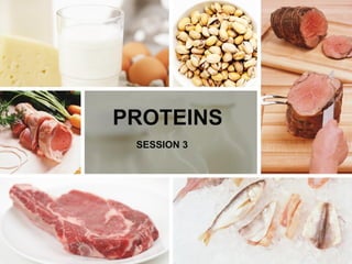
  • 2. What are proteins? • Proteins are large, complex molecules that play many critical roles in the body. • They do most of the work in cells and are required for the structure, function, and regulation of the body’s tissues and organs. • Proteins are made up of hundreds or thousands of smaller units called amino acids, which are attached to one another in long chains. • There are 20 different types of amino acids that can be combined to make a protein. • The sequence of amino acids determines each protein’s unique 3- dimensional structure and its specific function. • Amino acids are coded by combinations of three DNA building blocks (nucleotides), determined by the sequence of genes.
  • 3. AMINO ACIDS • Cells produce proteins with strikingly different properties and activities by joining the same 20 amino acids in many different combinations and sequences. • This indicates that the properties of proteins are determined by the physical and chemical properties of their monomer units, the amino acids. • Amino acids are the basic structural units of proteins consisting of an amino group, (-NH2) a carboxyl (-COOH) group a hydrogen (H) atom and a (variable) distinctive (R) group. • All of the substituents in amino acid are attached (bonded) to a central α carbon atom. • This carbon atom is called α because it is bonded to the carboxyl (acidic) group.
  • 4.  In neutral solution (PH = 7), both the α- amino and α carboxyl group are ionized resulting the charged form of an amino acids called zwitterion (dipolar) as shown in the next slide.  Out of the 20 amino acids, proline is not an α amino acid rather an α - imino acid. Except for glycine, all amino acids contain at least one asymmetric carbon atom (the α - carbon atom).
  • 5.
  • 6. Stereochemistry Stereochemistry mainly emphasizes the configuration of amino acids at the α carbon atom, having either D or L- isomers
  • 7. Classification of Amino Acids They are classified based on; • Structure • Electrochemical classification • Biological and physiological • Metabolic fate
  • 8. Structural classification: This classification is based on the side chain radicals (R-groups) R groups with Aliphatic side chains e.g glycine, alanine, valine, leucine, isoleucine Sulfur group: cysteine, methionine Aromatic group; phenylalanine, tyrosine, tryptophan, Imino group; proline
  • 9. Electrochemical Amino acids could also be classified based on their acid – base properties Basic amino acids 1. Lysine 2. Arginine Neutral amino acids 1. Asparagine 2. Serine 3. Threonine 4. Glutamine Acidic amino acids 1. Glutamic acid 2. Aspartic acid
  • 10. Biological This classification is based on the functional property of amino acids for the organism. • Essential: Amino acids which are not synthesized in the body and must be provided in the diet to meet an animal’s metabolic needs are called essential amino acids. • Non essential: These amino acids are need not be provided through diet, because they can be biosynthesized in adequate amounts within the organism. • Semi-essential amino acids: they can be synthesized within the organism but their synthesis is not in sufficient amounts. In that they should also be provided in the diet. Essential Non-essential Semi-essential Arginine, histidine, leucine, isoleucine, lysine, methionine, phenylalanine, Threonine, Tryptophan, Valine Alanine, Asparagine, aspartine, cysteine, glutamic acid, glutamine, glycine, proline, serine, tyrosine Histidine, Arginine
  • 11. Metabolic fate of each amino acid Amino acids can be classified here as Glucogenic (potentially be converted to glucose), ketogenic (potentially be converted to ketone bodies) and both glucogenic and ketogenic. Glucogenic amino acids: Those amino acids in which their carbon skeleton gets degraded to pyrurate, α ketoglutarate, succinyl CoA, fumrate and oxaloacetate and then converted to Glucose and Glycogen, are called as Glucogenic amino acids. These include:- Alanine, cysteine, glycine, Arginine, glutamine, Isoleucine, tyrosine Ketogenic amino acids: Those amino acids in which their carbon skeleton is degraded to Acetoacetyl CoA, or acetyl CoA. then converted to acetone and β- hydroxy butyrate which are the main ketone bodies are called ketogenic amino acids. These includes:- Phenylalanine, tyrosine, tryptophan, isoleucine, leucine, and lysine. These amino acids have ability to form ketone bodies which is particularly evident in untreated diabetes mellitus in which large amounts of ketone bodies are produced by the liver (i.e. not only from fatty acids but also from ketogenic amino acids)
  • 12. Ketogenic and glucogenic amino acids: The division between ketogenic and glucogenic amino acids is not sharp for amino acids (Tryptophan, phenylalanine, tyrosine and Isoleucine are both ketogenic and glucogenic). Some of the amino acids that can be converted in to pyruvate, particularly (Alanine, Cysteine and serine, can also potentially form acetoacetate via acetyl CoA especially in severe starvation and untreated diabetes mellitus.
  • 13.
  • 14. Peptides • Proteins are macromolecules with a backbone formed by polymerization of amino acids in a polyamide structure. • These amide bonds in protein, known as peptide bonds formed by linkage of α - carboxyl group of one amino acid with α- amino groups of the next amino acid by amide bonds. • During the formation of a peptide bond, a molecule of water is eliminated as shown below:- • A peptide chain consisting of two amino acid residues is called a dipeptide, three amino acids tripeptide (e. g Glutathione)
  • 15.
  • 16. Glutathione • Glutathione is a tripeptide formed from amino acids glutamate, cysteine and Glycine, linked together in that order. The glutamate is linked to cysteine through the γ- carboxyl group and α - amino group of cysteine. • Here, the carboxyl group is first activated by ATP to form an acyl-phosphate derivative which is then attacked by cysteine amino group then undergoes condensation with glycine.
  • 17. • The role of GSH as a reductant is extremely important particularly in the highly oxidizing environment of the erythrocyte. • The sulfhydryl of GSH can be used to reduce peroxides formed during oxygen transport. The resulting oxidized form of GSH consists of two molecules disulfide bonded together (abbreviated GSSG). • The enzyme glutathione reductase utilizes NADPH as a cofactor to reduce GSSG back to two moles of GSH. • Hence, the pentose phosphate pathway is an extremely important pathway of erythrocytes for the continuing production of the NADPH needed by glutathione reductase. • In fact as much as 10% of glucose consumption, by erythrocytes, may be mediated by the pentose phosphate pathway.
  • 19. PROTEINS • Proteins are macromolecules with a backbone formed by polymerization of amino acids in a polyamide structure. Class Assignment 2: 1. List functions of proteins (3 marks) 2. Using the flow chart on slide 18, describe the classification of proteins (7 marks)
  • 20. Protein Organization Primary structure The simplest level of protein structure, primary structure, is simply the sequence of amino acids in a polypeptide chain. For example, the hormone insulin has two polypeptide chains, A and B, shown in diagram below. (The insulin molecule shown here is cow insulin, although its structure is similar to that of human insulin.) Each chain has its own set of amino acids, assembled in a particular order. For instance, the sequence of the A chain starts with glycine at the N- terminus and ends with asparagine at the C-terminus, and is different from the sequence of the B chain.
  • 21.
  • 22. • The sequence of a protein is determined by the DNA of the gene that encodes the protein (or that encodes a portion of the protein, for multi- subunit proteins). • A change in the gene's DNA sequence may lead to a change in the amino acid sequence of the protein. • Even changing just one amino acid in a protein’s sequence can affect the protein’s overall structure and function. • For instance, a single amino acid change is associated with sickle cell anemia, an inherited disease that affects red blood cells. • In sickle cell anemia, one of the polypeptide chains that make up hemoglobin, the protein that carries oxygen in the blood, has a slight sequence change. • The glutamic acid that is normally the sixth amino acid of the hemoglobin β chain (one of two types of protein chains that make up hemoglobin) is replaced by a valine.
  • 23. Secondary Structure • The next level of protein structure, secondary structure, refers to local folded structures that form within a polypeptide due to interactions between atoms of the backbone. • (The backbone just refers to the polypeptide chain apart from the R groups – so all we mean here is that secondary structure does not involve R group atoms.) • The most common types of secondary structures are the α helix and the β pleated sheet. • Both structures are held in shape by hydrogen bonds, which form between the carbonyl O of one amino acid and the amino H of another.
  • 24.
  • 25. Tertiary structure • The overall three-dimensional structure of a polypeptide is called its tertiary structure. The tertiary structure is primarily due to interactions between the R groups of the amino acids that make up the protein. • R group interactions that contribute to tertiary structure include hydrogen bonding, ionic bonding, dipole-dipole interactions, and London dispersion forces – basically, the whole gamut of non-covalent bonds. • For example, R groups with like charges repel one another, while those with opposite charges can form an ionic bond. • Similarly, polar R groups can form hydrogen bonds and other dipole-dipole interactions. • Also important to tertiary structure are hydrophobic interactions, in which amino acids with nonpolar, hydrophobic R groups cluster together on the inside of the protein, leaving hydrophilic amino acids on the outside to interact with surrounding water molecules.
  • 26. • Finally, there’s one special type of covalent bond that can contribute to tertiary structure: the disulfide bond. • Disulfide bonds, covalent linkages between the sulfur-containing side chains of cysteines, are much stronger than the other types of bonds that contribute to tertiary structure. • They act like molecular "safety pins," keeping parts of the polypeptide firmly attached to one another.
  • 27. Quaternary structure • Many proteins are made up of a single polypeptide chain and have only three levels of structure (the ones we’ve just discussed). • However, some proteins are made up of multiple polypeptide chains, also known as subunits. When these subunits come together, they give the protein its quaternary structure. • As mentioned earlier, hemoglobin carries oxygen in the blood and is made up of four subunits, two each of the α and β types. • Another example is DNA polymerase, an enzyme that synthesizes new strands of DNA and is composed of ten subunits^55start superscript, 5, end superscript. • In general, the same types of interactions that contribute to tertiary structure (mostly weak interactions, such as hydrogen bonding) also hold the subunits together to give quaternary structure
  • 28.
  • 29. Protein Denaturation (Reading Assignment) • The phenomenon of disorganization of native protein structure is known as denaturation. • Denaturation results in the loss of secondary, tertiary and quaternary structureof proteins. This involves a change in physical, chemical and biological properties of protein molecule Agents of denaturation • Physical agents: Heat, violent shaking, X-ravs,UV radiation. • Chemical agents : Acids, alkalies, organic solvents(ether,alcohol), salts of heavy metals (Pb, Hg), urea,salicylate.
  • 30. Characteristics of denaturation a) The native helical structure of protein is lost b) The primary structure of a protein with peptide linkages remains intact i.e., peptide bonds are not hydrolysed. c) The protein loses its biologicalactivity. d) Denatured protein becomes insoluble in the solventin which it was originally soluble. e) The viscosity ol denatured protein (solution) increaseswhile its surface tension decreases. f) Denaturation is associated with increase in ionizable and sulfhydryl groups of protein. This is due to loss of hydrogen and disulfide bonds. g) Denatured protein is more easily digested. This is due to increased exposure of peptide bonds to enzymes. Cooking causes protein denaturation and, therefore, cooked food (protein)is more easily digested
  • 31. h) Denaturation is usually irreversible. For instance, omelet can be prepared from an egg (protein-albumin) but the reversal is not possible. i) Careful denaturation is sometimes reversible (known as renaturation). Hemoglobin undergoes denaturation in the presence of salicylate. By removal of salicylate, hemoglobin is renatured. j) Denatured protein cannot be crystallized
  • 32. • Flocculation: • lt is the process of protein precipitation at isoelectric pH. • The precipitate is referred to as flocculum. Casein (milk protein) can be easily precipitated when adjusted to isoelectric pH (4.6 by dilute acetic acid. • Flocculation is reversible. • On application of heat, flocculum can be converted into an irreversible mass, coagulum Coagulation: • The term 'coagulum' refers to a semi-solid viscous precipitate of protein. reversible denaturation results in coagulation. • Coagulation is optimum and requires lowest temperature at isoelectric pH. Albumins and globulins (to a lesser extent) are coagulable proteins. • Heat coagulation test is commonly used to detect the presence of albumin in urine.
  • 33. Protein Separation Methods (Reading Assessment) Group 1: Lydia, Brian, David (Protein Denaturation) Group 2: Maxwell, Lennick, Gabriel, Peter, Reuben, Dawn, Kevin, Eric, Ian (Protein separation methods) Oral assessment (5 marks)