Lab talk 190210 efficacy studies on radioligand hits_beginnings of fret assay_principles_induction of rel 1
•Download as PPT, PDF•
2 likes•59 views

1. Titration of REL 1 antagonist hits identified by radio mimetic assay to quantify efficacy (IC50) 2. Exposition of HT FRET assay principles for large scale compound library screens to empirically identify REL 1 antagonist hits

Recommended
Recommended
More Related Content
What's hot
What's hot (20)
Similar to Lab talk 190210 efficacy studies on radioligand hits_beginnings of fret assay_principles_induction of rel 1
Similar to Lab talk 190210 efficacy studies on radioligand hits_beginnings of fret assay_principles_induction of rel 1 (20)
More from Laurence Dawkins-Hall
More from Laurence Dawkins-Hall (20)
Recently uploaded
Recently uploaded (20)
Lab talk 190210 efficacy studies on radioligand hits_beginnings of fret assay_principles_induction of rel 1
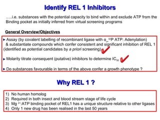
- 1. Identify REL 1 InhibitorsIdentify REL 1 Inhibitors Assay (by covalent labelling of recombinant ligase with α_32 P ATP: Adenylation) & substantiate compounds which confer consistent and significant inhibition of REL 1 (identified as potential candidates by a priori screening) Molarity titrate consequent (putative) inhibitors to determine IC50 Do substances favourable in terms of the above confer a growth phenotype ? Why REL 1 ?Why REL 1 ? 1) No human homolog 2) Required in both insect and blood stream stage of life cycle 3) Mg 2+ :ATP binding pocket of REL1 has a unique structure relative to other ligases 4) Only 1 new drug has been realised in the last 50 years General Overview/ObjectivesGeneral Overview/Objectives …..i.e. substances with the potential capacity to bind within and exclude ATP from the Binding pocket as initially inferred from virtual screening programs
- 2. 20S Editosome: Core Catalytic Complex20S Editosome: Core Catalytic Complex These 4 enzymes constitute 20S Core catalytic Editing comples Insertion and deletion editing structurally and functionally compartmentalised: In particular, Yeast 2 hybrid and coimmunoppt have identified an ExoUase-REL 1 subcomplex The same is true for TUTase-REL 2 in context of insertion editing REL 2 !REL 2 ! REL 1 !REL 1 !
- 3. REL 1-AMP* intermediateREL 1-AMP* intermediate Auto Adenylation In initial step enzymes auto adenylates by covalent condensation between lysine residue in ATP binding pocket and ATP Covalent adenylation co ordinated by Mg 2+ ions in ATP pocket Subsequently, AMP moiety transferred to 5’ phosphate of 3’ RNA species Finally, 3’ OH displaces AMP and phosphodiester bond is restored
- 4. Skeleton MethodSkeleton Method Set up radio ligation reactions, cf. REL 1; α32 P_ATP; compound in DMSO For each compound set up duplicate sets of reactions per assay Include DMSO only negative control and 1 x published + ive control (e.g. #16209) Resolve labelled products on Novex bis tris acrylamide gels (10%) Run to a specified point based on dye front (~ 45 minutes) Open gel stack and cut away gel front below 40kD mark Fix gel with Methanol acetic acid and subsequently rehydrate/ equilibrate with glycerol_fix solution Vacuum dry gel for 1 hour Expose dryed gel to Phospho imaging cassette o/n Visualise radio ligation using Typhoon Obtain 3 independent data sets from each compound cohort Quantify data using Image quant:
- 5. CompoundCompound ClassClass SetSet # substances# substances Drug discovery algorithmDrug discovery algorithm 1. NCI batch #1 #1 16 total PNAS Compounds screened with ‘Auto dock 4’ Ranked Verified with so-called ‘relaxed complex scheme’ Identified leads ‘discovered’ by screening multiple data bases for compounds with similar scaffolds to PNAS compounds 2. FDA #2 1 in house (sigma) 11 more predicted but financially prohibitive 1 pending (Mar 2010?) 3. NCI batch #2 #1 8 total 4. Hit 2 lead #3 15 total 5. Sigma #3 2 total Identify compounds that bind to ATP binding pocket of REL 1, viz. :Identify compounds that bind to ATP binding pocket of REL 1, viz. : 1 Strong lead !1 Strong lead ! 3 Strong leads !3 Strong leads ! Also,Tetracycline needs to be evaluatedAlso,Tetracycline needs to be evaluated 2 Nominal_100uM2 Nominal_100uM
- 6. CompoundCompound TypeType CompoundCompound 1010µµMM 100100µMµM TitratedTitrated N=N= ICIC5050 RangeRange Actual Computed ICActual Computed IC5050 GraphPad PrismGraphPad Prism NCI BatchNCI Batch #2#2 # 45609 ! 95 % (78%) (88%) 55 3-1uM # 162535 !!# 162535 !! 85_90 % (85%) (~98%)* (90 %) (>99.9%)* 99 ~1uM (N=1) 3-1uM (N=6) <1uM (N=2) # 1698 ! 80 % (50%) (90%) 66 10uM- 3uM (N=4) ~10uM (N=2) NCI BatchNCI Batch #1#1 # 117079 30_80% >99% (~80 %) (~50-70%) 44 100uM- 30uM (N=3) 30uM (N=1) # 125908 20_70% ~95 % (~50-70%) 44 100uM- 30uM (N=4) # 45201 NA ~70% Ignore !Ignore ! Sigma rareSigma rare LibraryLibrary MordantMordant Black !!Black !! (~98%)* (~99.8%)* 55 ~1uM (N=1) >1uM (N=4) Values derive from titrations’ Identified using Auto Dock 4 relaxed complex scheme *0.25ul undiluted prep #1_080802 1ul 50% diluted prep #3_050203 2.16µM +/- 1.20µM (N=4) 1.53µM +/- 1.17µM (N=6) 8.36µM +/- 1.71µM (N=4) 78.54µM +/- 1.58µM (N=3) 46.61µM +/- 3.02µM (N=4) 1.59µM +/- 1.10µM (N=3)
- 7. NCI Batch #2_10uMNCI Batch #2_10uM
- 8. = Remainder of original REL 1 prepRemainder of original REL 1 prep = Fresh REL 1 prep (May 2Fresh REL 1 prep (May 2ndnd 2003)2003) # 125908 # 117079 Strong inhibition in common with # 0011 # 45201 Evidence of efficacy @100uM in common with # 0011 # 1# 1 NCI Batch #1NCI Batch #1 100uM100uM
- 9. 34A10S10S-1-1 50-100S50-100S-1-1 20S20S-1-1 100S100S-1-1 34B Titration of leads from initial screensTitration of leads from initial screens
- 10. % (Putative) Inhibition of Ligation (Adenylation) by #45609 (solute) relative to DMSO (solvent)_# 45609 Molarity Titration DMSO DMSO 100uM 100uM 30uM30uM 10uM 10uM 3uM 3uM 1uM 1uM 300nM 300nM 100nM 100nM 30nM 30nM 0 20 40 60 80 100 120 # 0035B # 0037A Molar Titration MeanSpecificSignal_%DMSO IC50 resides here 199,000,000 counts ! 65,000,000 counts ! 0.9% 1.6% 7.9% 36% 68% IC50 resides here 0.7% 1.8% 6.2% 33% 67% Both assay sets generated with undiluted enzyme prep #1 & concord Thus, these assays were pooled for purposes of generating IC50 data via GraphPad Prism 0.25ul undiluted Prep #1_080802
- 11. % (Putative) Inhibition of Ligation (Adenylation) by # 45609 (solute) relative to DMSO (solvent)_# 45609 Molarity Titration: Mean data assays # 0035B & # 0037A DMSO 100uM 30uM 10uM 3uM 1uM 300nM 100nM 30nM 0 20 40 60 80 100 120 #0035B_# 0037A Molarity Titration: 100uM-30nM MeanSpecificSignal/DMSO_% DMSO 100uM 30uM 10uM 3uM 1uM 300nM 100nM 30nM 0.8% +/- 0.1% 1.7% +/- 0.2% 7.3% +/- 1.4% 34.4% +/- 2.0% 67.4% +/- 3.1% IC50 ~3-1uM IC50 (GraphPad v.5) 2.16 +/- 1.20µM R2 0.9983
- 12. Dose-Response # 45609 -2 -1 0 1 2 3 0 50 100 150 Log(10) Concentration [µM] Activity[%] Data from titrations subject to non linear regression analysis and log transformation Actual data fit to predicted (regression) fit indicated by Pearson correlation coefficient IC50 (GraphPad v.5) 2.16 +/- 1.20µM R2 0.9983
- 13. % (Putative) Inhibition of Ligation (Adenylation) by #45609 (solute) relative to DMSO (solvent)_# 45609 Molarity Titration DMSO DMSO 100uM 100uM 30uM30uM 10uM 10uM 3uM 3uM 1uM 1uM 300nM 300nM 100nM 100nM 30nM 30nM 0 20 40 60 80 100 120 140 # 0035B # 0016B Molar Titration MeanSpecificSignal_%DMSO IC50 resides here 199,000,000 counts ! 10,000,000 counts ! 0.9% 1.6% 7.9% 36% 68% IC50 resides here 12% 16% 22% 38% 74% 0.25ul undiluted Prep #1_080802 0.5ul 50% diluted Prep Optimal sets generated with undiluted enzyme prep #1 In contrast earlier data derived from glycerol diluted prep yielded lower S:N
- 14. % (Putative) Inhibition of Ligation (Adenylation) by #1698 (solute) relative to DMSO (solvent)_# 1698 Molarity Titration DMSO DMSO 100uM 100uM 30uM 30uM 10uM 10uM 3uM 3uM 1uM 1uM 300nM 300nM 100nM 100nM 30nM 30nM 0 20 40 60 80 100 120 # 0018B # 0035/36A 100uM-30nM MeanSpecificSignal/DMSO_% DMSO 100uM 30uM 10uM 3uM 1uM 300nM 100nM 30nM # 1698 27.1+/- 8.8% 52.7+/- 0.3% 39.1+/- 1.8% 60.5+/- 3.5% IC50 ~ 10uM IC50 ~ 10-3uM 0.25ul Undiluted Prep #1_080802 IC50 (GraphPad v.5) 8.36 +/- 1.71µM R2 0.9955 Submitted to GraphPad
- 15. Dose-Response # 1698 -2 -1 0 1 2 3 0 50 100 150 Log(10) concentration [µM] Activity[%] IC50 (GraphPad v.5) 8.36 +/- 1.71µM R2 0.9955
- 16. % (Putative) Inhibition of Ligation (Adenylation) by #162535 (solute) relative to DMSO (solvent)_# 162535 Molarity Titration DMSO DMSO DMSO 100uM 100uM 100uM 30uM 30uM 30uM 10uM 10uM 10uM 3uM 3uM 3uM 1uM 1uM 1uM 300nM 300nM 300nM 100nM 100nM 100nM 30nM 30nM 30nM 0 20 40 60 80 100 120 140 160 180 # 0036B # 0037B # 0034B Molarity Titration: 100uM-30nM MeanSpecificsignal/DMSO_% DMSO 100uM 30uM 10uM 3uM 1uM 300nM 100nM 30nM # 162535 0.25ul Undiluted Prep #1_080802 0.25ul Undiluted Prep #1_080802 21% 42% 42% 90% 36% 74%141,000,000 90,000,0000.4%+/-0.03% 0.8%+/-0.05% 4.3%+/-0.3% 0.5%+/-0.2% 1.4%+/-0.4% 7.0%+/-2.4% 0.7%+/-0.1% 1.9%+/- 9.5%+/-0.5% 64% IC50 ~ 1uM-300nM ! 0.25ul Undiluted Prep #1_080802 IC50 ~ 3uM-1uM ! 200,000,000 All data sets generated with undiluted enzyme prep #1 Two sets concord but one is more disparate with respect to predicted IC50 However, because all 3 represent replicate cohorts all data pooled for purposes of IC50 elucidation
- 17. % (Putative) Inhibition of Ligation (Adenylation) by #162535 (solute) relative to DMSO (solvent)_# 162535 Molarity Titration DMSO 100uM 30uM 10uM 3uM 1uM 300nM 100nM 30nM 0 20 40 60 80 100 120 140 160 Assays #0034B_0036B_0037B Molarity Titration:100uM-30nM MeanSpecificSignal/DMSO_% DMSO 100uM 30uM 10uM 3uM 1uM 300nM 100nM 30nM 0.5% +/- 0.2% 1.4% +/- 0.6% 7.0% +/- 2.6% 33.0% +/- 10.9% 68.5% +/- 23.2% IC50 ~ 3-1uM ● Inclusion of assay # 0036B reduces putative IC50 but increases variance (relative to assays # 0034B_36B alone) ● Nevertheless, putative IC50 still resides between 3µM-1µM ● In context of assay # 0036B (N=2) putative IC50 < 1µM and variance lowest of all 0.25ul Undiluted Prep #1_080802 IC50 (GraphPad v.5) 1.53 +/- 1.17µM R2 0.9991 Error bars reflect disparity between assay #0036B and other two assays
- 18. Dose-Response #162535 -2 -1 0 1 2 3 0 50 100 150 Log(10) concentration [µM] activity[%] IC50 (GraphPad v.5) 1.53 +/- 1.17µM R2 0.9991 Error bars reflect disparity between assay # 0036B and other two assays Nevertheless regression fit to actual data, i.e. R2 excellent
- 19. % (Putative) Inhibition of Ligation (Adenylation) by Mordant Black (solute) relative to DMSO (solvent)_# Mordant Black Molarity Titration 0 20 40 60 80 100 120 140 1ul Prep #1_080802 0.25ul Prep #1_080802 SpecificSignal/DMSO_% 26.6+/-0.2% 23.7% 51.9% ● For 0.25ul Prep #1_080802, N=3 = 1 #0029A_1 2 #0034A_1 3 #0034A_2 ● For 1ul Prep #1_080802, N=1 = 1 #0033A ● For Prep #3_050203 N=1 1 #0029A_3 ● For assay #0029A putative IC50 ~1uM ● How ever, for assays w here N=3, putative IC50 >1uM ● For assays w here N=3, data convergence better relative to #0033A betw een 100uM-3uM 0.25ul undiluted REL 1 4.4+/-0.2% 1.1+/-0.1% 0.7+/-0.1% 60.8+/-0.7% 53.7% 15.4 1.8% 0.4% 0.1% 1ul Prep #3_050203# 0033A # 0034A_1 # 0034A_2 # 0029A_1 # 0029A_3 100uM 30nM IC50 (GraphPad v.5) 1.59 +/- 1.10µM R2 0.9998 Submitted to GraphPad Formely Mordant black assays completed but undecided as to which data sets to pool for IC50 determination According with other assays just those data sets derived from Prep #1 were submitted
- 20. Dose-Response Mordant Black -2 -1 0 1 2 3 0 50 100 150 Log(10) Concentration [µM] activity[%] IC50 (GraphPad v.5) 1.59 +/- 1.10µM R2 0.9998 I’m Happy !
- 21. % (Putative) Inhibition of REL 1 Ligation (Adenylation) by #117079 (solute) relative to DMSO (solvent)_# 117079 Molarity Titration DMSO DMSO 100uM 100uM 30uM 30uM 10uM 10uM 3uM 3uM 1uM 1uM 300nM 300nM 100nM 100nM 30nM 30nM 0.0 20.0 40.0 60.0 80.0 100.0 120.0 140.0 BAC Prep #3_050203 BAC Prep #1_080802 #117079 Molarity Titration: 100uM-30nM SpecificSignal/DMSO_% 18.6%18.6% 54%54% 23.4%23.4% 75.6%75.6% 1ul Enzyme1ul Enzyme 0.25ul Enzyme0.25ul Enzyme Predicted IC50 Prep #3 ~30uM Predicted IC50 for prep #100uM-30uM The latter concurs with previous assays50S50S-1-1 100S100S-1-1
- 22. % (Putative) Inhibition of REL 1 Ligation (Adenylation) by #117079 (solute) relative to DMSO (solvent): #117079 Molarity Titration. Assay #0032A/B DMSO 1mM 300uM 100uM 30uM 10uM 3uM 1uM 0 20 40 60 80 100 120 N = 3 Molarity Titration #117079: 1mM - 1uM MeanSpecificSignal/DMSO_% DMSO 1mM 300uM 100uM 30uM 10uM 3uM 1uM IC50 (GraphPad v.5) 78.54 +/- 1.58µM R2 0.9918 1ul Prep #3_050203
- 23. Dose-Response # 117079 0 1 2 3 4 0 50 100 150 Log (10)concentration [µM] Activity[%] IC50 (GraphPad v.5) 78.54 +/- 1.58µM R2 0.9918
- 24. % (Putative) Inhibition of REL 1 Ligation (Adenylation) by #125908 (solute) relative to DMSO (solvent): #125908 Molarity Titration. Assay #0031A/B DMSO 1mM 300uM 100uM 30uM 10uM 3uM 1uM 0 20 40 60 80 100 120 140 N = 4 Molarity Titration #125908: 1mM-1uM MeanSpecificSignal/DMSO_% DMSO 1mM 300uM 100uM 30uM 10uM 3uM 1uM 1ul Prep #3_050203 IC50 (GraphPad v.5) 46.61 +/- 3.02µM R2 0.9675
- 25. Dose-Response # 125908 0 1 2 3 4 0 50 100 150 Log (10)concentration [µM] Activity[%] IC50 (GraphPad v.5) 46.61 +/- 3.02µM R2 0.9675
- 26. #16209 #45609 IC50 = 1.01 uM IC50 = 2.16 +/- 1.20uM #1698 IC50 = 8.36 +/-1.71 uM #162535 IC50 = 1.53 +/- 1.17 uM LogP = -1.043 LogP = 0.492 Log P values for # 45609 More encouraging than for #16209 This bodes well for successful EC50 determination by Alamar Blue viability assay
- 27. A fluorescence-based high-throughput ligase assay Donor (D) = FAM, Acceptor (A) = Cy5, 250 nM Assay will utilise a 104bp RNA fragment (CYb) in concert with labelled 20mers Coupling efficiency varies with fluorophore distance 70-100A is where FRET is achieved Forster distance = Separation at which 50% photon transfer achieved
- 28. Assay EstablishmentAssay Establishment 1) Purify catalytic domain (L151-324 ) of REL 1 (in association with TEV cleavable His Tag A2) by FPLC utilising NTA columns in concert with imidazole gradient 2) Express 104bp RNA (CYb) species from linear pBlueScript clone by In Vitro transcription 3) Anneal labelled 20mers 4) Test E Coli purified REL1 batch versus adenylation verified leads 5) Independently verify FRET by radioactive ligation assays Assay OptimisationAssay Optimisation Various parameters associated with ‘Mix & measure’ assay will be optimised, e.g. Gap distance: 16 donor/acceptor fluorophore combinations assayed over 0-12bp hiatus RNA Substrate: Is CYb best ? Fluorophore species: TAMRA, Cy3, Cy5 etc Dynamic detection/sensitivity range: Serial substrate dilutions versus detection DMSO Concentration: 0.1% - 10% Assay ImplementationAssay Implementation 1) ~3000 SMDC compounds to screen 2) Initial primary screen @ 10uM + 1% DMSO 3) Compounds >/- 3 x SD from (collective) median inhibition will be deemed ‘active’ and subject to counter screens
- 29. #1#1 #2#2 #3#3 #4#4 50KD 40KD 30KD 20KD PP PP PP P?P?SS PP PP PP P?P?SS SS SS SS SS S?S? S?S? + + + +- - - - Catalytic domainCatalytic domain REL1REL1 Specific and high level expression of L1 (catalytic REL 1 domain) in pellet No obvious sign of soluble protein ! Is this due to a lack of A2 (N-terminal TEV His tag) rendering L1 soluble ? +/+/-- 0.5mM IPTG0.5mM IPTGWestern blotting with: α REL 1Ab α His tag Ab
Editor's Notes
- medicinal chemists: no way this is a drug! so much for rational drug design mention charge as problem for crossing membranes as well as blood brain barrier LogP values plan to replace sulfomic acid with sulfonamide