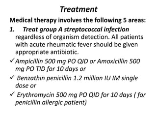
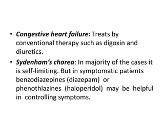
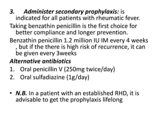
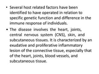
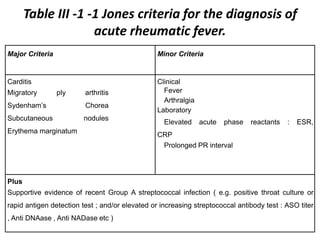
Rheumatic Fever
• Learning objectives: at the end of this lesson the student will be able to :
•
• 1. Define rheumatic fever
•
• 2. Understand the etiologic agents of rheumatic fever
•
• 3. Understand the Epidemiology of rheumatic fever
•
• 4. Describe the pathogenesis of rheumatic fever
•
• 5. Identify the clinical manifestation of rheumatic fever
•
• 6. Understand the diagnostic approach of rheumatic fever
•
• 7. Manage patients having rheumatic fever
•
• 8. Design strategies for prevention of rheumatic fever
Background:
• Rheumatic fever causes chronic progressive
damage to the heart and its valves
• The association between sore throat and
rheumatic fever was not made until 1880. The
dramatic decline in the incidence of rheumatic
fever in the developed world is thought to be
largely owing to antibiotic treatment of
streptococcal infection, though it stated to
decline before the era of antibiotic, probably due
improvement of socioeconomic status.
Pathophysiology
Acute rheumatic fever is a sequel of a previous
group A streptococcal infection, usually of the
upper respiratory tract. One beta-streptococcal
serotype (e.g., M types 3, 5, 18,
• 19, 24) is linked directly to acute rheumatic fever.
Rheumatogenicity of GAS is important factor as
not all GAS pharyngitis is associated with
development of rheumatic fever.
• Rheumatic fever follows Lancefield β hemolytic
streptococcus pharyngitis within the
• interval of 2-3 weeks.
• The mechanism is elusive, but the followings
are proposed ones:
• Dysfunction of the immune Response
• Antigenic Mimicry
– Similarity between the carbohydrate moiety if
GAS and glycoprotein of heart valve
– Molecular similarity between some
Streptococcal antigens and sarcolema or other
moiety of human myocardial cells.
• Several host related factors have been
identified to have operated in relation to
specific genetic function and difference in the
immune response of individuals.
• The disease involves the heart, joints,
central nervous system (CNS), skin, and
subcutaneous tissues. It is characterized by an
exudative and proliferative inflammatory
lesion of the connective tissue, especially that
of the heart, joints, blood vessels, and
subcutaneous tissue.
• Clinical Manifestation
•
• Acute rheumatic fever is associated with 2 distinct
patterns of presentation.
• The first pattern of presentation is sudden onset. It
typically begins as polyarthritis 2-6 weeks after
streptococcal pharyngitis, and it is usually
characterized by fever and toxicity.
• The second pattern is insidious or subclinical and the
initial abnormality is mild carditis.
• Age at onset influences the order of complications.
Younger children tend to develop carditis first, while
older patients tend to develop arthritis first.
Diagnosis:
• Diagnosis of acute rheumatic fever requires a
high index of suspicion.
• Jones criteria developed by the American
Heart Association is used to make the
diagnosis.
Table III -1 -1 Jones criteria for the diagnosis of
acute rheumatic fever.
Major Criteria Minor Criteria
Carditis
Migratory ply arthritis
Sydenham’s Chorea
Subcutaneous nodules
Erythema marginatum
Clinical
Fever
Arthralgia
Laboratory
Elevated acute phase reactants : ESR,
CRP
Prolonged PR interval
Plus
Supportive evidence of recent Group A streptococcal infection ( e.g. positive throat culture or
rapid antigen detection test ; and/or elevated or increasing streptococcal antibody test : ASO titer
, Anti DNAase , Anti NADase etc )
In addition to evidence of a previous
streptococcal infection, the diagnosis of acute
rheumatic fever requires 2 major Jones
criteria or 1 major plus 2 minor Jones criteria.
• 1) Carditis, (pancarditis here), occurs in as many
as 40- 60% of patients and may manifest as:
a) New murmur b) Cardiomegaly
c) Congestive heart failure
d) Pericarditis with or without a pericardial rub and
resolve without squeal of constriction.
e) Valvular disease: mitral and aortic valves are
commonly affected. Healing of rheumatic
valvulitis will lead into fibrous thickening and
adhesion, resulting in progressive valvular
damage. But, about 80% of mild valvulitis would
resolve. There is a risk of developing endocarditis
on a damaged valve.
2) Migratory polyarthritis occurs in 75% of
cases and involves many joints at a time. The
larger joints are mainly affected.
3) Subcutaneous nodules: occur in 10% of
patients and are edematous fragmented
collagen fibers. They are firm painless nodules
on the extensor surfaces of wrists, elbows,
and knees.
4) Erythema marginatum occurs in about 5% of
cases. The rash is serpiginous and long lasting.
5) Sydenham’s chorea (i.e., St Vitus’ dance) is
a characteristic movement disorder that
occurs in 5-10% of cases. Sydenham’s chorea
consists of rapid purposeless movements of
the face and upper extremities. Onset may be
delayed for several months to years and may
cease when the patient is asleep.
Laboratory Studies:
• No specific confirmatory laboratory tests exist.
However, several laboratory findings indicate
continuing rheumatic inflammation. Some are
part of the Jones minor criteria.
• Supportive evidences
o Streptococcal antibody tests disclose preceding
streptococcal infection
– ASO titer: positive in 80% of cases
– Anti DNAase β & Anti hyaluronidase is positive in 95
% of cases
 Isolate group A streptococci via throat culture
which has 20-40% yield.
Laboratory minor criteria
Acute phase reactants (e.g. raised ESR and C-
reactive protein [CRP])
Leukocytosis may be seen.
• Anemia usually is caused by suppression of
erythropoiesis.
• ECG: PR interval prolongation is seen in 25%
of all cases but is neither specific to nor
diagnostic.
Treatment
Medical therapy involves the following 5 areas:
1. Treat group A streptococcal infection
regardless of organism detection. All patients
with acute rheumatic fever should be given
appropriate antibiotic.
Ampicillin 500 mg PO QID or Amoxicillin 500
mg PO TID for 10 days or
 Benzathin penicillin 1.2 million IU IM single
dose or
 Erythromycin 500 mg PO QID for 10 days ( for
penicillin allergic patient)
2. Therapy for manifestation of acute rheumatic fever
 Arthritis:
 ASA is given at dose 2 gm four times per day for 4-6
weeks, no indication for steroids.
 Carditis
 Severe Carditis with congestive heart failure should be
treated with
A. Prednisolone 60 to 80 mg /day, to be tapered as patient
improves
B. Start ASA during tapering phase to be given for 4-6weeks
C. But both have no influence on the future development of
valvular heart disease (VHD).
• Congestive heart failure: Treats by
conventional therapy such as digoxin and
diuretics.
• Sydenham’s chorea: In majority of the cases it
is self-limiting. But in symptomatic patients
benzodiazepines (diazepam) or
phenothiazines (haloperidol) may be helpful
in controlling symptoms.
3. Administer secondary prophylaxis: is
indicated for all patients with rheumatic fever.
Taking benzathin penicillin is the first choice for
better compliance and longer prevention.
Benzathin penicillin 1.2 million IU IM every 4 weeks
, but if the there is high risk of recurrence, it can
be given every 3weeks
Alternative antibiotics
1. Oral penicillin V (250mg twice/day)
2. Oral sulfadiazine (1g/day)
• N.B. In a patient with an established RHD, it is
advisable to get the prophylaxis lifelong
L1.. rheumatic fever

L1.. rheumatic fever

  • 1.
    Rheumatic Fever • Learningobjectives: at the end of this lesson the student will be able to : • • 1. Define rheumatic fever • • 2. Understand the etiologic agents of rheumatic fever • • 3. Understand the Epidemiology of rheumatic fever • • 4. Describe the pathogenesis of rheumatic fever • • 5. Identify the clinical manifestation of rheumatic fever • • 6. Understand the diagnostic approach of rheumatic fever • • 7. Manage patients having rheumatic fever • • 8. Design strategies for prevention of rheumatic fever
  • 2.
    Background: • Rheumatic fevercauses chronic progressive damage to the heart and its valves • The association between sore throat and rheumatic fever was not made until 1880. The dramatic decline in the incidence of rheumatic fever in the developed world is thought to be largely owing to antibiotic treatment of streptococcal infection, though it stated to decline before the era of antibiotic, probably due improvement of socioeconomic status.
  • 3.
    Pathophysiology Acute rheumatic feveris a sequel of a previous group A streptococcal infection, usually of the upper respiratory tract. One beta-streptococcal serotype (e.g., M types 3, 5, 18, • 19, 24) is linked directly to acute rheumatic fever. Rheumatogenicity of GAS is important factor as not all GAS pharyngitis is associated with development of rheumatic fever. • Rheumatic fever follows Lancefield β hemolytic streptococcus pharyngitis within the • interval of 2-3 weeks.
  • 4.
    • The mechanismis elusive, but the followings are proposed ones: • Dysfunction of the immune Response • Antigenic Mimicry – Similarity between the carbohydrate moiety if GAS and glycoprotein of heart valve – Molecular similarity between some Streptococcal antigens and sarcolema or other moiety of human myocardial cells.
  • 5.
    • Several hostrelated factors have been identified to have operated in relation to specific genetic function and difference in the immune response of individuals. • The disease involves the heart, joints, central nervous system (CNS), skin, and subcutaneous tissues. It is characterized by an exudative and proliferative inflammatory lesion of the connective tissue, especially that of the heart, joints, blood vessels, and subcutaneous tissue.
  • 6.
    • Clinical Manifestation • •Acute rheumatic fever is associated with 2 distinct patterns of presentation. • The first pattern of presentation is sudden onset. It typically begins as polyarthritis 2-6 weeks after streptococcal pharyngitis, and it is usually characterized by fever and toxicity. • The second pattern is insidious or subclinical and the initial abnormality is mild carditis. • Age at onset influences the order of complications. Younger children tend to develop carditis first, while older patients tend to develop arthritis first.
  • 7.
    Diagnosis: • Diagnosis ofacute rheumatic fever requires a high index of suspicion. • Jones criteria developed by the American Heart Association is used to make the diagnosis.
  • 8.
    Table III -1-1 Jones criteria for the diagnosis of acute rheumatic fever. Major Criteria Minor Criteria Carditis Migratory ply arthritis Sydenham’s Chorea Subcutaneous nodules Erythema marginatum Clinical Fever Arthralgia Laboratory Elevated acute phase reactants : ESR, CRP Prolonged PR interval Plus Supportive evidence of recent Group A streptococcal infection ( e.g. positive throat culture or rapid antigen detection test ; and/or elevated or increasing streptococcal antibody test : ASO titer , Anti DNAase , Anti NADase etc )
  • 9.
    In addition toevidence of a previous streptococcal infection, the diagnosis of acute rheumatic fever requires 2 major Jones criteria or 1 major plus 2 minor Jones criteria.
  • 10.
    • 1) Carditis,(pancarditis here), occurs in as many as 40- 60% of patients and may manifest as: a) New murmur b) Cardiomegaly c) Congestive heart failure d) Pericarditis with or without a pericardial rub and resolve without squeal of constriction. e) Valvular disease: mitral and aortic valves are commonly affected. Healing of rheumatic valvulitis will lead into fibrous thickening and adhesion, resulting in progressive valvular damage. But, about 80% of mild valvulitis would resolve. There is a risk of developing endocarditis on a damaged valve.
  • 11.
    2) Migratory polyarthritisoccurs in 75% of cases and involves many joints at a time. The larger joints are mainly affected. 3) Subcutaneous nodules: occur in 10% of patients and are edematous fragmented collagen fibers. They are firm painless nodules on the extensor surfaces of wrists, elbows, and knees.
  • 12.
    4) Erythema marginatumoccurs in about 5% of cases. The rash is serpiginous and long lasting. 5) Sydenham’s chorea (i.e., St Vitus’ dance) is a characteristic movement disorder that occurs in 5-10% of cases. Sydenham’s chorea consists of rapid purposeless movements of the face and upper extremities. Onset may be delayed for several months to years and may cease when the patient is asleep.
  • 13.
    Laboratory Studies: • Nospecific confirmatory laboratory tests exist. However, several laboratory findings indicate continuing rheumatic inflammation. Some are part of the Jones minor criteria. • Supportive evidences o Streptococcal antibody tests disclose preceding streptococcal infection – ASO titer: positive in 80% of cases – Anti DNAase β & Anti hyaluronidase is positive in 95 % of cases  Isolate group A streptococci via throat culture which has 20-40% yield.
  • 14.
    Laboratory minor criteria Acutephase reactants (e.g. raised ESR and C- reactive protein [CRP]) Leukocytosis may be seen. • Anemia usually is caused by suppression of erythropoiesis. • ECG: PR interval prolongation is seen in 25% of all cases but is neither specific to nor diagnostic.
  • 15.
    Treatment Medical therapy involvesthe following 5 areas: 1. Treat group A streptococcal infection regardless of organism detection. All patients with acute rheumatic fever should be given appropriate antibiotic. Ampicillin 500 mg PO QID or Amoxicillin 500 mg PO TID for 10 days or  Benzathin penicillin 1.2 million IU IM single dose or  Erythromycin 500 mg PO QID for 10 days ( for penicillin allergic patient)
  • 16.
    2. Therapy formanifestation of acute rheumatic fever  Arthritis:  ASA is given at dose 2 gm four times per day for 4-6 weeks, no indication for steroids.  Carditis  Severe Carditis with congestive heart failure should be treated with A. Prednisolone 60 to 80 mg /day, to be tapered as patient improves B. Start ASA during tapering phase to be given for 4-6weeks C. But both have no influence on the future development of valvular heart disease (VHD).
  • 17.
    • Congestive heartfailure: Treats by conventional therapy such as digoxin and diuretics. • Sydenham’s chorea: In majority of the cases it is self-limiting. But in symptomatic patients benzodiazepines (diazepam) or phenothiazines (haloperidol) may be helpful in controlling symptoms.
  • 18.
    3. Administer secondaryprophylaxis: is indicated for all patients with rheumatic fever. Taking benzathin penicillin is the first choice for better compliance and longer prevention. Benzathin penicillin 1.2 million IU IM every 4 weeks , but if the there is high risk of recurrence, it can be given every 3weeks Alternative antibiotics 1. Oral penicillin V (250mg twice/day) 2. Oral sulfadiazine (1g/day) • N.B. In a patient with an established RHD, it is advisable to get the prophylaxis lifelong