Without food,water and oxygen, human beings could
Without food, water and oxygen, human beings could
not survive.
not survive.
The digestive system is a set of organs which change
The digestive system is a set of organs which change
what we eat into substances that can be used in the
what we eat into substances that can be used in the
body.
body.
These substances can be used for energy, growth and
These substances can be used for energy, growth and
repair.
repair.
3.
THE DIGESTIVE SYSTEM
THEDIGESTIVE SYSTEM
The
The alimentary canal
alimentary canal is a tube that runs from the
is a tube that runs from the
mouth to the anus
mouth to the anus
It begins at the oral cavity, runs through thorax as
It begins at the oral cavity, runs through thorax as
oesophagus, which enters into stomach, from stomach
oesophagus, which enters into stomach, from stomach
starts small intestine and then the last part is the
starts small intestine and then the last part is the
large intestine
large intestine
As food passes through the alimentary canal it is
As food passes through the alimentary canal it is
changed and the nourishment is taken into the blood
changed and the nourishment is taken into the blood
Waste passes out the end of the canal
Waste passes out the end of the canal
Certain organs and glands add juices to the canal at
Certain organs and glands add juices to the canal at
various points
various points
4.
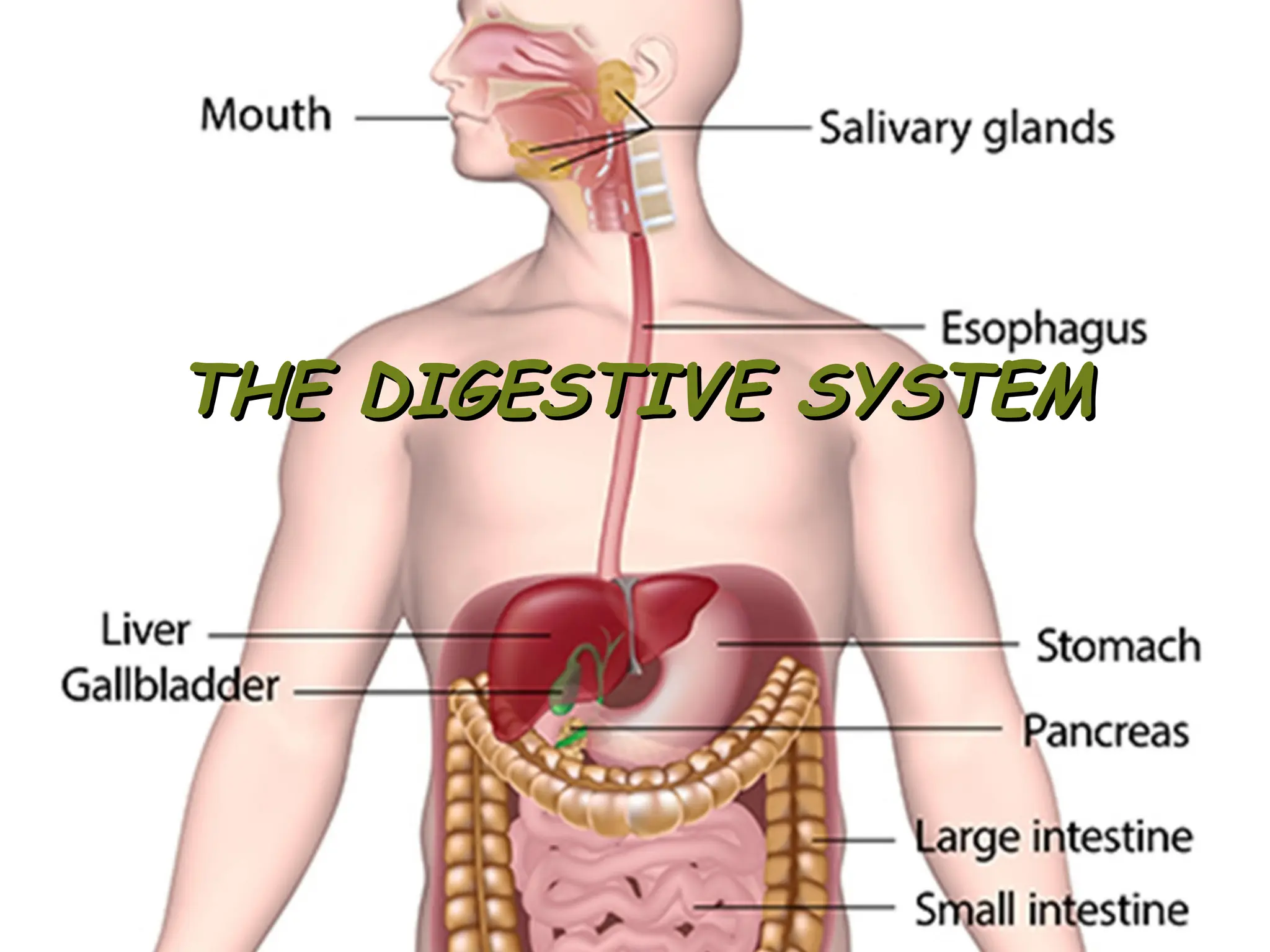
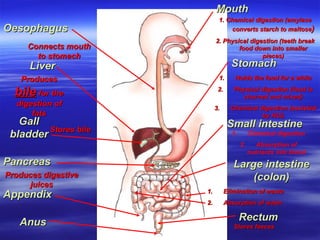
Mouth
Mouth
1. Chemical digestion(amylase
1. Chemical digestion (amylase
converts starch to maltose
converts starch to maltose)
)
2. Physical digestion (teeth break
2. Physical digestion (teeth break
food down into smaller
food down into smaller
pieces)
pieces)
Oesophagus
Oesophagus
Connects mouth
Connects mouth
to stomach
to stomach
Stomach
Stomach
Liver
Liver
Produces
Produces
bile
bile for the
for the
digestion of
digestion of
fats
fats
Gall
Gall
bladder
bladder Stores bile
Stores bile
1.
1. Holds the food for a while
Holds the food for a while
2.
2. Physical digestion (food is
Physical digestion (food is
churned and mixed)
churned and mixed)
3.
3. Chemical digestion (assisted
Chemical digestion (assisted
by HCl)
by HCl)
Pancreas
Pancreas
Produces digestive
Produces digestive
juices
juices
Small intestine
Small intestine
1. Chemical digestion
2. Absorption of
nutrients into blood
Appendix
Appendix
Large intestine
Large intestine
(colon)
(colon)
1.
1. Elimination of waste
Elimination of waste
2.
2. Absorption of water
Absorption of water
Rectum
Rectum
Stores faeces
Stores faeces
Anus
Anus
5.
The activities ofthe digestive system can be grouped
The activities of the digestive system can be grouped
under five main headings.
under five main headings.
1.
1.Ingestion.
Ingestion. Taking of food into the alimentary tract, i.e.
Taking of food into the alimentary tract, i.e.
eating and drinking.
eating and drinking.
2.
2.Propulsion.
Propulsion. Mixes and moves the contents along the
Mixes and moves the contents along the
alimentary tract.
alimentary tract.
3.
3.Digestion.
Digestion. Includes mechanical and chemical.
Includes mechanical and chemical.
4.
4.Absorption.
Absorption. Digested food substances pass through the
Digested food substances pass through the
walls of small intestines into the blood and lymph
walls of small intestines into the blood and lymph
capillaries for use by body cells.
capillaries for use by body cells.
5.
5.Elimination.
Elimination. Food substances that cannot be digested and
Food substances that cannot be digested and
absorbed are excreted as
absorbed are excreted as faeces
faeces by the process of
by the process of
defaecation
defaecation.
.
6.
Food can bebroken down (digested) in one of two
Food can be broken down (digested) in one of two
ways:
ways:
1.
1. Physical Digestion
Physical Digestion
This is where large pieces of food are broken down
This is where large pieces of food are broken down
into smaller pieces of the same food
into smaller pieces of the same food
2.
2. Chemical Digestion
Chemical Digestion
This is where food is broken down into a different
This is where food is broken down into a different
substance that can easily pass into the blood
substance that can easily pass into the blood
Digestion
7.
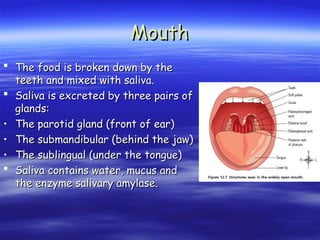
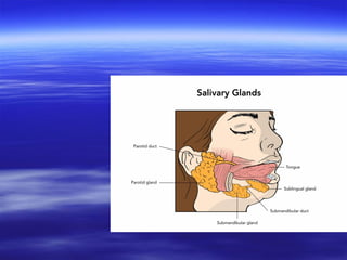
Mouth
Mouth
The foodis broken down by the
The food is broken down by the
teeth and mixed with saliva.
teeth and mixed with saliva.
Saliva is excreted by three pairs of
Saliva is excreted by three pairs of
glands:
glands:
• The parotid gland (front of ear)
The parotid gland (front of ear)
• The submandibular (behind the jaw)
The submandibular (behind the jaw)
• The sublingual (under the tongue)
The sublingual (under the tongue)
Saliva contains water, mucus and
Saliva contains water, mucus and
the enzyme salivary amylase.
the enzyme salivary amylase.
9.
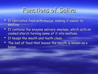
Functions of Saliva
Functionsof Saliva
It lubricates food with mucus, making it easier to
It lubricates food with mucus, making it easier to
swallow.
swallow.
It contains the enzyme salivary amylase, which acts on
It contains the enzyme salivary amylase, which acts on
cooked starch turning some of it into maltose.
cooked starch turning some of it into maltose.
It keeps the mouth and teeth clean.
It keeps the mouth and teeth clean.
The ball of food that leaves the mouth is known as a
The ball of food that leaves the mouth is known as a
bolus.
bolus.
10.
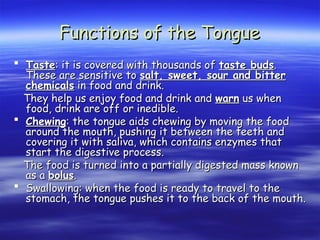
Functions of theTongue
Functions of the Tongue
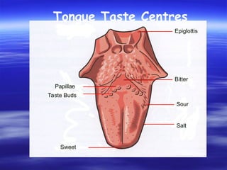
Taste
Taste: it is covered with thousands of
: it is covered with thousands of taste buds
taste buds.
.
These are sensitive to
These are sensitive to salt, sweet, sour and bitter
salt, sweet, sour and bitter
chemicals
chemicals in food and drink.
in food and drink.
They help us enjoy food and drink and
They help us enjoy food and drink and warn
warn us when
us when
food, drink are off or inedible.
food, drink are off or inedible.
Chewing
Chewing: the tongue aids chewing by moving the food
: the tongue aids chewing by moving the food
around the mouth, pushing it between the teeth and
around the mouth, pushing it between the teeth and
covering it with saliva, which contains enzymes that
covering it with saliva, which contains enzymes that
start the digestive process.
start the digestive process.
The food is turned into a partially digested mass known
The food is turned into a partially digested mass known
as a
as a bolus
bolus.
.
Swallowing: when the food is ready to travel to the
Swallowing: when the food is ready to travel to the
stomach, the tongue pushes it to the back of the mouth.
stomach, the tongue pushes it to the back of the mouth.
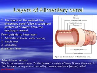
Layers of Alimentarycanal
Layers of Alimentary canal
The layers of the walls of the
The layers of the walls of the
alimentary canal follow a consistent
alimentary canal follow a consistent
pattern of 4 layers, from the
pattern of 4 layers, from the
esophagus onward.
esophagus onward.
From outside to inner layer
From outside to inner layer
1.
1. Adventitia or serosa – outer covering
Adventitia or serosa – outer covering
2.
2. Muscle layer
Muscle layer
3.
3. Submucosa
Submucosa
4.
4. Mucosa – lining.
Mucosa – lining.
Adventitia or serosa:
This is the outermost layer. In the thorax it consists of loose fibrous tissue and in
the abdomen the organs are covered by a serous membrane (serosa) called
peritoneum.
13.
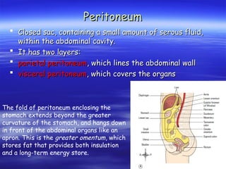
Peritoneum
Peritoneum
Closed sac,containing a small amount of serous fluid,
Closed sac, containing a small amount of serous fluid,
within the abdominal cavity.
within the abdominal cavity.
It has two layers:
It has two layers:
parietal peritoneum
parietal peritoneum, which lines the abdominal wall
, which lines the abdominal wall
visceral peritoneum
visceral peritoneum, which covers the organs
, which covers the organs
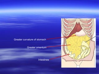
The fold of peritoneum enclosing the
stomach extends beyond the greater
curvature of the stomach, and hangs down
in front of the abdominal organs like an
apron. This is the greater omentum, which
stores fat that provides both insulation
and a long-term energy store.
14.
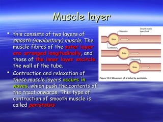
Muscle layer
Muscle layer
this consists of two layers of
this consists of two layers of
smooth (involuntary) muscle
smooth (involuntary) muscle. The
. The
muscle fibres of the
muscle fibres of the outer layer
outer layer
are arranged longitudinally
are arranged longitudinally, and
, and
those of
those of the inner layer encircle
the inner layer encircle
the wall of the tube.
the wall of the tube.
Contraction and relaxation of
Contraction and relaxation of
these muscle layers
these muscle layers occurs in
occurs in
waves
waves, which push the contents of
, which push the contents of
the tract onwards. This type of
the tract onwards. This type of
contraction of smooth muscle is
contraction of smooth muscle is
called
called peristalsis
peristalsis
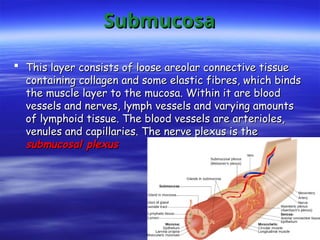
Submucosa
Submucosa
This layerconsists of loose areolar connective tissue
This layer consists of loose areolar connective tissue
containing collagen and some elastic fibres, which binds
containing collagen and some elastic fibres, which binds
the muscle layer to the mucosa. Within it are blood
the muscle layer to the mucosa. Within it are blood
vessels and nerves, lymph vessels and varying amounts
vessels and nerves, lymph vessels and varying amounts
of lymphoid tissue. The blood vessels are arterioles,
of lymphoid tissue. The blood vessels are arterioles,
venules and capillaries. The nerve plexus is the
venules and capillaries. The nerve plexus is the
submucosal plexus
submucosal plexus
17.
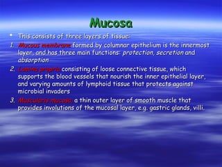
Mucosa
Mucosa
This consistsof three layers of tissue:
This consists of three layers of tissue:
1.
1. Mucous membrane
Mucous membrane formed by columnar epithelium is the innermost
formed by columnar epithelium is the innermost
layer, and has three main functions:
layer, and has three main functions: protection
protection,
, secretion
secretion and
and
absorption
absorption
2.
2. Lamina propria
Lamina propria consisting of loose connective tissue, which
consisting of loose connective tissue, which
supports the blood vessels that nourish the inner epithelial layer,
supports the blood vessels that nourish the inner epithelial layer,
and varying amounts of lymphoid tissue that protects against
and varying amounts of lymphoid tissue that protects against
microbial invaders
microbial invaders
3.
3. Muscularis mucosa
Muscularis mucosa,
, a thin outer layer of smooth muscle that
a thin outer layer of smooth muscle that
provides involutions of the mucosal layer, e.g. gastric glands, villi.
provides involutions of the mucosal layer, e.g. gastric glands, villi.
18.
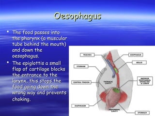
Oesophagus
Oesophagus
The foodpasses into
The food passes into
the pharynx (a muscular
the pharynx (a muscular
tube behind the mouth)
tube behind the mouth)
and down the
and down the
oesophagus.
oesophagus.
The epiglottis a small
The epiglottis a small
flap of cartilage blocks
flap of cartilage blocks
the entrance to the
the entrance to the
larynx, this stops the
larynx, this stops the
food going down the
food going down the
wrong way and prevents
wrong way and prevents
choking
choking.
.
19.
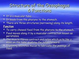
Structure of theOesophagus
Structure of the Oesophagus
& Functions
& Functions
It is a muscular tube.
It is a muscular tube.
It leads from the pharynx to the stomach.
It leads from the pharynx to the stomach.
There are three strictures (narrowing) along its length.
There are three strictures (narrowing) along its length.
Function
Function
To carry chewed food from the pharynx to the stomach.
To carry chewed food from the pharynx to the stomach.
Food moves along it by a muscular contraction known as
Food moves along it by a muscular contraction known as
peristalsis.
peristalsis.
The muscle fibres contract and relax which acts like a
The muscle fibres contract and relax which acts like a
wave on the tube, pushing the bolus forward.
wave on the tube, pushing the bolus forward.
It’s lining secretes mucus to lubricate the passage of
It’s lining secretes mucus to lubricate the passage of
food.
food.
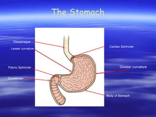
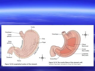
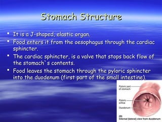
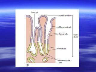
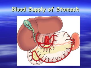
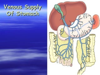
Stomach Structure
Stomach Structure
It is a J-shaped, elastic organ.
It is a J-shaped, elastic organ.
Food enters it from the oesophagus through the cardiac
Food enters it from the oesophagus through the cardiac
sphincter.
sphincter.
The cardiac sphincter, is a valve that stops back flow of
The cardiac sphincter, is a valve that stops back flow of
the stomach`s contents.
the stomach`s contents.
Food leaves the stomach through the pyloric sphincter
Food leaves the stomach through the pyloric sphincter
into the duodenum (first part of the small intestine).
into the duodenum (first part of the small intestine).
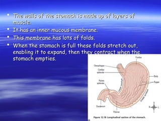
23.
The wallsof the stomach is made up of layers of
The walls of the stomach is made up of layers of
muscle.
muscle.
It has an inner mucous membrane.
It has an inner mucous membrane.
This membrane has lots of folds.
This membrane has lots of folds.
When the stomach is full these folds stretch out,
When the stomach is full these folds stretch out,
enabling it to expand, then they contract when the
enabling it to expand, then they contract when the
stomach empties.
stomach empties.
24.
Functions of TheStomach
Functions of The Stomach
It digests protein through the action of enzymes.
It digests protein through the action of enzymes.
It churns food with the gastric juices.
It churns food with the gastric juices.
It helps lubricate the food by producing mucus.
It helps lubricate the food by producing mucus.
It absorbs alcohol.
It absorbs alcohol.
It kills bacteria by producing hydrochloric acid.
It kills bacteria by producing hydrochloric acid.
It is involved in iron absorption
It is involved in iron absorption
25.
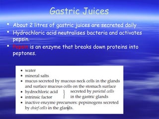
Gastric Juices
About2 litres of gastric juices are secreted daily
Hydrochloric acid neutralises bacteria and activates
pepsin.
Pepsin is an enzyme that breaks down proteins into
peptones.
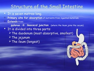
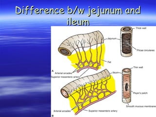
Structure of theSmall Intestine
It is seven metres long.
Primary site for absorption of nutrients from ingested materials
Extends from:
pylorus ileocecal junction (where the ileum joins the cecum)
It is divided into three parts:
The duodenum (most absorptive, smallest)
The jejunum
The ileum (longest)
30.
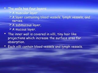
The wallshas four layers:
A muscular layer
A layer containing blood vessels, lymph vessels, and
nerves.
A submucous layer,
A mucous layer.
The inner wall is covered in villi, tiny hair like
projections which increase the surface area for
absorption.
Each villi contain blood vessels and lymph vessels.
31.
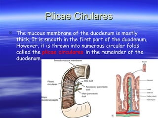
Plicae Cirulares
Plicae Cirulares
The mucous membrane of the duodenum is mostly
thick. It is smooth in the first part of the duodenum.
However, it is thrown into numerous circular folds
called the plicae circulares in the remainder of the
duodenum.
32.
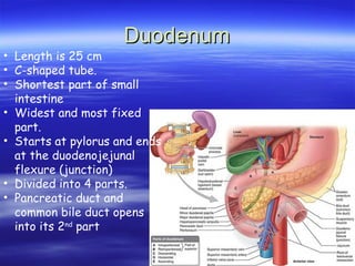
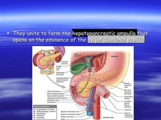
Duodenum
Duodenum
• Length is25 cm
• C-shaped tube.
• Shortest part of small
intestine
• Widest and most fixed
part.
• Starts at pylorus and ends
at the duodenojejunal
flexure (junction)
• Divided into 4 parts.
• Pancreatic duct and
common bile duct opens
into its 2nd
part
34.
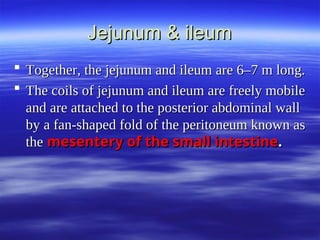
Jejunum & ileum
Jejunum& ileum
Together, the jejunum and ileum are 6–7 m long.
Together, the jejunum and ileum are 6–7 m long.
The coils of jejunum and ileum are freely mobile
The coils of jejunum and ileum are freely mobile
and are attached to the posterior abdominal wall
and are attached to the posterior abdominal wall
by a fan-shaped fold of the peritoneum known as
by a fan-shaped fold of the peritoneum known as
the
the mesentery of the small intestine
mesentery of the small intestine.
.
35.
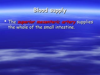
Blood supply
Blood supply
The
The superior mesenteric artery
superior mesenteric artery supplies
supplies
the whole of the small intestine. Venous
the whole of the small intestine. Venous
drainage is by the
drainage is by the superior mesenteric
superior mesenteric
vein
vein that joins other veins to form the
that joins other veins to form the
portal vein.
portal vein.
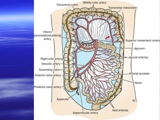
36.
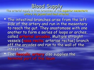
The intestinalbranches arise from the left
The intestinal branches arise from the left
side of the artery and run in the mesentery
side of the artery and run in the mesentery
to reach the gut. They anastomose with one
to reach the gut. They anastomose with one
another to form a series of loops or arches,
another to form a series of loops or arches,
called
called arterial arcades
arterial arcades. Multiple straight
. Multiple straight
vessels (
vessels (vasa recta
vasa recta; arteriae rectae) branch
; arteriae rectae) branch
off the arcades and run to the wall of the
off the arcades and run to the wall of the
intestine.
intestine.
The
The ileocolic artery
ileocolic artery also supplies the
also supplies the
terminal part of the ileum
terminal part of the ileum
Blood Supply
Blood Supply
The arterial supply is from branches of the
The arterial supply is from branches of the superior mesenteric
superior mesenteric
artery via jejunal and ileal arteries
artery via jejunal and ileal arteries
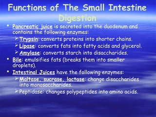
Functions of TheSmall Intestine
Digestion
Pancreatic juice is secreted into the duodenum and
contains the following enzymes:
Trypsin: converts proteins into shorter chains.
Lipase: converts fats into fatty acids and glycerol.
Amylase: converts starch into disaccharides.
Bile: emulsifies fats (breaks them into smaller
droplets).
Intestinal Juices have the following enzymes:
Maltase, sucrase, lactase: change disaccharides
into monosaccharides.
Peptidase: changes polypeptides into amino acids.
39.
Blood supply
Blood supply
The
The superior mesenteric artery
superior mesenteric artery supplies
supplies
the whole of the small intestine.
the whole of the small intestine.
41.
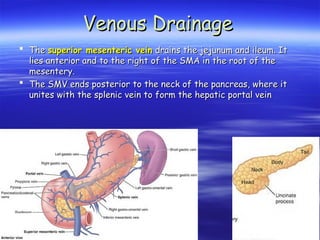
The
The superiormesenteric vein
superior mesenteric vein drains the jejunum and ileum. It
drains the jejunum and ileum. It
lies anterior and to the right of the SMA in the root of the
lies anterior and to the right of the SMA in the root of the
mesentery.
mesentery.
The SMV ends posterior to the neck of the pancreas, where it
The SMV ends posterior to the neck of the pancreas, where it
unites with the splenic vein to form the hepatic portal vein
unites with the splenic vein to form the hepatic portal vein
Venous Drainage
Venous Drainage
42.
Absorption
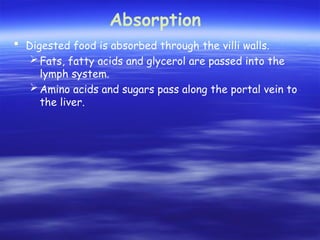
Digested foodis absorbed through the villi walls.
Fats, fatty acids and glycerol are passed into the
lymph system.
Amino acids and sugars pass along the portal vein to
the liver.
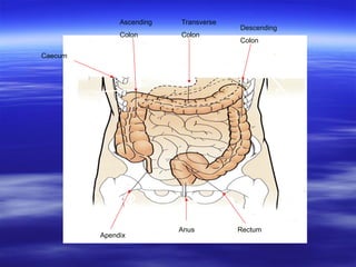
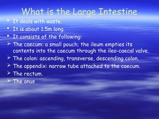
What is theLarge Intestine
It deals with waste.
It is about 1.5m long.
It consists of the following:
The caecum: a small pouch; the ileum empties its
contents into the caecum through the ileo-caecal valve.
The colon: ascending, transverse, descending colon.
The appendix: narrow tube attached to the caecum.
The rectum.
The anus
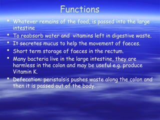
45.
Functions
Whatever remainsof the food, is passed into the large
intestine
To reabsorb water and vitamins left in digestive waste.
It secretes mucus to help the movement of faeces.
Short term storage of faeces in the rectum.
Many bacteria live in the large intestine, they are
harmless in the colon and may be useful e.g. produce
Vitamin K.
Defecation: peristalsis pushes waste along the colon and
then it is passed out of the body.
46.
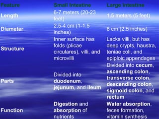
Feature Small IntestineLarge Intestine
Length
6-7 meters (20-23
feet)
1.5 meters (5 feet)
Diameter
2.5-4 cm (1-1.5
inches)
6 cm (2.5 inches)
Structure
Inner surface has
folds (plicae
circulares), villi, and
microvilli
Lacks villi, but has
deep crypts, haustra,
teniae coli, and
epiploic appendages
Parts
Divided into
duodenum,
jejunum, and ileum
Divided into cecum,
ascending colon,
transverse colon,
descending colon,
sigmoid colon, and
rectum
Function
Digestion and
absorption of
nutrients
Water absorption,
feces formation,
vitamin synthesis
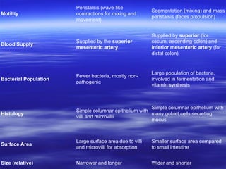
47.
Motility
Peristalsis (wave-like
contractions formixing and
movement)
Segmentation (mixing) and mass
peristalsis (feces propulsion)
Blood Supply
Supplied by the superior
mesenteric artery
Supplied by superior (for
cecum, ascending colon) and
inferior mesenteric artery (for
distal colon)
Bacterial Population
Fewer bacteria, mostly non-
pathogenic
Large population of bacteria,
involved in fermentation and
vitamin synthesis
Histology
Simple columnar epithelium with
villi and microvilli
Simple columnar epithelium with
many goblet cells secreting
mucus
Surface Area
Large surface area due to villi
and microvilli for absorption
Smaller surface area compared
to small intestine
Size (relative) Narrower and longer Wider and shorter