SlideShare a Scribd company logo
1 of 75
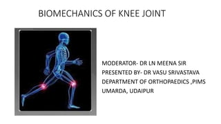
BIOMECHANICS OF KNEE JOINT
MODERATOR- DR LN MEENA SIR
PRESENTED BY- DR VASU SRIVASTAVA
DEPARTMENT OF ORTHOPAEDICS ,PIMS
UMARDA, UDAIPUR
KNEE JOINT-
1.Tibiofemoral component
2.Patellofemoral component
TIBIOFEMORAL COMPONANT/JOINT-
•It is a double condyloid joint
•Has 3 degree of freedom-
sagittal plane- flexion/ extension
transverse plane- medial rotation/ lateral rotation
frontal plane- abduction/ adduction
TIBIOFEMORAL JOINT IS COMPOSED OF-
• DISTAL FEMUR-
from lateral and medial side it is convex shape
Medial condyle is larger than the lateral condyle
• PROXIMAL TIBIA-
It has medial condyle or platue and lateral condyle or platue
Medial condyle is larger than the lateral condyle
Lateral condyle is projected anteriorly
In between the condyle there is intercondyler tubercle
The tibial condyles are slightly concave to 5-7 degree ,inclined inferiorly
GENU VALGUM AND GENU VARUM
• LONGITUDINAL AXIS- LINE PARALLEL TO THE LONG BONE FROM THE CENTRE.
• MECHANICAL AXIS- LINE JOINING FROM THE CENTRE OF HEAD OF FEMUR TO
KNEE JOINT TO CENTRE OF CALCANEUM.
• IT IS A WEIGHT BEARING LINE
• So the angle formed between the longitudinal axis and the mechanical axis is
known as the physiological valgus angle ,which is normally 5-7 degree.
• Both these axis further forms a medial tibiofemoral angle, which is normally
185 degree.
• So, if there is increase in this medial tibio femoral angle to >185 degree ,it is
known as genu valgum.
• And if medial tibio-femoral angle is <185 degree ,it is known as genu varum
Genu valgum
• increases in compressive force in
the lateral compartment and
tensile force in the medial
compartment.
• Leads to joint laxity in the lateral
compartment.
• Knock knee.
• Adduction movement increases in
the knee
Genu varum
• Increase in the compressive force
in the medial compartment of joint
and tensile force in the lateral
compartment
• Leads to bow knees
• In the single leg stance –the weight bearing axis from the centre is
shifted to medial side,
• So the compressive force is more towards the medial condyle of
tibiofemoral component , so this is the reason that there is much
more chance of osteoarthritic changes in the medial condyle side.
Menisci of knee joint
• Cushions of the knee joint.
• Made of fibrocartilage tissue
• Disc shape.
• Medial meniscus is semi-lunar shape
• Lateral meniscus is circular in shape
• Each meniscus has anterior horn or anterior end and
posterior horn or posterior end.
• There is no meniscus over the intercondylar
tubercle.
• Thick periphery and thin centrally
• Lateral meniscus is shorter than the medial meniscus
• Function- increases the articular area.
increase joint congruency
increase joint stability
shock absorber
Distribute weight bearing force
STABILITY OF KNEE JOINT
• Stability of the knee joint depends upon the-
1.capsule
2.collateral ligaments
3.cruciate ligaments
4.various muscles associated with the knee joint movements
CAPSULE OF KNEE JOINT
THE CAPSULE HAS 2 LAYERS-
1. SUPERFICIAL LAYER 2. DEEP LAYER
SUPERFICIAL LAYER/ FIBROUS LAYER
• Provide passive stability
• Has extensor retinaculum
medial patellar retinaculum
lateral patellar retinaculum
Includes 6 ligaments-
1.Medial patella femoral ligament
2.Lateral patellofemoral ligament
3.Medial patello meniscus ligament
4.Lateral patello meniscus ligament
5. Medial patello tibial ligament
6. Lateral patello tibial ligament
DEEP LAYER / SYNOVIAL LAYER
• Related to medial and lateral femoral
condyle and intercondylar area.
• Some time associated with PLICA
syndrome-
• PLICA – at the time of gestation, some
part of patella do not get fuse ,which
causes irritation in the superior,
medial and lateral side of the patella.
• Most common is medial patella PLICA
• Syndrome can also occur on trauma
/fall.
• It is a reminant of synovial membrane
COLLATERAL LIGAMENT
Medial collateral ligament
• Arise from adductor tubercle
• Has 2 fibers-
Superficial or anterior fiber-
insertion is just distal to pes
anserinus
Deep or posterior fiber- insertion is
proximal surface of shaft of tibia
and is attached to capsule
• Function-
1.restricting the valgus force.
2.restricting the ACL while flexion
and prevents excessive lateral
rotation of tibia.
Lateral collateral ligament
• A/K as fibular collateral ligament
• Origin- lateral femoral epicondyle
• Insertion- head of fibula
• Not attached to capsule
• Function-
Restricting the varus force
Lateral
collateral
ligament
ANTERIOR CRUCIATE LIGAMENT
ORIGIN- postero-medial aspect of lateral femoral condyle
Insertion- medial tubercle of the spine
• Fibers of ACL are inferiorly, medially and anteriorly.
• Has 2 bundles of fibers-
1.antero-medial bundle
2.postero-lateral bundle
• Function-
1.resist the anterior translation of tibia on femur.
• Role of antero-medial bundle- during flexion, the anteromedial bundle get tight and
prevents the anterior translation of tibia on femur.
• Role of postero-lateral bundle- during extension, the postero-lateral bundle get tight and
prevents the anterior translation of tibia on femur.
Least amount of anterior translation during extension because postero-lateral bundle is
very strong.
• In 30 degree of flexion there is max. anterior translation of tibia.
2. prevents hyper-extension of knee by postero-lateral bundle.
3. help in providing rotational stability i.e medial/ lateral rotation or varus/valgus
MUSCLES ASSOCIATED WITH ANT. CRUCIATE LIGAMENT-
1. Quadricep muscle- extension. So anterior translation is prevented
by posterolateral bundle of ACL.
2. GASTROCNEMUS- prevent anterior translation
3. Hamstring and soleus- associated with posterior translation
POSTERIOR CRUCIATE LIGAMENT
• Arise from the anteromedial aspect of medial femoral condyle and attached to
the posterior horn of both menisci or 1cm distal to joint line in tibial platue.
• Direction of fiber- inferiorly and posteriorly
• Posterior cruciate ligament is shorter than ant. Cruciate ligament but the
surface area of post. Cruciate ligament is 150% more than ant. Cruciate
ligament.
Bundles of post. Cruciate ligament-
1. anterolateral bundle- a)weak during extension
b)on flexion (80-90degree) maximum tightness in post.
Cruciate ligament.
2. Posteromedial bundle- a)tight during extension
b) on flexion ,the posteromedial bundle get tightness.
FUNCTIONS OF POS. CRUCIATE LIGAMENT-
1) restriction to posterior translation of tibia on femur
2) provide valgus and varus stability
3) tibial rotational stability by postero medial bundle
Role of muscles in post. Cruciate ligament-
1) popletius – it helps in posterior translation of tibia if post. Cruciate ligament
is absent.
2) hamstrings- even though it is a knee flexor, but prevents the posterior
translation of tibia.
3) gastrocnemius – increase the strain on post. Cruciate ligament at more than
40 degree of flexion.
KNEE JOINT KINAMATICS
KNEE JOINT HAS FOLLOWING MOVEMENTS-
1)Flexion and extension
2)medial rotation and lateral rotation
3)abduction and addiction
1) Flexion and extension -
• It is the principle motion of knee complex
• Movement occurs in transcondylar axis. but in knee trans-epicondylar is not constant,it
changes with the movement.
Can have 2 catogery motion
1)fixed tibia, only femur is in movement state
2)fixed femur, only tibia is in movement state
(when tibia is fixed and only femur is in movement state ,eg- down squatting position) ,
During flexion
so, femur is going for flexion ,the arthrokinamatic movement the femur rolls posteriorly
and slide or glide anteriorly.
so, at 0-25 degree of flexion ,there is only posterior rolling function and at more then 25
degree of flexion, the femur motion is accompanied by both the anterior sliding motion
and post. rolling motion.
During extension-
During extension the femur is showing anterior rolling and slide posteriorly
-after 25 degree of extension ,sliding starts, so also known as pure spin movement.
During extension- anterior rolling and
slide posteriorly
During flexion- rolling posteriorly
and gliding/sliding anteriorly
2) fixed femur, only tibia is in movement state(eg. standing squatting position)
Tibia flexion- tibial cavity is concave i.e concave on convex
Movement- posterior rolling and sliding/gliding posteriorly
Posterior cruciate ligament is helpful in preventing posterior rolling from
dislocation.
On tibial extension- anterior rolling and anterior gliding/sliding
Role of Ante. Cruciate ligament and posterior
cruciate ligament in knee flexion and extension
1. when flexion increases, ACL get stretched out and prevent the ant. Translation
of tibia and help from dislocating.
2. on extension ,PCL get stretched and prevent posterior translation
Role of menisci in flexion and extension movement
• Menisci is wedge shape i.e thin from the centre and
thick from the pheriphery ,so it helps in uphill
movement of femur.
• During flexion the shear force transmit anteriorly.
• During extension ,the shear force transmit
posteriorly
2. Medial and lateral rotation of knee joint
• Occurs in transverse plane and longitudinal axis.
• Longitudinal axis passes through medial tibial condyle or medial tibial
intercondyloid tubercle. So the movement around the pivot is in medial
tibial condyle or med. Tibial intercondyloid tubercle and movement in
lateral condyle will be more.
(Pivot or axis through which the movement take place will be restricted ,so
medial condyle will have less rotation.)
So,
lateral rotation- medial condyle will go anteriorly and lateral condyle will go
posteriorly.
medial rotation- the lateral tibial condyle will have arc more anteriorly and
medial tibial condyle les movement posteriorly
SCREW HOME MECHANISM
• Screw home mechanism (SHM) of knee joint is a critical mechanism that play
an important role in terminal extension of the knee.
• There is an observable rotation of the knee during flexion and extension.
• This rotation is important for healthy movement of the knee.
• During the last 30 degrees of knee extension, the tibia (open chain) or femur
(closed chain) must externally or internally rotate, respectively, about 10
degrees.
• This slight rotation is due to inequality of the articular surface of femur
condyles.
• Rotation must occur to achieve full extension and then flexion from full
extension
• The "screw-home" mechanism is considered to be a key element to
knee stability for standing upright. The tibia rotates internally during
the swing phase and externally during the stance phase.
• External rotation occurs during the terminal degrees of knee
extension and results in tightening of both cruciate ligaments, which
locks the knee.
• The tibia is then in the position of maximal stability with respect to
the femur.
• Last 30 degrees of Extension causes a Medial rotation of Femur on
Tibia will keep joint in closed packed position. The Knee is Unlocked
by Lateral rotation of Femur.
• In open Kinematic chain Tibia laterally rotates on Femur during last 5
degrees of Extension to produce LOCKING. Unlocking by Medial
rotation
Role of menisci and ligaments on medial and lateral
rotation-
ROLE OF MENISCI-
• On medial rotation of tibia on femur- lateral menisci move anteriorly due to
shape.
• On lateral rotation of tibia on femur – medial menisci move posteriorly
ROLE OF LIGAMENTS-
• As in flexion to extension –most of the ligaments get lax or loose, so they do
not restrict the movement, but in med/lat. rotation the ligaments causes
restriction of movements.
Upto 0-90 degree flexion- max. value of medial and lateral rotation.
Upto 0-20 degree - lateral rotation and upto 0- 15 degree –medial rotation.
• So, lateral rotation is more than the medial rotation in flexion position.
So, upto 0-90 degree –medial/lat. rotation increases and 90-130 degree-
med/lat. rotation decreases
3.Abduction and adduction of knee
ABDUCTION – VALGUS MOVEMENT ADDUCTION- VARUS MOVEMENT
occurs in frontal plane i.e AP AXIS movements
5-8 degree of abduction or adduction are possible in extension, as the
ligaments are very tough
13-20 degree of movement of abduction / adduction are possible in flexion.
In abduction- tibia is going outward ,so joint is associated with valgus force
and increase in compression in the lateral compartment of condyle
In adduction – tibia is coming inwards ,causing varus force , and increase in
compression in medial compartment of condyle.
LOCKING AND UNLOCKING OF KNEE
• One of the most imp. topic in knee biomechanics
• Comes under coupled motion
• Coupled motion- motion that occurs in one axis and that motion occurs in
association with another axis.eg.- a motion occurring in x axis and associated
with y axis.
• IMPORTANCE-
• As medial condyle of femur is slightly distal than the lateral condyle ,so this
distal association creates a physiological association with the tibia creating a
physiological valgus.
• Normally tibia is a bit laterally, on flexion of tibia, the tibia comes bit midline of
the body. So, knee flexion is accompanied by varus and abduction movement
and extension is linked to valgus and adduction.
• Flexion occurs in sagittal axis and varus / valgus occurs in frontal axis,so there is
involvement of 2 axis, that’s why knee has coupled motion.
Coupled motion in knee joint
FLEXION / EXTENSION ACCOMPANIED BY LATERAL AND MEDIAL ROTATION
• This motion occurs in last 30 degree of flexion to extension of tibia (fixed femur),
leads to lateral rotation of tibia.
Medial tibial condyle having rolling and ant. Gliding -
the medial tibial condyle of tibia has greater degree of surface area, so associated
with rolling and ant. Gliding and associated with lateral rotation on medial side and
the lateral condyle act as PIVOT, so this is known as screw hole mechanism,
where lateral condyle act as a screw and medial condyle side rotating and gliding.
• This complete process is known as locking of knee or automatic
rotation or terminal rotation or screw home mechanism.
UNLOCKING OF KNEE-
• On flexion of tibia (with fixed femur)- the knee has slight medial rotation i.e
flexion accompanied by medial rotation.
• Popliteus muscle is directly related to unlocking and locking because of its
action.
The popliteus assists in knee flexion. Its function is dependent on whether the
lower extremity is in a weight-bearing or non-weight-bearing state; it is considered
the primary internal rotator of the tibia in the non-weight-bearing state.
• "Locking" the knee occurs with extension during weight-bearing. This describes
the femur medially rotating on the tibia, allowing for full extension without
muscular expenditure.
• When "unlocking" the knee, the popliteus contracts causing flexion and lateral
rotation of the femur on the tibia. This is why some refer to the popliteus as the
"key" to the locked knee
Screw home mechanism in non weight bearing
• LOCKING-
- extension is combined with lateral rotation
-flexion is combined with medial rotation
here tibia is fixed on moving femur so, at last 30 degree of extension from flexion
,small lateral condyle of tibia will have rolling and gliding posteriorly or opposite
direction.
So ,rolling upward and gliding posteriorly and medial rotation of femur
Screw home mechanism in weight bearing-
Tibia is fixed and femur is rotating (in extension ,while standing from
squatting position)
Extension is combined with medial rotation of femur
Flexion is combined with lateral rotation of femur
BIOMECHANICS OF THE KNEE

More Related Content

What's hot

Ankle joint
Ankle jointAnkle joint
Ankle jointRahul Jha
 
Biomechanics of the Hip Joint
Biomechanics of the Hip JointBiomechanics of the Hip Joint
Biomechanics of the Hip JointUsheem Syed
 
Biomechanics of Lumbar spine
Biomechanics of Lumbar spineBiomechanics of Lumbar spine
Biomechanics of Lumbar spineSagarGajra1
 
Arches of foot
Arches  of  footArches  of  foot
Arches of footharsha2063
 
Foot biomechanics
Foot biomechanicsFoot biomechanics
Foot biomechanicsdeepakanap
 
The Knee Complex
The Knee ComplexThe Knee Complex
The Knee ComplexPhilip Keriaka
 
Kinetics of knee joint
Kinetics of knee jointKinetics of knee joint
Kinetics of knee jointAkhilaNatesan
 
Biomechanics of thoracic spine ppt
Biomechanics of thoracic spine pptBiomechanics of thoracic spine ppt
Biomechanics of thoracic spine pptMuskan Rastogi
 
Biomech lumbar spine
Biomech lumbar spineBiomech lumbar spine
Biomech lumbar spineMuhammadasif909
 
Deep layer of (Iliotibial) IT band
Deep layer of (Iliotibial) IT bandDeep layer of (Iliotibial) IT band
Deep layer of (Iliotibial) IT bandAjith lolita
 
Biomechanics of hip complex 1
Biomechanics of hip complex 1Biomechanics of hip complex 1
Biomechanics of hip complex 1Dibyendunarayan Bid
 
Arches of the Foot
Arches of the FootArches of the Foot
Arches of the FootShama
 
Extensor apparatus hand
Extensor apparatus hand Extensor apparatus hand
Extensor apparatus hand orthoprince
 
Knee biomechanics
Knee biomechanicsKnee biomechanics
Knee biomechanicsSreeraj S R
 
Biomechanich of the spine ppt (2)
Biomechanich of the spine ppt (2)Biomechanich of the spine ppt (2)
Biomechanich of the spine ppt (2)Dr.Debanjan Mondal(PT)
 
Biomechanics of knee complex 6 tibiofemoral jt function
Biomechanics of knee complex 6 tibiofemoral jt functionBiomechanics of knee complex 6 tibiofemoral jt function
Biomechanics of knee complex 6 tibiofemoral jt functionDibyendunarayan Bid
 

What's hot (20)

Ankle and foot complex
Ankle and foot complexAnkle and foot complex
Ankle and foot complex
 
Ankle joint
Ankle jointAnkle joint
Ankle joint
 
Biomechanics of the Hip Joint
Biomechanics of the Hip JointBiomechanics of the Hip Joint
Biomechanics of the Hip Joint
 
Biomechanics of Lumbar spine
Biomechanics of Lumbar spineBiomechanics of Lumbar spine
Biomechanics of Lumbar spine
 
Arches of foot
Arches  of  footArches  of  foot
Arches of foot
 
Foot biomechanics
Foot biomechanicsFoot biomechanics
Foot biomechanics
 
The Knee Complex
The Knee ComplexThe Knee Complex
The Knee Complex
 
Kinetics of knee joint
Kinetics of knee jointKinetics of knee joint
Kinetics of knee joint
 
Biomechanics of thoracic spine ppt
Biomechanics of thoracic spine pptBiomechanics of thoracic spine ppt
Biomechanics of thoracic spine ppt
 
Biomechanics of the shoulder
Biomechanics of the shoulder Biomechanics of the shoulder
Biomechanics of the shoulder
 
Biomech lumbar spine
Biomech lumbar spineBiomech lumbar spine
Biomech lumbar spine
 
Bio-mechanics of the ankle joint
Bio-mechanics of the ankle jointBio-mechanics of the ankle joint
Bio-mechanics of the ankle joint
 
Deep layer of (Iliotibial) IT band
Deep layer of (Iliotibial) IT bandDeep layer of (Iliotibial) IT band
Deep layer of (Iliotibial) IT band
 
Gait
GaitGait
Gait
 
Biomechanics of hip complex 1
Biomechanics of hip complex 1Biomechanics of hip complex 1
Biomechanics of hip complex 1
 
Arches of the Foot
Arches of the FootArches of the Foot
Arches of the Foot
 
Extensor apparatus hand
Extensor apparatus hand Extensor apparatus hand
Extensor apparatus hand
 
Knee biomechanics
Knee biomechanicsKnee biomechanics
Knee biomechanics
 
Biomechanich of the spine ppt (2)
Biomechanich of the spine ppt (2)Biomechanich of the spine ppt (2)
Biomechanich of the spine ppt (2)
 
Biomechanics of knee complex 6 tibiofemoral jt function
Biomechanics of knee complex 6 tibiofemoral jt functionBiomechanics of knee complex 6 tibiofemoral jt function
Biomechanics of knee complex 6 tibiofemoral jt function
 

Similar to BIOMECHANICS OF THE KNEE

Anatomy of knee
Anatomy of kneeAnatomy of knee
Anatomy of kneeDibinKThomas3
 
BIOMECHANICS & PATHOMECHANICS OF KNEE JOINT AND PATELLOFEMORAL JOINT
BIOMECHANICS & PATHOMECHANICS OF KNEE JOINT AND PATELLOFEMORAL JOINTBIOMECHANICS & PATHOMECHANICS OF KNEE JOINT AND PATELLOFEMORAL JOINT
BIOMECHANICS & PATHOMECHANICS OF KNEE JOINT AND PATELLOFEMORAL JOINTDr. Taniya Verma ( PT) Gold medalist
 
knee joint biomechanics 2nd BPTH Kinesiology
knee joint biomechanics 2nd BPTH Kinesiologyknee joint biomechanics 2nd BPTH Kinesiology
knee joint biomechanics 2nd BPTH KinesiologyNIKITAWAGHMARE6
 
anatomy and biomechanics of Shoulder joint
anatomy and biomechanics of Shoulder jointanatomy and biomechanics of Shoulder joint
anatomy and biomechanics of Shoulder jointHarsha Nandini
 
Knee joint.pptx
Knee joint.pptxKnee joint.pptx
Knee joint.pptxApoorvGarg28
 
Knee joint biomechanics
Knee joint biomechanicsKnee joint biomechanics
Knee joint biomechanicsBhavnaRajpurohit
 
Biomechanics of ankle_joint
Biomechanics of ankle_jointBiomechanics of ankle_joint
Biomechanics of ankle_jointNityal Kumar
 
Knee joint complex biomechanics
Knee joint complex biomechanicsKnee joint complex biomechanics
Knee joint complex biomechanicsAkhilaNatesan
 
anatomyofthekneejoint-170329185745.pdf
anatomyofthekneejoint-170329185745.pdfanatomyofthekneejoint-170329185745.pdf
anatomyofthekneejoint-170329185745.pdfUmaMaheshwariJ3
 
Anatomy of the knee joint
Anatomy of the knee jointAnatomy of the knee joint
Anatomy of the knee jointRajesh Raj
 
Pathomechanics Knee.pptx
Pathomechanics Knee.pptxPathomechanics Knee.pptx
Pathomechanics Knee.pptxVenkatSingh
 
Knee instability
Knee instabilityKnee instability
Knee instabilitypunithpc605
 
Ligamnet around knee and injury and management
Ligamnet around knee and injury and managementLigamnet around knee and injury and management
Ligamnet around knee and injury and managementBirajkc5
 
hip biomechanics, a simple presentation for beginners
hip biomechanics, a simple presentation for beginnership biomechanics, a simple presentation for beginners
hip biomechanics, a simple presentation for beginnersSupreetBajwa2
 
Applied Biomechanics of Cervical Spine
Applied Biomechanics of Cervical SpineApplied Biomechanics of Cervical Spine
Applied Biomechanics of Cervical SpineDr. POONAM N. BANTHIA
 
shoulderanatomy-160501022628.pptx
shoulderanatomy-160501022628.pptxshoulderanatomy-160501022628.pptx
shoulderanatomy-160501022628.pptxVaisHali822687
 
KNEE JOINT GK.pptx
KNEE JOINT GK.pptxKNEE JOINT GK.pptx
KNEE JOINT GK.pptxRifaRahmalia2
 

Similar to BIOMECHANICS OF THE KNEE (20)

Biomechanics
BiomechanicsBiomechanics
Biomechanics
 
knee ..pptx
knee ..pptxknee ..pptx
knee ..pptx
 
Anatomy of knee
Anatomy of kneeAnatomy of knee
Anatomy of knee
 
BIOMECHANICS & PATHOMECHANICS OF KNEE JOINT AND PATELLOFEMORAL JOINT
BIOMECHANICS & PATHOMECHANICS OF KNEE JOINT AND PATELLOFEMORAL JOINTBIOMECHANICS & PATHOMECHANICS OF KNEE JOINT AND PATELLOFEMORAL JOINT
BIOMECHANICS & PATHOMECHANICS OF KNEE JOINT AND PATELLOFEMORAL JOINT
 
knee joint biomechanics 2nd BPTH Kinesiology
knee joint biomechanics 2nd BPTH Kinesiologyknee joint biomechanics 2nd BPTH Kinesiology
knee joint biomechanics 2nd BPTH Kinesiology
 
anatomy and biomechanics of Shoulder joint
anatomy and biomechanics of Shoulder jointanatomy and biomechanics of Shoulder joint
anatomy and biomechanics of Shoulder joint
 
Knee joint.pptx
Knee joint.pptxKnee joint.pptx
Knee joint.pptx
 
Knee joint biomechanics
Knee joint biomechanicsKnee joint biomechanics
Knee joint biomechanics
 
Knee joint.pptx
Knee joint.pptxKnee joint.pptx
Knee joint.pptx
 
Biomechanics of ankle_joint
Biomechanics of ankle_jointBiomechanics of ankle_joint
Biomechanics of ankle_joint
 
Knee joint complex biomechanics
Knee joint complex biomechanicsKnee joint complex biomechanics
Knee joint complex biomechanics
 
anatomyofthekneejoint-170329185745.pdf
anatomyofthekneejoint-170329185745.pdfanatomyofthekneejoint-170329185745.pdf
anatomyofthekneejoint-170329185745.pdf
 
Anatomy of the knee joint
Anatomy of the knee jointAnatomy of the knee joint
Anatomy of the knee joint
 
Pathomechanics Knee.pptx
Pathomechanics Knee.pptxPathomechanics Knee.pptx
Pathomechanics Knee.pptx
 
Knee instability
Knee instabilityKnee instability
Knee instability
 
Ligamnet around knee and injury and management
Ligamnet around knee and injury and managementLigamnet around knee and injury and management
Ligamnet around knee and injury and management
 
hip biomechanics, a simple presentation for beginners
hip biomechanics, a simple presentation for beginnership biomechanics, a simple presentation for beginners
hip biomechanics, a simple presentation for beginners
 
Applied Biomechanics of Cervical Spine
Applied Biomechanics of Cervical SpineApplied Biomechanics of Cervical Spine
Applied Biomechanics of Cervical Spine
 
shoulderanatomy-160501022628.pptx
shoulderanatomy-160501022628.pptxshoulderanatomy-160501022628.pptx
shoulderanatomy-160501022628.pptx
 
KNEE JOINT GK.pptx
KNEE JOINT GK.pptxKNEE JOINT GK.pptx
KNEE JOINT GK.pptx
 

More from Vasu Srivastava

SCOLIOSIS PPT DR VASU SRIVASTAVA.pptx
SCOLIOSIS PPT DR VASU SRIVASTAVA.pptxSCOLIOSIS PPT DR VASU SRIVASTAVA.pptx
SCOLIOSIS PPT DR VASU SRIVASTAVA.pptxVasu Srivastava
 
MEDIAL EPICONDYLE FRACTURE BY DR. VASU SRIVASTAVA
MEDIAL EPICONDYLE FRACTURE BY DR. VASU SRIVASTAVAMEDIAL EPICONDYLE FRACTURE BY DR. VASU SRIVASTAVA
MEDIAL EPICONDYLE FRACTURE BY DR. VASU SRIVASTAVAVasu Srivastava
 
OSTEOLYSIS AND LOOSENING OF total hip arthroplasty IMPLANTS.pptx by dr vasu ...
OSTEOLYSIS AND LOOSENING OF total hip arthroplasty  IMPLANTS.pptx by dr vasu ...OSTEOLYSIS AND LOOSENING OF total hip arthroplasty  IMPLANTS.pptx by dr vasu ...
OSTEOLYSIS AND LOOSENING OF total hip arthroplasty IMPLANTS.pptx by dr vasu ...Vasu Srivastava
 
ACUTE OSTEOMYELITIS PPT.pptx
ACUTE OSTEOMYELITIS PPT.pptxACUTE OSTEOMYELITIS PPT.pptx
ACUTE OSTEOMYELITIS PPT.pptxVasu Srivastava
 
SPIROCHAETAL INFECTION in orthopaedics.pptx
SPIROCHAETAL INFECTION in orthopaedics.pptxSPIROCHAETAL INFECTION in orthopaedics.pptx
SPIROCHAETAL INFECTION in orthopaedics.pptxVasu Srivastava
 
MEDIAL EPICONDYLE VASU.pptx
MEDIAL EPICONDYLE VASU.pptxMEDIAL EPICONDYLE VASU.pptx
MEDIAL EPICONDYLE VASU.pptxVasu Srivastava
 

More from Vasu Srivastava (6)

SCOLIOSIS PPT DR VASU SRIVASTAVA.pptx
SCOLIOSIS PPT DR VASU SRIVASTAVA.pptxSCOLIOSIS PPT DR VASU SRIVASTAVA.pptx
SCOLIOSIS PPT DR VASU SRIVASTAVA.pptx
 
MEDIAL EPICONDYLE FRACTURE BY DR. VASU SRIVASTAVA
MEDIAL EPICONDYLE FRACTURE BY DR. VASU SRIVASTAVAMEDIAL EPICONDYLE FRACTURE BY DR. VASU SRIVASTAVA
MEDIAL EPICONDYLE FRACTURE BY DR. VASU SRIVASTAVA
 
OSTEOLYSIS AND LOOSENING OF total hip arthroplasty IMPLANTS.pptx by dr vasu ...
OSTEOLYSIS AND LOOSENING OF total hip arthroplasty  IMPLANTS.pptx by dr vasu ...OSTEOLYSIS AND LOOSENING OF total hip arthroplasty  IMPLANTS.pptx by dr vasu ...
OSTEOLYSIS AND LOOSENING OF total hip arthroplasty IMPLANTS.pptx by dr vasu ...
 
ACUTE OSTEOMYELITIS PPT.pptx
ACUTE OSTEOMYELITIS PPT.pptxACUTE OSTEOMYELITIS PPT.pptx
ACUTE OSTEOMYELITIS PPT.pptx
 
SPIROCHAETAL INFECTION in orthopaedics.pptx
SPIROCHAETAL INFECTION in orthopaedics.pptxSPIROCHAETAL INFECTION in orthopaedics.pptx
SPIROCHAETAL INFECTION in orthopaedics.pptx
 
MEDIAL EPICONDYLE VASU.pptx
MEDIAL EPICONDYLE VASU.pptxMEDIAL EPICONDYLE VASU.pptx
MEDIAL EPICONDYLE VASU.pptx
 

Recently uploaded

Accessible design: Minimum effort, maximum impact
Accessible design: Minimum effort, maximum impactAccessible design: Minimum effort, maximum impact
Accessible design: Minimum effort, maximum impactdawncurless
 
Contemporary philippine arts from the regions_PPT_Module_12 [Autosaved] (1).pptx
Contemporary philippine arts from the regions_PPT_Module_12 [Autosaved] (1).pptxContemporary philippine arts from the regions_PPT_Module_12 [Autosaved] (1).pptx
Contemporary philippine arts from the regions_PPT_Module_12 [Autosaved] (1).pptxRoyAbrique
 
SOCIAL AND HISTORICAL CONTEXT - LFTVD.pptx
SOCIAL AND HISTORICAL CONTEXT - LFTVD.pptxSOCIAL AND HISTORICAL CONTEXT - LFTVD.pptx
SOCIAL AND HISTORICAL CONTEXT - LFTVD.pptxiammrhaywood
 
Introduction to AI in Higher Education_draft.pptx
Introduction to AI in Higher Education_draft.pptxIntroduction to AI in Higher Education_draft.pptx
Introduction to AI in Higher Education_draft.pptxpboyjonauth
 
Introduction to ArtificiaI Intelligence in Higher Education
Introduction to ArtificiaI Intelligence in Higher EducationIntroduction to ArtificiaI Intelligence in Higher Education
Introduction to ArtificiaI Intelligence in Higher Educationpboyjonauth
 
Incoming and Outgoing Shipments in 1 STEP Using Odoo 17
Incoming and Outgoing Shipments in 1 STEP Using Odoo 17Incoming and Outgoing Shipments in 1 STEP Using Odoo 17
Incoming and Outgoing Shipments in 1 STEP Using Odoo 17Celine George
 
How to Configure Email Server in Odoo 17
How to Configure Email Server in Odoo 17How to Configure Email Server in Odoo 17
How to Configure Email Server in Odoo 17Celine George
 
Mastering the Unannounced Regulatory Inspection
Mastering the Unannounced Regulatory InspectionMastering the Unannounced Regulatory Inspection
Mastering the Unannounced Regulatory InspectionSafetyChain Software
 
mini mental status format.docx
mini    mental       status     format.docxmini    mental       status     format.docx
mini mental status format.docxPoojaSen20
 
Crayon Activity Handout For the Crayon A
Crayon Activity Handout For the Crayon ACrayon Activity Handout For the Crayon A
Crayon Activity Handout For the Crayon AUnboundStockton
 
A Critique of the Proposed National Education Policy Reform
A Critique of the Proposed National Education Policy ReformA Critique of the Proposed National Education Policy Reform
A Critique of the Proposed National Education Policy ReformChameera Dedduwage
 
call girls in Kamla Market (DELHI) 🔝 >༒9953330565🔝 genuine Escort Service 🔝✔️✔️
call girls in Kamla Market (DELHI) 🔝 >༒9953330565🔝 genuine Escort Service 🔝✔️✔️call girls in Kamla Market (DELHI) 🔝 >༒9953330565🔝 genuine Escort Service 🔝✔️✔️
call girls in Kamla Market (DELHI) 🔝 >༒9953330565🔝 genuine Escort Service 🔝✔️✔️9953056974 Low Rate Call Girls In Saket, Delhi NCR
 
ECONOMIC CONTEXT - LONG FORM TV DRAMA - PPT
ECONOMIC CONTEXT - LONG FORM TV DRAMA - PPTECONOMIC CONTEXT - LONG FORM TV DRAMA - PPT
ECONOMIC CONTEXT - LONG FORM TV DRAMA - PPTiammrhaywood
 
The Most Excellent Way | 1 Corinthians 13
The Most Excellent Way | 1 Corinthians 13The Most Excellent Way | 1 Corinthians 13
The Most Excellent Way | 1 Corinthians 13Steve Thomason
 
Presentation by Andreas Schleicher Tackling the School Absenteeism Crisis 30 ...
Presentation by Andreas Schleicher Tackling the School Absenteeism Crisis 30 ...Presentation by Andreas Schleicher Tackling the School Absenteeism Crisis 30 ...
Presentation by Andreas Schleicher Tackling the School Absenteeism Crisis 30 ...EduSkills OECD
 
_Math 4-Q4 Week 5.pptx Steps in Collecting Data
_Math 4-Q4 Week 5.pptx Steps in Collecting Data_Math 4-Q4 Week 5.pptx Steps in Collecting Data
_Math 4-Q4 Week 5.pptx Steps in Collecting DataJhengPantaleon
 
Alper Gobel In Media Res Media Component
Alper Gobel In Media Res Media ComponentAlper Gobel In Media Res Media Component
Alper Gobel In Media Res Media ComponentInMediaRes1
 
Kisan Call Centre - To harness potential of ICT in Agriculture by answer farm...
Kisan Call Centre - To harness potential of ICT in Agriculture by answer farm...Kisan Call Centre - To harness potential of ICT in Agriculture by answer farm...
Kisan Call Centre - To harness potential of ICT in Agriculture by answer farm...Krashi Coaching
 

Recently uploaded (20)

Accessible design: Minimum effort, maximum impact
Accessible design: Minimum effort, maximum impactAccessible design: Minimum effort, maximum impact
Accessible design: Minimum effort, maximum impact
 
Contemporary philippine arts from the regions_PPT_Module_12 [Autosaved] (1).pptx
Contemporary philippine arts from the regions_PPT_Module_12 [Autosaved] (1).pptxContemporary philippine arts from the regions_PPT_Module_12 [Autosaved] (1).pptx
Contemporary philippine arts from the regions_PPT_Module_12 [Autosaved] (1).pptx
 
SOCIAL AND HISTORICAL CONTEXT - LFTVD.pptx
SOCIAL AND HISTORICAL CONTEXT - LFTVD.pptxSOCIAL AND HISTORICAL CONTEXT - LFTVD.pptx
SOCIAL AND HISTORICAL CONTEXT - LFTVD.pptx
 
TataKelola dan KamSiber Kecerdasan Buatan v022.pdf
TataKelola dan KamSiber Kecerdasan Buatan v022.pdfTataKelola dan KamSiber Kecerdasan Buatan v022.pdf
TataKelola dan KamSiber Kecerdasan Buatan v022.pdf
 
Introduction to AI in Higher Education_draft.pptx
Introduction to AI in Higher Education_draft.pptxIntroduction to AI in Higher Education_draft.pptx
Introduction to AI in Higher Education_draft.pptx
 
Introduction to ArtificiaI Intelligence in Higher Education
Introduction to ArtificiaI Intelligence in Higher EducationIntroduction to ArtificiaI Intelligence in Higher Education
Introduction to ArtificiaI Intelligence in Higher Education
 
Incoming and Outgoing Shipments in 1 STEP Using Odoo 17
Incoming and Outgoing Shipments in 1 STEP Using Odoo 17Incoming and Outgoing Shipments in 1 STEP Using Odoo 17
Incoming and Outgoing Shipments in 1 STEP Using Odoo 17
 
How to Configure Email Server in Odoo 17
How to Configure Email Server in Odoo 17How to Configure Email Server in Odoo 17
How to Configure Email Server in Odoo 17
 
Mastering the Unannounced Regulatory Inspection
Mastering the Unannounced Regulatory InspectionMastering the Unannounced Regulatory Inspection
Mastering the Unannounced Regulatory Inspection
 
mini mental status format.docx
mini    mental       status     format.docxmini    mental       status     format.docx
mini mental status format.docx
 
Crayon Activity Handout For the Crayon A
Crayon Activity Handout For the Crayon ACrayon Activity Handout For the Crayon A
Crayon Activity Handout For the Crayon A
 
A Critique of the Proposed National Education Policy Reform
A Critique of the Proposed National Education Policy ReformA Critique of the Proposed National Education Policy Reform
A Critique of the Proposed National Education Policy Reform
 
call girls in Kamla Market (DELHI) 🔝 >༒9953330565🔝 genuine Escort Service 🔝✔️✔️
call girls in Kamla Market (DELHI) 🔝 >༒9953330565🔝 genuine Escort Service 🔝✔️✔️call girls in Kamla Market (DELHI) 🔝 >༒9953330565🔝 genuine Escort Service 🔝✔️✔️
call girls in Kamla Market (DELHI) 🔝 >༒9953330565🔝 genuine Escort Service 🔝✔️✔️
 
ECONOMIC CONTEXT - LONG FORM TV DRAMA - PPT
ECONOMIC CONTEXT - LONG FORM TV DRAMA - PPTECONOMIC CONTEXT - LONG FORM TV DRAMA - PPT
ECONOMIC CONTEXT - LONG FORM TV DRAMA - PPT
 
The Most Excellent Way | 1 Corinthians 13
The Most Excellent Way | 1 Corinthians 13The Most Excellent Way | 1 Corinthians 13
The Most Excellent Way | 1 Corinthians 13
 
Presentation by Andreas Schleicher Tackling the School Absenteeism Crisis 30 ...
Presentation by Andreas Schleicher Tackling the School Absenteeism Crisis 30 ...Presentation by Andreas Schleicher Tackling the School Absenteeism Crisis 30 ...
Presentation by Andreas Schleicher Tackling the School Absenteeism Crisis 30 ...
 
CĂłdigo Creativo y Arte de Software | Unidad 1
CĂłdigo Creativo y Arte de Software | Unidad 1CĂłdigo Creativo y Arte de Software | Unidad 1
CĂłdigo Creativo y Arte de Software | Unidad 1
 
_Math 4-Q4 Week 5.pptx Steps in Collecting Data
_Math 4-Q4 Week 5.pptx Steps in Collecting Data_Math 4-Q4 Week 5.pptx Steps in Collecting Data
_Math 4-Q4 Week 5.pptx Steps in Collecting Data
 
Alper Gobel In Media Res Media Component
Alper Gobel In Media Res Media ComponentAlper Gobel In Media Res Media Component
Alper Gobel In Media Res Media Component
 
Kisan Call Centre - To harness potential of ICT in Agriculture by answer farm...
Kisan Call Centre - To harness potential of ICT in Agriculture by answer farm...Kisan Call Centre - To harness potential of ICT in Agriculture by answer farm...
Kisan Call Centre - To harness potential of ICT in Agriculture by answer farm...
 

BIOMECHANICS OF THE KNEE

  • 1. BIOMECHANICS OF KNEE JOINT MODERATOR- DR LN MEENA SIR PRESENTED BY- DR VASU SRIVASTAVA DEPARTMENT OF ORTHOPAEDICS ,PIMS UMARDA, UDAIPUR
  • 3.
  • 4. TIBIOFEMORAL COMPONANT/JOINT- •It is a double condyloid joint •Has 3 degree of freedom- sagittal plane- flexion/ extension transverse plane- medial rotation/ lateral rotation frontal plane- abduction/ adduction
  • 5. TIBIOFEMORAL JOINT IS COMPOSED OF- • DISTAL FEMUR- from lateral and medial side it is convex shape Medial condyle is larger than the lateral condyle • PROXIMAL TIBIA- It has medial condyle or platue and lateral condyle or platue Medial condyle is larger than the lateral condyle Lateral condyle is projected anteriorly In between the condyle there is intercondyler tubercle The tibial condyles are slightly concave to 5-7 degree ,inclined inferiorly
  • 6.
  • 7. GENU VALGUM AND GENU VARUM • LONGITUDINAL AXIS- LINE PARALLEL TO THE LONG BONE FROM THE CENTRE. • MECHANICAL AXIS- LINE JOINING FROM THE CENTRE OF HEAD OF FEMUR TO KNEE JOINT TO CENTRE OF CALCANEUM. • IT IS A WEIGHT BEARING LINE • So the angle formed between the longitudinal axis and the mechanical axis is known as the physiological valgus angle ,which is normally 5-7 degree. • Both these axis further forms a medial tibiofemoral angle, which is normally 185 degree. • So, if there is increase in this medial tibio femoral angle to >185 degree ,it is known as genu valgum. • And if medial tibio-femoral angle is <185 degree ,it is known as genu varum
  • 8.
  • 9. Genu valgum • increases in compressive force in the lateral compartment and tensile force in the medial compartment. • Leads to joint laxity in the lateral compartment. • Knock knee. • Adduction movement increases in the knee Genu varum • Increase in the compressive force in the medial compartment of joint and tensile force in the lateral compartment • Leads to bow knees
  • 10. • In the single leg stance –the weight bearing axis from the centre is shifted to medial side, • So the compressive force is more towards the medial condyle of tibiofemoral component , so this is the reason that there is much more chance of osteoarthritic changes in the medial condyle side.
  • 11. Menisci of knee joint • Cushions of the knee joint. • Made of fibrocartilage tissue • Disc shape. • Medial meniscus is semi-lunar shape • Lateral meniscus is circular in shape • Each meniscus has anterior horn or anterior end and posterior horn or posterior end. • There is no meniscus over the intercondylar tubercle. • Thick periphery and thin centrally • Lateral meniscus is shorter than the medial meniscus • Function- increases the articular area. increase joint congruency increase joint stability shock absorber Distribute weight bearing force
  • 12.
  • 13.
  • 14. STABILITY OF KNEE JOINT • Stability of the knee joint depends upon the- 1.capsule 2.collateral ligaments 3.cruciate ligaments 4.various muscles associated with the knee joint movements
  • 16.
  • 17. THE CAPSULE HAS 2 LAYERS- 1. SUPERFICIAL LAYER 2. DEEP LAYER SUPERFICIAL LAYER/ FIBROUS LAYER • Provide passive stability • Has extensor retinaculum medial patellar retinaculum lateral patellar retinaculum Includes 6 ligaments- 1.Medial patella femoral ligament 2.Lateral patellofemoral ligament 3.Medial patello meniscus ligament 4.Lateral patello meniscus ligament 5. Medial patello tibial ligament 6. Lateral patello tibial ligament DEEP LAYER / SYNOVIAL LAYER • Related to medial and lateral femoral condyle and intercondylar area. • Some time associated with PLICA syndrome- • PLICA – at the time of gestation, some part of patella do not get fuse ,which causes irritation in the superior, medial and lateral side of the patella. • Most common is medial patella PLICA • Syndrome can also occur on trauma /fall. • It is a reminant of synovial membrane
  • 18.
  • 19. COLLATERAL LIGAMENT Medial collateral ligament • Arise from adductor tubercle • Has 2 fibers- Superficial or anterior fiber- insertion is just distal to pes anserinus Deep or posterior fiber- insertion is proximal surface of shaft of tibia and is attached to capsule • Function- 1.restricting the valgus force. 2.restricting the ACL while flexion and prevents excessive lateral rotation of tibia. Lateral collateral ligament • A/K as fibular collateral ligament • Origin- lateral femoral epicondyle • Insertion- head of fibula • Not attached to capsule • Function- Restricting the varus force
  • 20.
  • 22. ANTERIOR CRUCIATE LIGAMENT ORIGIN- postero-medial aspect of lateral femoral condyle Insertion- medial tubercle of the spine • Fibers of ACL are inferiorly, medially and anteriorly. • Has 2 bundles of fibers- 1.antero-medial bundle 2.postero-lateral bundle • Function- 1.resist the anterior translation of tibia on femur. • Role of antero-medial bundle- during flexion, the anteromedial bundle get tight and prevents the anterior translation of tibia on femur. • Role of postero-lateral bundle- during extension, the postero-lateral bundle get tight and prevents the anterior translation of tibia on femur. Least amount of anterior translation during extension because postero-lateral bundle is very strong. • In 30 degree of flexion there is max. anterior translation of tibia. 2. prevents hyper-extension of knee by postero-lateral bundle. 3. help in providing rotational stability i.e medial/ lateral rotation or varus/valgus
  • 23.
  • 24. MUSCLES ASSOCIATED WITH ANT. CRUCIATE LIGAMENT- 1. Quadricep muscle- extension. So anterior translation is prevented by posterolateral bundle of ACL. 2. GASTROCNEMUS- prevent anterior translation 3. Hamstring and soleus- associated with posterior translation
  • 25.
  • 26. POSTERIOR CRUCIATE LIGAMENT • Arise from the anteromedial aspect of medial femoral condyle and attached to the posterior horn of both menisci or 1cm distal to joint line in tibial platue. • Direction of fiber- inferiorly and posteriorly • Posterior cruciate ligament is shorter than ant. Cruciate ligament but the surface area of post. Cruciate ligament is 150% more than ant. Cruciate ligament. Bundles of post. Cruciate ligament- 1. anterolateral bundle- a)weak during extension b)on flexion (80-90degree) maximum tightness in post. Cruciate ligament. 2. Posteromedial bundle- a)tight during extension b) on flexion ,the posteromedial bundle get tightness.
  • 27.
  • 28. FUNCTIONS OF POS. CRUCIATE LIGAMENT- 1) restriction to posterior translation of tibia on femur 2) provide valgus and varus stability 3) tibial rotational stability by postero medial bundle Role of muscles in post. Cruciate ligament- 1) popletius – it helps in posterior translation of tibia if post. Cruciate ligament is absent. 2) hamstrings- even though it is a knee flexor, but prevents the posterior translation of tibia. 3) gastrocnemius – increase the strain on post. Cruciate ligament at more than 40 degree of flexion.
  • 29. KNEE JOINT KINAMATICS KNEE JOINT HAS FOLLOWING MOVEMENTS- 1)Flexion and extension 2)medial rotation and lateral rotation 3)abduction and addiction
  • 30. 1) Flexion and extension - • It is the principle motion of knee complex • Movement occurs in transcondylar axis. but in knee trans-epicondylar is not constant,it changes with the movement. Can have 2 catogery motion 1)fixed tibia, only femur is in movement state 2)fixed femur, only tibia is in movement state (when tibia is fixed and only femur is in movement state ,eg- down squatting position) , During flexion so, femur is going for flexion ,the arthrokinamatic movement the femur rolls posteriorly and slide or glide anteriorly. so, at 0-25 degree of flexion ,there is only posterior rolling function and at more then 25 degree of flexion, the femur motion is accompanied by both the anterior sliding motion and post. rolling motion. During extension- During extension the femur is showing anterior rolling and slide posteriorly -after 25 degree of extension ,sliding starts, so also known as pure spin movement.
  • 31.
  • 32.
  • 33. During extension- anterior rolling and slide posteriorly During flexion- rolling posteriorly and gliding/sliding anteriorly
  • 34. 2) fixed femur, only tibia is in movement state(eg. standing squatting position) Tibia flexion- tibial cavity is concave i.e concave on convex Movement- posterior rolling and sliding/gliding posteriorly Posterior cruciate ligament is helpful in preventing posterior rolling from dislocation. On tibial extension- anterior rolling and anterior gliding/sliding
  • 35. Role of Ante. Cruciate ligament and posterior cruciate ligament in knee flexion and extension 1. when flexion increases, ACL get stretched out and prevent the ant. Translation of tibia and help from dislocating. 2. on extension ,PCL get stretched and prevent posterior translation
  • 36. Role of menisci in flexion and extension movement • Menisci is wedge shape i.e thin from the centre and thick from the pheriphery ,so it helps in uphill movement of femur. • During flexion the shear force transmit anteriorly. • During extension ,the shear force transmit posteriorly
  • 37.
  • 38.
  • 39.
  • 40.
  • 41. 2. Medial and lateral rotation of knee joint • Occurs in transverse plane and longitudinal axis. • Longitudinal axis passes through medial tibial condyle or medial tibial intercondyloid tubercle. So the movement around the pivot is in medial tibial condyle or med. Tibial intercondyloid tubercle and movement in lateral condyle will be more. (Pivot or axis through which the movement take place will be restricted ,so medial condyle will have less rotation.) So, lateral rotation- medial condyle will go anteriorly and lateral condyle will go posteriorly. medial rotation- the lateral tibial condyle will have arc more anteriorly and medial tibial condyle les movement posteriorly
  • 42. SCREW HOME MECHANISM • Screw home mechanism (SHM) of knee joint is a critical mechanism that play an important role in terminal extension of the knee. • There is an observable rotation of the knee during flexion and extension. • This rotation is important for healthy movement of the knee. • During the last 30 degrees of knee extension, the tibia (open chain) or femur (closed chain) must externally or internally rotate, respectively, about 10 degrees. • This slight rotation is due to inequality of the articular surface of femur condyles. • Rotation must occur to achieve full extension and then flexion from full extension
  • 43.
  • 44. • The "screw-home" mechanism is considered to be a key element to knee stability for standing upright. The tibia rotates internally during the swing phase and externally during the stance phase. • External rotation occurs during the terminal degrees of knee extension and results in tightening of both cruciate ligaments, which locks the knee. • The tibia is then in the position of maximal stability with respect to the femur. • Last 30 degrees of Extension causes a Medial rotation of Femur on Tibia will keep joint in closed packed position. The Knee is Unlocked by Lateral rotation of Femur. • In open Kinematic chain Tibia laterally rotates on Femur during last 5 degrees of Extension to produce LOCKING. Unlocking by Medial rotation
  • 45.
  • 46. Role of menisci and ligaments on medial and lateral rotation- ROLE OF MENISCI- • On medial rotation of tibia on femur- lateral menisci move anteriorly due to shape. • On lateral rotation of tibia on femur – medial menisci move posteriorly ROLE OF LIGAMENTS- • As in flexion to extension –most of the ligaments get lax or loose, so they do not restrict the movement, but in med/lat. rotation the ligaments causes restriction of movements. Upto 0-90 degree flexion- max. value of medial and lateral rotation. Upto 0-20 degree - lateral rotation and upto 0- 15 degree –medial rotation. • So, lateral rotation is more than the medial rotation in flexion position. So, upto 0-90 degree –medial/lat. rotation increases and 90-130 degree- med/lat. rotation decreases
  • 47. 3.Abduction and adduction of knee ABDUCTION – VALGUS MOVEMENT ADDUCTION- VARUS MOVEMENT occurs in frontal plane i.e AP AXIS movements 5-8 degree of abduction or adduction are possible in extension, as the ligaments are very tough 13-20 degree of movement of abduction / adduction are possible in flexion. In abduction- tibia is going outward ,so joint is associated with valgus force and increase in compression in the lateral compartment of condyle In adduction – tibia is coming inwards ,causing varus force , and increase in compression in medial compartment of condyle.
  • 48. LOCKING AND UNLOCKING OF KNEE • One of the most imp. topic in knee biomechanics • Comes under coupled motion • Coupled motion- motion that occurs in one axis and that motion occurs in association with another axis.eg.- a motion occurring in x axis and associated with y axis. • IMPORTANCE- • As medial condyle of femur is slightly distal than the lateral condyle ,so this distal association creates a physiological association with the tibia creating a physiological valgus. • Normally tibia is a bit laterally, on flexion of tibia, the tibia comes bit midline of the body. So, knee flexion is accompanied by varus and abduction movement and extension is linked to valgus and adduction. • Flexion occurs in sagittal axis and varus / valgus occurs in frontal axis,so there is involvement of 2 axis, that’s why knee has coupled motion.
  • 49. Coupled motion in knee joint FLEXION / EXTENSION ACCOMPANIED BY LATERAL AND MEDIAL ROTATION • This motion occurs in last 30 degree of flexion to extension of tibia (fixed femur), leads to lateral rotation of tibia. Medial tibial condyle having rolling and ant. Gliding - the medial tibial condyle of tibia has greater degree of surface area, so associated with rolling and ant. Gliding and associated with lateral rotation on medial side and the lateral condyle act as PIVOT, so this is known as screw hole mechanism, where lateral condyle act as a screw and medial condyle side rotating and gliding. • This complete process is known as locking of knee or automatic rotation or terminal rotation or screw home mechanism.
  • 50.
  • 51.
  • 52. UNLOCKING OF KNEE- • On flexion of tibia (with fixed femur)- the knee has slight medial rotation i.e flexion accompanied by medial rotation. • Popliteus muscle is directly related to unlocking and locking because of its action. The popliteus assists in knee flexion. Its function is dependent on whether the lower extremity is in a weight-bearing or non-weight-bearing state; it is considered the primary internal rotator of the tibia in the non-weight-bearing state. • "Locking" the knee occurs with extension during weight-bearing. This describes the femur medially rotating on the tibia, allowing for full extension without muscular expenditure. • When "unlocking" the knee, the popliteus contracts causing flexion and lateral rotation of the femur on the tibia. This is why some refer to the popliteus as the "key" to the locked knee
  • 53. Screw home mechanism in non weight bearing • LOCKING- - extension is combined with lateral rotation -flexion is combined with medial rotation here tibia is fixed on moving femur so, at last 30 degree of extension from flexion ,small lateral condyle of tibia will have rolling and gliding posteriorly or opposite direction. So ,rolling upward and gliding posteriorly and medial rotation of femur
  • 54. Screw home mechanism in weight bearing- Tibia is fixed and femur is rotating (in extension ,while standing from squatting position) Extension is combined with medial rotation of femur Flexion is combined with lateral rotation of femur