1. The study used high-resolution fluorescence recovery after photobleaching (FRAP) to examine the dynamics and organization of AMPA receptors (AMPARs) within the postsynaptic density (PSD) of single dendritic spines in cultured hippocampal neurons.
2. They found that under basal conditions, AMPARs showed limited lateral diffusion within the PSD, but clustered together in a matrix that continuously reshaped in an actin-dependent manner.
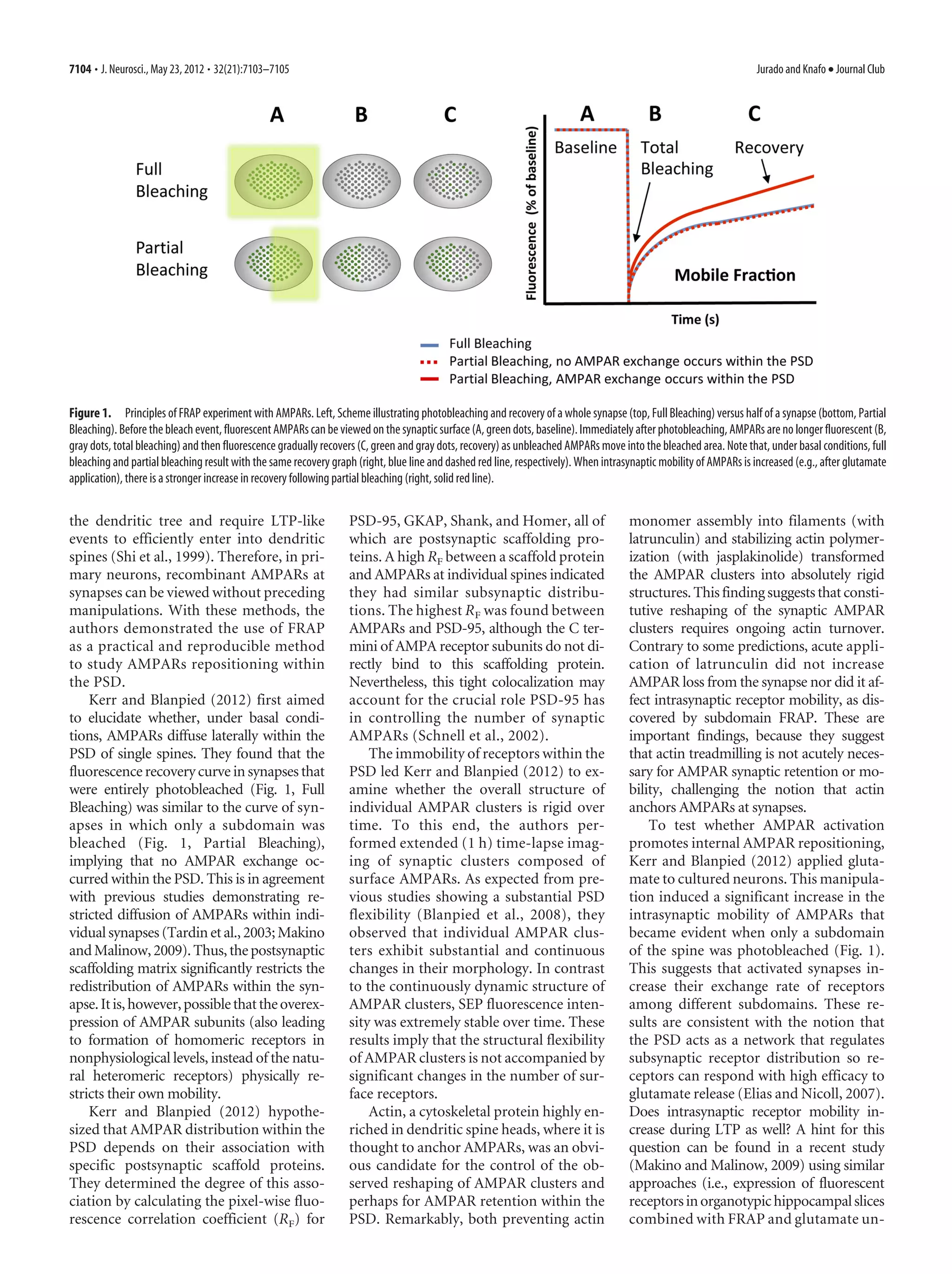
3. Application of glutamate increased the intrasynaptic mobility of AMPARs, suggesting activated synapses promote exchange of receptors among subdomains. This supports the idea that the PSD regulates subsynaptic receptor distribution.
1) A study in Drosophila found that overexpression of the fatty acid binding protein Fabp7 correlates with enhanced long-term memory formation. Specifically, increased nuclear localization of Fabp7 protein correlates with increased long-term memory.
2) The study here found that flies overexpressing Fabp7 also show enhanced formation of anesthesia-resistant memory (ARM) under certain temperature conditions.
3) The level of cytoplasmic versus nuclear Fabp7 protein expression correlates with the ratio of ARM performance to long-term memory performance. Increased cytoplasmic localization correlates with higher ARM to LTM ratios. This suggests Fabp7 may regulate different stages of memory formation through its subcellular localization.
This study examines the effects of the antibiotic chloramphenicol (CAP) on brain metabolism in freely moving rats using picosecond time-resolved fluorescence spectroscopy. The researchers found that CAP, in addition to inhibiting mitochondrial protein synthesis, also influences redox processes in the respiratory chain by producing a marked increase in fluorescent signal in the nucleus raphe dorsalis, indicating a rise in NADH concentration. This suggests CAP efficiently inhibits complex I of the respiratory chain, which could explain why it suppresses paradoxical sleep. The approach provides a novel method for evaluating drug effects on deep brain structures in vivo.
The document summarizes a study that investigated the effects of low molecular weight components from the venom of the Chilean Black Widow spider (Latrodectus mactans) on synaptic activity. Whole-cell patch clamp techniques were used to study the effects of purified venom on rat hippocampal neuronal cultures. The venom increased neuronal membrane resistance, prolonged action potentials, and increased spontaneous synaptic activity in a concentration- and time-dependent manner. This demonstrates that the venom has pharmacological effects through low molecular weight fragments, despite L. mactans venom not containing alpha-latrotoxin like the European Black Widow spider venom.
Next-Generation Sequencing and its Applications in RNA-Seqb0rAAs
Next-generation sequencing technologies like ion semiconductor sequencing and pyrosequencing have enabled applications of RNA-Seq. RNA-Seq involves sequencing cDNA to analyze transcriptomes, identify differentially expressed genes between conditions, and reconstruct transcripts through methods like genome-guided assembly or by building de Bruijn graphs from k-mers.
The document discusses RNA-seq analysis. It begins with an introduction to Mikael Huss, a bioinformatics scientist, and provides an overview of how genomics, RNA profiles, protein profiles, and interactomics relate within systems biology. The document then discusses how gene expression analysis can provide insights into basic research questions regarding tissue and cell identity, as well as insights into diseases by identifying genes that are over- or under-expressed in patients. Finally, it provides a brief overview of the typical workflow for RNA-seq analysis, which involves mapping RNA sequencing reads to a reference genome or transcriptome.
This document discusses a model proposed by Penrose and Hameroff that assigns a cognitive role to quantum computations occurring in microtubules within brain neurons. It summarizes the key points of criticism against this model, particularly regarding the issue of environmental decoherence destroying any quantum superpositions before they could be relevant for cognition. The document then examines these criticisms in more detail, finding issues with the assumptions and calculations in the previous analyses. It argues that under a corrected analysis, microtubules could potentially maintain quantum coherence for microseconds to milliseconds, long enough to be neurophysiologically significant. It also proposes additional mechanisms by which microtubules may be further isolated from decoherence effects in their biological environment.
Issues in brainmapping...EEG and brainmap spectral profiles in cortical lesio...Professor Yasser Metwally
Issues in brainmapping...EEG and brainmap spectral profiles in cortical lesions, subcortical white matter lesions and subcortical gray matter (diencephalic) lesions
DNA profiling is a forensic technique that uses a person's unique DNA to identify them. It examines DNA found at crime scenes. Two main techniques are used: Restriction fragment length polymorphism cuts DNA into fragments of varying lengths, which are then compared to suspects' DNA. Short tandem repeat profiling makes copies of DNA sections and examines repetitive patterns that differ between people. DNA profiling is a powerful forensic tool that can include or exclude suspects by matching DNA evidence to their profiles.
The document presents a computational model of episodic memory encoding in the dentate gyrus region of the hippocampus using an ART neural network. The dentate gyrus is proposed to encode episodes in binary patterns in a way that achieves high levels of pattern encoding and separation. The model incorporates a method for encoding episodes as binary patterns that are stored sparsely. Simulation results show that the model achieves good separation of different episodes depending on the vigilance parameter, which is assumed to correlate with attention during episode perception.
Kouvaros S and Papatheodoropoulos C, (2016). Major dorsoventral differences i...Stylianos Kouvaros
1) The study examines differences in modulation of the local hippocampal CA1 network between the dorsal and ventral hippocampus by NMDA, mGlu5, adenosine A2A, and cannabinoid CB1 receptors.
2) Activation of NMDA receptors reduced excitatory transmission more in the ventral hippocampus than in the dorsal hippocampus by an adenosine A1 receptor-independent mechanism. In the dorsal hippocampus, NMDA receptors reduced inhibition and enhanced postsynaptic excitability.
3) Co-activation of mGlu5 and NMDA receptors strongly potentiated their effects in the dorsal hippocampus but had no potentiating effect in the ventral hippocampus. This potentiation in the dorsal hippocampus required adenos
This document compares the visual organs of insects and mammals. It discusses the similarities and differences in their phototransduction pathways and anatomical structures. Some key points:
- Both use opsins bound to retinal as the photosensitive pigment, but insects use different opsins than mammals and transduce light signals through different G proteins and second messengers.
- Compound eyes of insects like Drosophila are made of many individual ommatidia that each detect light from a different angle, while mammals have a single pupil and non-uniform retina.
- Photoreceptor cells in both hyperpolarize in response to light via different downstream mechanisms, but both require regeneration of the unexcited pigment form for
Light Regulates Plant Alternative Splicing through the Control of Transcripti...ShreyaMandal4
This document discusses how light regulates alternative splicing in plants through controlling transcription elongation. It presents a study that investigated the effects of light-dark conditions and histone deacetylase inhibitors on alternative splicing in Arabidopsis seedlings. The study found that light increases RNA polymerase II elongation, which regulates alternative splicing. Light-dark conditions affected alternative splicing but not total mRNA levels. The results suggest that kinetic coupling between transcription and alternative splicing is an important mechanism for plants to respond to environmental cues like light.
This study provides evidence that correlated neural activity can propagate through multiple stages of a neural circuit involved in song production in songbirds. The researchers recorded neural activity simultaneously from two or three song nuclei - LMAN, RA, and HVc. They found correlated activity between neuron firing in these nuclei, both during spontaneous activity and in response to auditory stimuli like the bird's own song. This correlated activity persisted even when the activity in one nucleus (HVc) was disrupted. This suggests the song circuit contains highly interconnected neurons that can preserve timing information about groups of neural firing through multiple synaptic connections. Since this song circuit is important for vocal learning, preserving correlated activity may be important for learning and producing sequenced motor behaviors.
The introduction of supernova system: a vector system for single-cell labelin...Div. of Neurogenet., NIG
Here, we introduce the “Supernova system”, which has been reported in the following two papers:
- NMDAR-Regulated Dynamics of Layer 4 Neuronal Dendrites during Thalamocortical Reorganization in Neonates. Mizuno et al., Neuron 2014.
- Supernova: A Versatile Vector System for Single-Cell Labeling and Gene Function Studies in vivo. Luo et al. Sci. Rep.2016.
Lab web site: https://www.nig.ac.jp/labs/NeurGen/
Supernova support site: http://snsupport.webcrow.jp/
contact: tiwasato(at)nig.ac.jp
This document discusses the role of AMPA receptor (AMPAR) surface diffusion in synaptic plasticity and memory. It reports that:
1) AMPAR surface diffusion is important for the establishment and maintenance of long-term potentiation (LTP) through replenishing synaptic AMPARs.
2) Blocking AMPAR surface diffusion through crosslinking attenuates LTP in hippocampal brain slices and impairs hippocampal-dependent memory formation.
3) Postsynaptic AMPAR surface diffusion is a critical trafficking mechanism for the expression of LTP and learning in the hippocampus.
This document is a thesis presented by Sebastian Aguiar to the Keck Science Department in partial fulfillment of a Bachelor of Arts degree. It aims to characterize a novel monoclonal antibody targeting the AMPA receptor subunits GluA1, GluA2, and GluA3. The thesis provides background on AMPA receptor structure, function, trafficking, role in synaptic plasticity, involvement in neuropathology, and pharmacology. It then describes the research questions and methods used to visualize the antibody's binding in rat, monkey and human brain tissue using confocal and electron microscopy.
Lydia Yeshitla, Research Scholar at the Neurobiology Section of UCSDLydia Yeshitla
1) The document describes an experiment cloning a pH-sensitive fluorescent protein (pHRed) onto the GLUA1 AMPA receptor subunit to track intracellular trafficking and degradation of AMPA receptors by lysosomes.
2) Restriction enzymes (AGE1 and BSRG1) were used to cut the DNA in order to ligate pHRed onto GLUA1 using PCR. This would allow detection of AMPA receptors in the acidic lysosome lumen.
3) Bacteria were transformed with the ligated pHRed-GluA1 DNA. Colonies were selected and the DNA was sequenced to validate that the cloning procedure was done correctly.
This document describes an in vitro model using mouse hippocampal brain slices to study the recovery of functional connectivity in neural tissue after damage. The study found that regularly stimulating severed hippocampal slices with low frequency electrical pulses promoted the recovery of synaptic connections between the slices over time. Specifically, they observed fiber volley potentials and excitatory postsynaptic potentials reemerging between 20-40 minutes and 1 hour after initiating stimulation, respectively, suggesting the restoration of neural connectivity. The authors hypothesize that electrical stimulation enhances recovery by maintaining neural membrane function and propose this model could help investigate strategies to promote recovery from brain injury.
This study investigated the use of quantum dots (qdots) to label and visualize two endogenous synaptic proteins, GABAA-1 receptors and glutamate transporters (VGLUT1), in the rat cerebellum. Qdots allowed for the clear visualization of these proteins in very small presynaptic structures like parallel fiber varicosities that are below the diffraction limit of conventional microscopy. Specifically, qdots formed clusters around interneurons and isolated clusters on interneuron dendrites, revealing the presence of GABAA receptors. They also labeled sub-micrometer parallel fibers and 1-2 micrometer presynaptic varicosities containing VGLUT1. While double labeling with two qdot colors was attempted, some receptor sites remained unlabeled
This document describes a research study that used laser capture microdissection to isolate distinct cell populations from mouse and human inner ear tissue for gene expression analysis. The researchers were able to extract RNA from formalin-fixed paraffin-embedded tissue sections, amplify it, and detect cell-type specific gene expression by RT-PCR. This technique allows targeted analysis of gene expression in specific inner ear structures, and could be applied to archived human temporal bone samples preserved in formalin. The researchers validated the method by detecting housekeeping genes and cell markers in laser-captured cells from mouse and human inner ear tissue.
This study analyzed dendritic spine morphology in the CA1 region of the hippocampus in a mouse model of Alzheimer's disease (AD). Three key findings were observed:
1) Dendritic spine necks in the stratum oriens layer were significantly shorter in AD mice compared to controls.
2) The frequency of dendritic spines with small head volumes increased in the stratum radiatum layer of AD mice.
3) These layer-specific changes to spine morphology in an AD mouse model may underlie the synaptic dysfunction and cognitive impairments seen in the disease. The changes reflect the effects of amyloid-beta overexpression on excitatory synapses in the hippocampus.
This document describes a study that used laser stimulation of neurons expressing channelrhodopsin-2 (ChR2) to study synaptic transmission in cultured hippocampal neurons. Recombinant adeno-associated virus (rAAV) was used to deliver the ChR2 gene to the neurons. Laser stimulation was able to activate action potentials in ChR2-expressing neurons. By voltage-clamping a neuron and scanning a laser, synaptic responses were observed at some locations, indicating spatial localization of stimulation. Pharmacological tests identified responses that were synaptic. While monosynaptic responses could not be entirely distinguished from polysynaptic ones, smaller amplitudes, simpler shapes, and latencies around 8 ms suggested monosynaptic interactions.
Septal cholinergic neuromodulation tunes the astrocyte dependent gating of hi...Masuma Sani
1. The study found daily fluctuations in levels of the NMDA receptor co-agonist D-serine in the hippocampus, with higher levels during waking and lower levels during sleep.
2. Optogenetic stimulation of cholinergic fibers in the hippocampus increased D-serine levels and NMDA receptor activity, indicating that acetylcholine regulates D-serine availability.
3. Astrocytes were found to release D-serine in a process requiring alpha7 nicotinic acetylcholine receptors, suggesting astrocytes link cholinergic activity to NMDA receptor function through D-serine.
Nano-Tecnología aplicada a la Medicina.
Conferencia realizada en Febrero de 2022 en el IES Jose María de Pereda de Santander. por el profesor Ivan Sasselli Ramos, en base a su estudio y el artículo publicado sobre este tema en la revista Science, 374 (6569), • DOI: 10.1126/science.abh3602 junto con los profesores Z. ÁlvarezA. N. Kolberg-EdelbrockI. R. y el propio Sasselli
NEUROSCIENCEDendritic action potentials and computation in.docxhallettfaustina
1) Researchers investigated the dendrites of layer 2/3 pyramidal neurons in the human cerebral cortex ex vivo and discovered a class of calcium-mediated dendritic action potentials (dCaAPs) not previously described.
2) dCaAPs were graded in amplitude and maximal for threshold stimuli but dampened for stronger stimuli, enabling linear classification of nonseparable inputs - a computation thought to require multilayered networks.
3) dCaAPs were mediated by calcium channels, had a stereotypical wide waveform, and their impact on the soma was variable, either coupling to trigger somatic action potentials or remaining uncoupled in the dendrite.
NEUROSCIENCEDendritic action potentials and computation in.docxvannagoforth
NEUROSCIENCE
Dendritic action potentials and computation in
human layer 2/3 cortical neurons
Albert Gidon1, Timothy Adam Zolnik1, Pawel Fidzinski2,3, Felix Bolduan4, Athanasia Papoutsi5,
Panayiota Poirazi5, Martin Holtkamp2, Imre Vida3,4, Matthew Evan Larkum1,3*
The active electrical properties of dendrites shape neuronal input and output and are fundamental to
brain function. However, our knowledge of active dendrites has been almost entirely acquired from
studies of rodents. In this work, we investigated the dendrites of layer 2 and 3 (L2/3) pyramidal neurons
of the human cerebral cortex ex vivo. In these neurons, we discovered a class of calcium-mediated
dendritic action potentials (dCaAPs) whose waveform and effects on neuronal output have not been
previously described. In contrast to typical all-or-none action potentials, dCaAPs were graded; their
amplitudes were maximal for threshold-level stimuli but dampened for stronger stimuli. These dCaAPs
enabled the dendrites of individual human neocortical pyramidal neurons to classify linearly nonseparable
inputs—a computation conventionally thought to require multilayered networks.
T
he expansion of the human brain during
evolution led to an extraordinarily thick
cortex (~3 mm), which is disproportion-
ately thickened in layers 2 and 3 (L2/3)
(1). Consequently, human cortical neu-
rons of L2/3 constitute large and elaborate
dendritic trees (2, 3), decorated by numer-
ous synaptic inputs (1). The active electrical
properties of these dendrites largely deter-
mine the repertoire of transformations of the
synaptic inputs to axonal action potentials
(APs) at the output. Thus, they constitute a key
element of the neuron’s computational power.
We used dual somatodendritic patch clamp
recordings and two-photon imaging to directly
investigate the active properties of L2/3 den-
drites in acute slices from surgically resected
brain tissue of the human neocortex from epi-
lepsy and tumor patients. Subthreshold (steady-
state) potentials attenuated from the dendrite
to the soma with a length constant (lsteady) of
195 mm (fig. S1; n = 23 cells). In the opposite
direction, the back-propagating action poten-
tials (bAPs) attenuated from the soma to the
dendrite with a lbAP of 290 mm (Fig. 1, A to C;
n = 31 cells). Both lbAP and lsteady were shorter
than the length of the apical dendrite (the
somata of these cells were located ~850 mm
below the pia mater, on average, and the apical
dendrite extended up to layer 1), which implies
that strong attenuation governs the electrical
activity to and from most synapses located on
the apical dendrite.
We filled cells with the calcium indicator
Oregon-green BAPTA-1 (100 mM) and mea-
sured the change in fluorescence (DF/F) under
a two-photon microscope while triggering APs
at the soma. Trains of somatic APs (10 APs) at
50 Hz failed to cause Ca2+ influx at distal apical
dendrites (fig. S2). AP trains with a higher fre-
quency (10 APs at 200 Hz) did invade most of
the ...
Rats were trained to recognize a sequence of five odors. Neuronal activity was recorded in the hippocampus as rats performed this task. The study found that approximately 26% of hippocampal neurons encoded temporal relationships between odors by changing firing rates depending on whether an odor was in or out of the learned sequence. Some neurons responded more to in-sequence odors, others to out-of-sequence odors. The neuronal ensemble activity reflected whether items were in the correct temporal order. This suggests hippocampal neurons encode temporal relationships between events, important for episodic memory.
The document reports on a study investigating the geometrical optimization, spectroscopic analysis, electronic structure, and nuclear magnetic resonance of (S)-(−)-N-(5-Nitro-2-pyridyl) alaninol (SN5N2PLA) using density functional theory calculations. The Fourier transform infrared and Raman spectra were recorded and vibrational assignments were analyzed. Ultraviolet-visible spectra were also recorded and electronic properties like HOMO-LUMO energies were calculated. Nuclear magnetic resonance chemical shifts were calculated and compared to experimental data. The first hyperpolarizability and other properties were computed to investigate the compound's potential as a nonlinear optical material.
This chapter discusses the amygdala in Alzheimer's disease (AD). It notes that the amygdala is severely affected by AD pathology and is involved in emotional processing. Damage to the amygdala in AD leads to neuropsychiatric symptoms like anxiety and personality changes in patients. Studies also show impaired emotional memory in AD patients that correlates with amygdala atrophy. Autopsies reveal neurofibrillary tangles, amyloid plaques and neuronal loss in the amygdala of AD patients. Imaging studies further demonstrate amygdala volume reductions in AD patients, even in early stages, associated with cognitive impairment.
Morphological alterations to neurons of the amygdalashiraknafo
This study examined the effects of amyloid-beta (Aβ) accumulation in the amygdala of an Alzheimer's disease mouse model. The mice showed impaired fear conditioning compared to controls. While there was no neuronal loss in the lateral nucleus of the amygdala (LA), the dendritic trees and spines of LA projection neurons were morphologically altered. Specifically, plaque-free neurons in the transgenic mice had decreased large dendritic spines compared to controls. This suggests that Aβ deposition leads to structural changes in the amygdala that may underlie impaired emotional memory seen in Alzheimer's disease.
Spines, plasticity, and cognition in alzheimer model mice.shiraknafo
This review article discusses research on transgenic Alzheimer's disease (AD) mouse models and how they are used to investigate dendritic spine structure, synaptic function, and cognition. Studies show that soluble amyloid-beta initiates synaptic dysfunction and loss, as well as tau pathology, contributing to both synaptic and neuronal loss. These changes in synapse structure and function, as well as outright synapse and neuron loss, underlie the neural system dysfunction that causes cognitive deficits in AD. Understanding how dementia develops in AD is crucial for developing effective therapies.
1. The study found that NMDA receptor activation triggers an association between the tumor suppressor protein PTEN and the synaptic scaffolding protein PSD-95.
2. This association requires a PDZ-binding motif on PTEN and leads to PTEN being recruited to and anchored at dendritic spines.
3. Enhancing PTEN's lipid phosphatase activity was found to specifically drive depression of AMPA receptor-mediated synaptic responses, and this activity was required for NMDA receptor-dependent long-term depression (LTD) but not other forms of plasticity.
This document summarizes a study that found phosphatidylinositol-(3,4,5)-trisphosphate (PIP3) is continuously required to maintain AMPA receptor clustering at the postsynaptic membrane, which is necessary for sustaining synaptic function. The study manipulated PIP3 levels in hippocampal neurons and found that reducing PIP3 impaired the accumulation of PSD-95 in spines and caused AMPA receptors to become more mobile and migrate away from the postsynaptic density, leading to synaptic depression over time. Therefore, constant low-level turnover of PIP3 at synapses is required to maintain AMPA receptor clustering and synaptic strength under normal conditions.
WIP is a negative regulator of neuronal maturation and synaptic activity. Mice lacking WIP (WIP-/-) have enlarged neuronal somas and overgrown neuritic and dendritic branches, especially during early development. WIP-/- neurons also have increased amplitude and frequency of miniature excitatory postsynaptic currents, indicating more mature synapses than in wild-type neurons. This reveals WIP as a previously unknown regulator of neuronal maturation and synaptic activity.
1) A peptide called FGL enhances learning and memory in rodents by stimulating the delivery of glutamate receptors to synapses, strengthening synaptic transmission. This effect is mediated through the activation of protein kinase C.
2) FGL treatment heightens the induction of long-term potentiation in response to synaptic activity, improving the encoding of information.
3) FGL is poised to begin clinical trials this year for Alzheimer's disease, as prior studies show it improves memory and reduces neuropathology in rodent models of AD.
1) The study assessed changes in hippocampal dendritic spines of APP/PS1 transgenic mice, a model of Alzheimer's disease.
2) It found a substantial decrease in the frequency of large dendritic spines in plaque-free regions of the dentate gyrus in these mice compared to controls.
3) Dendrites passing through amyloid plaques also showed alterations in spine density and morphology, with lower spine density within plaques and higher density on dendrites contacting plaques.
This document summarizes research investigating the cellular and molecular mechanisms underlying cognitive enhancement induced by a peptide called FGL. The key findings are:
1. FGL activates fibroblast growth factor receptor (FGFR) signaling pathways in vivo and in vitro.
2. FGL enhances long-term potentiation (LTP) in hippocampal slices by facilitating delivery of AMPA receptors to synapses through activation of NMDA receptors.
3. Both the LTP enhancement and cognitive improvement produced by FGL are mediated by initial protein kinase C (PKC) activation followed by sustained calcium/calmodulin-dependent protein kinase II (CaMKII) activation.
4. These results link facilitation
1) The document discusses common intracellular signaling pathways that are involved in both growth/differentiation processes early in development and synaptic plasticity in mature neurons.
2) Two examples are described in detail: the Ras-PI3K pathway and signaling elicited by neural cell adhesion molecules (NCAMs) interacting with growth factor receptors.
3) For the Ras-PI3K pathway, calcium entry via NMDA receptors can activate Ras, which then activates PI3K to produce PIP3 and drive LTP. Alternatively, NMDA receptors can activate Rap proteins to induce PTEN, reduce PIP3, and drive LTD.
This article reviews studies on transgenic Alzheimer's disease (AD) mouse models that investigate dendritic spine structure, synaptic function, and cognition. These studies show that soluble amyloid-beta initiates synaptic dysfunction and loss, as well as pathological changes in tau, which contribute to both synaptic and neuronal loss. In vivo imaging reveals plaque-associated spine loss and structural plasticity deficits in AD mouse models. Synaptic plasticity is also severely impaired in AD mouse models, with deficits in basal synaptic transmission and long-term potentiation emerging with age. Together, these changes in synapse structure and function are thought to underlie the cognitive deficits observed in AD.
2. 7104 • J. Neurosci., May 23, 2012 • 32(21):7103–7105 Jurado and Knafo • Journal Club
Figure 1. Principles of FRAP experiment with AMPARs. Left, Scheme illustrating photobleaching and recovery of a whole synapse (top, Full Bleaching) versus half of a synapse (bottom, Partial
Bleaching). Before the bleach event, fluorescent AMPARs can be viewed on the synaptic surface (A, green dots, baseline). Immediately after photobleaching, AMPARs are no longer fluorescent (B,
gray dots, total bleaching) and then fluorescence gradually recovers (C, green and gray dots, recovery) as unbleached AMPARs move into the bleached area. Note that, under basal conditions, full
bleaching and partial bleaching result with the same recovery graph (right, blue line and dashed red line, respectively). When intrasynaptic mobility of AMPARs is increased (e.g., after glutamate
application), there is a stronger increase in recovery following partial bleaching (right, solid red line).
the dendritic tree and require LTP-like PSD-95, GKAP, Shank, and Homer, all of monomer assembly into filaments (with
events to efficiently enter into dendritic which are postsynaptic scaffolding pro- latrunculin) and stabilizing actin polymer-
spines (Shi et al., 1999). Therefore, in pri- teins. A high RF between a scaffold protein ization (with jasplakinolide) transformed
mary neurons, recombinant AMPARs at and AMPARs at individual spines indicated the AMPAR clusters into absolutely rigid
synapses can be viewed without preceding they had similar subsynaptic distribu- structures. This finding suggests that consti-
manipulations. With these methods, the tions. The highest RF was found between tutive reshaping of the synaptic AMPAR
authors demonstrated the use of FRAP AMPARs and PSD-95, although the C ter- clusters requires ongoing actin turnover.
as a practical and reproducible method mini of AMPA receptor subunits do not di- Contrary to some predictions, acute appli-
to study AMPARs repositioning within rectly bind to this scaffolding protein. cation of latrunculin did not increase
the PSD. Nevertheless, this tight colocalization may AMPAR loss from the synapse nor did it af-
Kerr and Blanpied (2012) first aimed account for the crucial role PSD-95 has fect intrasynaptic receptor mobility, as dis-
to elucidate whether, under basal condi- in controlling the number of synaptic covered by subdomain FRAP. These are
tions, AMPARs diffuse laterally within the AMPARs (Schnell et al., 2002). important findings, because they suggest
PSD of single spines. They found that the The immobility of receptors within the that actin treadmilling is not acutely neces-
fluorescence recovery curve in synapses that PSD led Kerr and Blanpied (2012) to ex- sary for AMPAR synaptic retention or mo-
were entirely photobleached (Fig. 1, Full amine whether the overall structure of bility, challenging the notion that actin
Bleaching) was similar to the curve of syn- individual AMPAR clusters is rigid over anchors AMPARs at synapses.
apses in which only a subdomain was time. To this end, the authors per- To test whether AMPAR activation
bleached (Fig. 1, Partial Bleaching), formed extended (1 h) time-lapse imag- promotes internal AMPAR repositioning,
implying that no AMPAR exchange oc- ing of synaptic clusters composed of Kerr and Blanpied (2012) applied gluta-
curred within the PSD. This is in agreement surface AMPARs. As expected from pre- mate to cultured neurons. This manipula-
with previous studies demonstrating re- vious studies showing a substantial PSD tion induced a significant increase in the
stricted diffusion of AMPARs within indi- flexibility (Blanpied et al., 2008), they intrasynaptic mobility of AMPARs that
vidual synapses (Tardin et al., 2003; Makino observed that individual AMPAR clus- became evident when only a subdomain
and Malinow, 2009). Thus, the postsynaptic ters exhibit substantial and continuous of the spine was photobleached (Fig. 1).
scaffolding matrix significantly restricts the changes in their morphology. In contrast This suggests that activated synapses in-
redistribution of AMPARs within the syn- to the continuously dynamic structure of crease their exchange rate of receptors
apse. It is, however, possible that the overex- AMPAR clusters, SEP fluorescence inten- among different subdomains. These re-
pression of AMPAR subunits (also leading sity was extremely stable over time. These sults are consistent with the notion that
to formation of homomeric receptors in results imply that the structural flexibility the PSD acts as a network that regulates
nonphysiological levels, instead of the natu- of AMPAR clusters is not accompanied by subsynaptic receptor distribution so re-
ral heteromeric receptors) physically re- significant changes in the number of sur- ceptors can respond with high efficacy to
stricts their own mobility. face receptors. glutamate release (Elias and Nicoll, 2007).
Kerr and Blanpied (2012) hypothe- Actin, a cytoskeletal protein highly en- Does intrasynaptic receptor mobility in-
sized that AMPAR distribution within the riched in dendritic spine heads, where it is crease during LTP as well? A hint for this
PSD depends on their association with thought to anchor AMPARs, was an obvi- question can be found in a recent study
specific postsynaptic scaffold proteins. ous candidate for the control of the ob- (Makino and Malinow, 2009) using similar
They determined the degree of this asso- served reshaping of AMPAR clusters and approaches (i.e., expression of fluorescent
ciation by calculating the pixel-wise fluo- perhaps for AMPAR retention within the receptors in organotypic hippocampal slices
rescence correlation coefficient (RF) for PSD. Remarkably, both preventing actin combined with FRAP and glutamate un-
3. Jurado and Knafo • Journal Club J. Neurosci., May 23, 2012 • 32(21):7103–7105 • 7105
caging). Makino and Malinow (2009) References fluorescence recovery after photobleaching.
suggested that the mobility of SEP- Beattie EC, Carroll RC, Yu X, Morishita W, Ya- J Supramol Struct 5:565(417)-576(428).
suda H, von Zastrow M, Malenka RC (2000) Kerr JM, Blanpied TA (2012) Subsynaptic
GluA1 subunits is significantly decreased af-
Regulation of AMPA receptor endocytosis by AMPA receptor distribution is acutely regu-
ter LTP induction to preserve the recently lated by actin-driven reorganization of the
a signaling mechanism shared with LTD. Nat
requited receptors and maintain synaptic Neurosci 3:1291–1300. postsynaptic density. J Neurosci 32:658 – 673.
potentiation. Blanpied TA, Kerr JM, Ehlers MD (2008) Struc- Makino H, Malinow R (2009) AMPA receptor
In summary, the study of Kerr and tural plasticity with preserved topology in the incorporation into synapses during LTP: the
Blanpied (2012) represents an important postsynaptic protein network. Proc Natl Acad role of lateral movement and exocytosis. Neu-
advance in the study of the microscale Sci U S A 105:12587–12592. ron 64:381–390.
Broutman G, Baudry M (2001) Involvement of Man HY, Ju W, Ahmadian G, Wang YT (2000)
organization and dynamics of the post- the secretory pathway for AMPA receptors in Intracellular trafficking of AMPA receptors
synaptic membrane. Using FRAP on NMDA-induced potentiation in hippocam- in synaptic plasticity. Cell Mol Life Sci
subdomains of spines in dissociated neu- pus. J Neurosci 21:27–34. 57:1526 –1534.
rons, they determined that AMPARs are Dahan M, Levi S, Luccardini C, Rostaing P,
´ Park M, Penick EC, Edwards JG, Kauer JA,
relatively immobile within the PSD while Riveau B, Triller A (2003) Diffusion dynam- Ehlers MD (2004) Recycling endosomes
ics of glycine receptors revealed by single- supply AMPA receptors for LTP. Science
displaying an overall motion as clusters
quantum dot tracking. Science 302:442– 445. 305:1972–1975.
in a matrix that constantly reshapes in Ehlers MD (2000) Reinsertion or degradation Schnell E, Sizemore M, Karimzadegan S, Chen L,
an actin-dependent manner. Their con- of AMPA receptors determined by activity- Bredt DS, Nicoll RA (2002) Direct interac-
clusions challenge previous models for dependent endocytic sorting. Neuron 28: tions between PSD-95 and stargazin control
AMPA receptor positioning and anchor- 511–525.
synaptic AMPA receptor number. Proc Natl
ing at the postsynaptic density, and offer Elias GM, Nicoll RA (2007) Synaptic trafficking
Acad Sci U S A 99:13902–13907.
of glutamate receptors by MAGUK scaffold-
critical insight into the inner organization ing proteins. Trends Cell Biol 17:343–352.
Shi SH, Hayashi Y, Petralia RS, Zaman SH,
of living synapses. Undoubtedly, the final Wenthold RJ, Svoboda K, Malinow R (1999)
Gerges NZ, Backos DS, Rupasinghe CN, Spaller
picture of AMPAR trafficking will require Rapid spine delivery and redistribution of
MR, Esteban JA (2006) Dual role of the exo-
cyst in AMPA receptor targeting and insertion AMPA receptors after synaptic NMDA recep-
the combination of complementary imag- tor activation. Science 284:1811–1816.
ing techniques. In the next years, FRAP will into the postsynaptic membrane. EMBO J
25:1623–1634. Shi S, Hayashi Y, Esteban JA, Malinow R (2001)
probably be combined with the use of Gruenberg J (2001) The endocytic pathway: a Subunit-specific rules governing AMPA re-
photo-switchable fluorescent proteins (flu- mosaic of domains. Nat Rev Mol Cell Biol ceptor trafficking to synapses in hippocampal
orescent proteins that change their excita- 2:721–730. pyramidal neurons. Cell 105:331–343.
tion and emission spectra when exposed to Heynen AJ, Quinlan EM, Bae DC, Bear MF Tardin C, Cognet L, Bats C, Lounis B, Choquet D
(2000) Bidirectional, activity-dependent reg- (2003) Direct imaging of lateral movements
specific light) to explore receptor mobility,
ulation of glutamate receptors in the adult of AMPA receptors inside synapses. EMBO J
and with high spatial and temporal resolu- 22:4656 – 4665.
hippocampus in vivo. Neuron 28:527–536.
tion imaging, such as Photoactivated Local- Jacobson K, Derzko Z, Wu ES, Hou Y, Poste G Vandenberghe W, Bredt DS (2004) Early events
ization Microscopy and stochastic optical (1976) Measurement of the lateral mobility of in glutamate receptor trafficking. Curr Opin
reconstruction microscopy. cell surface components in single, living cells by Cell Biol 16:134 –139.