2. peptide that connects the motor domain to the coiled-coil stalk
(Fig. 1, E2) (24, 25). This structural transition, designated neck-
linker docking, shifts the lagging unbound head forward 16 nm
to the next microtubule-binding site toward the microtubule
plus end (Fig. 1, E3) (24, 26). Subsequently, ADP is released
from this new leading head resulting in both heads bound to the
microtubule (Fig. 1, E4 and E5) (27).
When both heads are bound to the microtubule, the neck-
linker domains are oriented in opposite directions (28, 29). The
neck-linker of the leading nucleotide-free head is oriented
backward, inhibiting ATP binding and hydrolysis by the front
head until the rear head detaches from the microtubule (Fig. 1,
E6) (30). The neck-linker of the lagging ATP-bound head
remains docked onto the catalytic core and directed forward.
ATP hydrolysis on the microtubule-bound lagging head results
in another structural rearrangement that leads to phosphate
release. Thereby, the lagging head transitions into a weakly
bound ADP state and detaches from the microtubule, thus
starting the cycle anew (Fig. 1, E7).
During the stepping cycle, each motor domain must remain
out-of-phase with the other. If both motor domains enter an
unbound state at the same time, the processive run ends. The
run length indicates the distance a motor steps along the micro-
tubule before dissociation and gives a measure of processivity.
Each kinesin family member has different average run lengths
ranging from Ͻ0.2 m, as in Eg5, to 2.1 m, as in conventional
kinesin-1, although this is construct-dependent for kinesin-1
and can range from 1.3 to 2.1 m (25, 31, 32). Despite the
structural conservation between different kinesin motors, there
are clear kinetic differences between the families.
One domain hypothesized to contribute to processivity is the
kinesin neck-linker, a small flexible peptide consisting of 12–18
amino acids (33–35). The neck-linker connects each motor
domain to the coiled-coil stalk where the junction between
these two entities has been assumed to occur at the same posi-
tion in the sequence as that observed in kinesin-1 (36). The
neck-linker itself undergoes a series of structural transitions as
outlined in the kinesin mechanochemical cycle. It is hypothe-
sized that longer neck-linkers increase the diffusional search
area and therefore could slow down stepping, allowing time for
the forward head to release from the microtubule (37).
Recently, alterations in neck-linker length were shown to affect
the kinetic cycle (38). Increasing neck-linker length resulted in
increased rear head binding, and decreasing neck-linker length
resulted in slower release of ADP from the unbound head. Both
effects result in slowing the productive kinetic cycle (38).
Because the neck-linker is involved in connecting the motor
domain and coiled-coil stalk, changes in the length of the neck-
linker are expected to alter communication between the two
motor heads.
Given that the overall size of the kinesin motor domain is
similar across all families and that they bind to microtubules in
a similar manner, it is surprising that the predicted length of the
neck-linker between families shows considerable variation
even though within each family the neck-linker/␣7 sequences
are almost completely conserved (36). This is especially puz-
zling because a multitude of studies show increasing the neck-
linker for a given kinesin by even one residue results in
decreases in run length and processivity (25, 31, 34, 39, 40). This
conundrum is particularly evident in members of the kinesin-2
family, KIF3AB and KIF3AC, which are unique in forming a
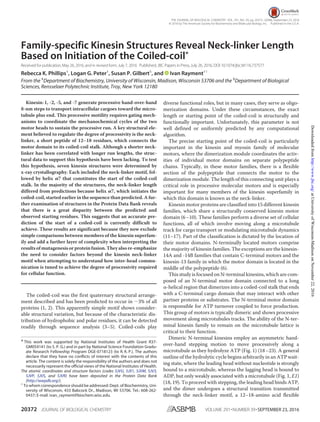
FIGURE 1. Schematic representation of the kinesin chemomechanical
cycle that illustrates key transitions that influence processivity. A pro-
cessive run is started by binding of either head followed by rapid ADP release
to form the E1 intermediate where the leading head that forms the initial
contact is microtubule-bound but nucleotide-free (Ø), whereas the trailing
head is detached with ADP tightly bound. ATP binding to the leading head
induces a series of structural transitions, including neck-linker docking that
allows the trailing ADP-head to move 16 nm ahead to its new microtubule-
binding site (E2–E4). ADP release from the new leading head (E4 and E5)
results in the E5 two-head bound state, thereby generating intermolecular
strain, which inhibits ATP binding at the now leading head. ATP hydrolysis at
the trailing head followed by phosphate (Pi) release generates a microtubule
weakly bound ADP state (E6 and E7). Detachment of the trailing head relieves
the intermolecular strain (E7), and initiates the next motor cycle.
Coiled-coil Initiation in Kinesin Motors
SEPTEMBER 23, 2016•VOLUME 291•NUMBER 39 JOURNAL OF BIOLOGICAL CHEMISTRY 20373
atUniversityofWisconsin-MadisononNovember22,2016http://www.jbc.org/Downloadedfrom
3. heterodimeric motor. KIF3AB and KIF3AC further interact
with an adaptor protein to bind a variety of cargoes for intrafla-
gellar and neuronal transport (11, 15, 41–45). KIF3AC in par-
ticular has been implicated in neuronal repair (11). Both
KIF3AB and KIF3AC are highly processive motors (46). Yet
their neck-linker is predicted to be three residues longer than
conventional kinesin, which would suggest that these motors
should not be so processive (36). KIF3AB and KIF3AC have
identical neck-linker lengths and nearly identical neck-linker
sequences, differing only at Thr-380 in KIF3C, which is an ala-
nine in KIF3A and KIF3B. Both motors are processive; KIF3AB
and KIF3AC have run lengths of 1.6 and 1.2 m, respectively
(37, 46). However, the kinetic parameters vary significantly
between the two motors (46–48). Furthermore, homodimeric
species KIF3AA, KIF3BB, and KIF3CC also exhibit vastly dif-
ferent processivity parameters. KIF3AA and KIF3BB are highly
processive, but KIF3CC travels at only 7.5 nm/s with an average
run length of just 0.6 m, which suggests that the neck-linker is
not the only determinant of processivity (46). These observa-
tions prompted an investigation of the true length of the neck-
linker in the best studied classes of N-terminal kinesin families.
The original estimates of the neck-linker length made the
assumption that the coiled-coil would begin on a hydrophobic
residue that lie in either the a or d position of the coiled-coil
heptad repeat (36). However, the predictions of the first residue
to adopt a helical conformation in any coiled-coil are ambigu-
ous, even though the body of a coiled-coil is well indicated by
current software (3, 4). Prediction software recognizes the hep-
tad repeat in a coiled-coil domain. However, because the begin-
ning and end of sequences may not follow that pattern, there is
insufficient information to make accurate predictions of the
start or of the end of a native coiled-coil. Given the considerable
variation in the amino acid sequence of neck-linkers and asso-
ciated coiled-coils, this raises the question whether the true
length of the neck-linker has been accurately identified across
the kinesin superfamily with the original assumption described
above.
There is only one structure for a dimeric N-terminal kinesin,
rat kinesin-1 (3KIN) because dimeric kinesins are difficult to
crystallize (14). The 3KIN structure provided the first picture of
the true ␣7 start and neck-linker length in the context of a
dimeric motor. Most kinesin motor structures are monomeric,
allowing the neck-linker to adopt varying conformations that
may not reflect that experienced by the dimeric motor in vivo.
For example, in the structure of the kinesin-2 KIF3B (3B6U) the
neck-linker includes a cis-proline; therefore, this structure is
unlikely to reflect the native neck-linker conformation. This
study is directed toward providing an experimental foundation
for determining the start site for the coiled-coil and by infer-
ence the length of the neck-linker. We determined the struc-
tures of the neck-linker and ␣7 helix from the following four
different kinesin families: kinesin-1, -2, -5, and -7. Kinesin-1 is
the canonical N-terminal processive kinesin motor. Kinesin-2
family members, KIF3A and KIF3C, are involved in long range
transport and neuronal repair (11, 15). Kinesin-5 family mem-
ber, Eg5, is unique within this subset as it is a bipolar tetrameric
kinesin whose role is to cross-link microtubules during cell
division (12, 13, 49). Kinesin-7 family member CENP-E is
responsible for transporting misaligned chromosomes during
congression in mitosis (16). In addition, we compared struc-
tures in the PDB2
that include a native start to their coiled-coil
with the predictions generated from either MARCOIL or
COILS-28 (3, 4). Overall, we find that structures of proteins are
necessary to determine the true start site of the coiled-coil
rather than relying on prediction software alone.
Results
Crystal Structures of Kinesin-1, -2, -5, and -7 Neck-linker ␣7
Helix Proteins—All neck-linker structures were homodimeric
and solved to a resolution of 2.3 Å or higher allowing for accu-
rate determination of secondary structure transitions. The
extent of the ordered structure in each construct is given in
Table 1. Several structures had multiple monomers in the
asymmetric unit. Individual monomers were similar as shown
in Table 1. For each neck-linker structure, the start of the
coiled-coil helix was determined using the Dictionary of Sec-
ondary Structure of Proteins algorithm (50). In each structure,
varying lengths of the neck-linker were ordered; thus we
focused on the initiation point of the ␣7 helix to determine
neck-linker length.
All of the structures were determined as fusions with the
C-terminal dimerization domain of EB1. Previous studies have
shown that inclusion of globular folding domains considerably
increases the ability to express and crystallize sections of coiled-
coil proteins and that they do not perturb the structure more
than one heptad from the point of fusion (51, 52).
2
The abbreviations used are: PDB, Protein Data Bank; MEPEG, methoxy
polyethylene glycol; PEE, pentaerythritol ethoxylate; PEG, polyethylene
glycol; TCEP, tris(2-carboxyethyl)phosphine; BTP, 1,3-bis[tris(hydro-
xymethyl)methylamino]propane.
TABLE 1
Average pairwise root means square differences between independent chains within each structural determination
First ordered residue is listed for the longest monomer and may not be ordered in other monomers in the asymmetric unit. r.m.s.d. is root mean square deviation. NA is not
applicable.
Construct
Monomers in
asymmetric unit Average r.m.s.d. First ordered residue
Å
CENP-E 6 0.862 Asn-336a
Eg5 4 0.450 Asn-336a
KIF3A-kinesin-1 8 0.739 Asn-352 (KIF3A)a
KIF3C 2 0.578 Asn-374a
KIF3A 1 NA Asp-354
Kinesin-1 2 0.529 Asn-340
Kinesin-1 ϩ DAL 1 NA Asp of DAL (Ala-345-2)
a
In these structures two residues that remain from the recombinant tobacco etch virus cleavage site were also observed.
Coiled-coil Initiation in Kinesin Motors
20374 JOURNAL OF BIOLOGICAL CHEMISTRY VOLUME 291•NUMBER 39•SEPTEMBER 23, 2016
atUniversityofWisconsin-MadisononNovember22,2016http://www.jbc.org/Downloadedfrom
4. The crystal structure of the kinesin-1 neck-linker (Fig. 2)
shows that the start of ␣7 and the end of the kinesin-1 neck-
linker observed here is identical to that seen in the structure of
the kinesin-1 dimer (PDB code 3KIN) (14). An overlay of the
kinesin-1 neck-linker structure and dimeric kinesin-1 is shown
in Fig. 3. Neck-linker residues Asn-340–Thr-344 were ordered
in the crystal structure and adopt a random coil conformation,
and the ␣7 helix begins at Ala-345 as expected. The consistency
of the kinesin-1 neck-linker structure with predictions and pre-
vious work supports the assertion that the structures from
other families likely represent the solution state of the junction
between their neck-linker and ␣7.
For the remaining neck-linker structures, the length of the
neck-linker and the ␣7 helix deduced from prediction algo-
rithms do not agree with the experimental structural data.
Although the neck-linker itself is flexible as evidenced by
docking, it is unlikely that the start residue for the ␣7 helix
changes. Studies using high resolution atomic force micros-
copy and computational modeling have shown that there is
no local conformational unwinding or “breathing” at helix
␣7 (53–55). Thus, the ␣7 start residues determined here are
most likely the same as in the full dimeric motor. The kine-
sin-2 family members, KIF3A and KIF3C (Fig. 2), have sig-
nificantly shorter neck-linkers and longer ␣7 helices than
earlier models based on the kinesin-1 family. Although pre-
dictions suggested that the ␣7 helix would begin at Leu-360
in KIF3A and at Leu-382 in KIF3C, in the crystal structures
␣7 starts five residues earlier at Pro-355 and Pro-377 in
KIF3A and KIF3C, respectively (Fig. 4). The discrepancy on
the start of ␣7 leads to a shortening of the neck-linker from
17 residues to 12. This appears to be in contrast with the
neck-linker observed in monomeric KIF3B structure (PDB
code 3B6U), which has a length of 16 residues. However,
the KIF3B structure (PDB code 3B6U) is monomeric where
it is unlikely that there were enough residues to form a
stable ␣-helix, leading to the discrepancy in the neck-linker
lengths. Although the native kinesin-2s are heterodimeric,
the crystallized constructs are homodimeric. It is not antic-
ipated that the length of the neck-linker or structures of the
coiled-coils will differ between the homodimer as compared
with the heterodimer, as both KIF3A and KIF3C resulted in a
neck-linker of 12 residues. Additionally, the buried residues
in the first heptad of the coiled-coil are similar in both KIF3A
and KIF3C, and thus it should not affect neck-linker length.
Furthermore, the native coiled-coil is stabilized by a hydro-
gen bond between a lysine and aspartate in both KIF3A and
KIF3C; thus this interaction should also be present in the
heterodimer (Fig. 4). There are sequence differences in ␣7
between KIF3A and KIF3C, but these occur in positions that
are solvent-exposed and do not interact with the adjacent
␣-helix (Fig. 4) and hence are not expected to greatly influ-
FIGURE2.Crystalstructuresofkinesin-1,-2,-5,and-7revealuniqueclass-
specific neck-linker ␣7 neck-coil domains. The neck-linker motif predicted
to occur based on the earlier studies of kinesin-1 is colored blue. Helix ␣7, as
observed in kinesin-1, is colored according to the kinesin family to which it
belongs. EB1, a coiled-coil fusion protein, is colored gray. There are varied
amounts of ordered neck-linker motifs. These structures show that the true
startofhelix␣7isvariableacrossthekinesinsuperfamily.Table4providesthe
protein sequence of each fusion protein and their coiled-coil registry. Figs.
2–6 were prepared in part with PyMOL.
FIGURE3.Overlayofthekinesin-1neck-linkerstructureanddimerickine-
sin-1 crystal structure (PDB code 3KIN) (14). The kinesin-1 neck-linker is
colored as in Fig. 2. Dimeric kinesin-1 is shown in light blue.
Coiled-coil Initiation in Kinesin Motors
SEPTEMBER 23, 2016•VOLUME 291•NUMBER 39 JOURNAL OF BIOLOGICAL CHEMISTRY 20375
atUniversityofWisconsin-MadisononNovember22,2016http://www.jbc.org/Downloadedfrom
5. ence the structure of the heterodimer. The structure of the
kinesin-2 neck-linkers in Fig. 2 show that the neck-linker
is shorter than that of kinesin-1, which has a 14-residue
neck-linker and the longest run length of all families tested
(39).
The Eg5 neck-linker crystal structure (Fig. 2) shows differ-
ences in length as well. The ␣7 helix begins at Lys-371 and not
Ile-375 as predicted, resulting in a neck-linker that was only 14
residues long. As in Eg5, the CENP-E neck-linker was predicted
to be 18 residues long. The coiled-coil of CENP-E begins at
Asp-341 rather than Leu-345, thus shortening the neck-linker
to 14 residues (Fig. 2). These crystal structures show that both
the CENP-E and Eg5 neck-linkers are the same length as that of
kinesin-1. A summary of the predicted neck-linker lengths and
FIGURE4.Selectedside-chaininteractionsinandthesequencedifferencesbetweenKIF3AandKIF3Cneck-linkersshowninstereo.KIF3AandKIF3Care
colored as in Fig. 2, with side chains shown as sticks and colored by element. In both KIF3A (A) and KIF3C (B), there is a hydrogen bonding interaction between
a lysine and an aspartate that stabilizes part of the coiled-coil. The sequences of the neck-linker and ␣7 are also shown (C) where the residues depicted in gray
were not included in the constructs but represent the full-length linker. Interestingly, none of the differences in sequence between KIF3A and KIF3C are
predicted to influence formation of a heterodimer.
Coiled-coil Initiation in Kinesin Motors
20376 JOURNAL OF BIOLOGICAL CHEMISTRY VOLUME 291•NUMBER 39•SEPTEMBER 23, 2016
atUniversityofWisconsin-MadisononNovember22,2016http://www.jbc.org/Downloadedfrom
6. actual neck-linker lengths along with the average run length is
listed in Table 2.
Crystal Structures of the Kinesin-1 Extended Neck-linker and
KIF3A-Kinesin-1 Hybrid—The coiled-coil stalk of the kinesin-1
motor has often been fused to the motor domain of other kine-
sin family members for single molecule studies where this
served as a template for understanding the effect of length dif-
ferences in the neck-linker seen across the entire kinesin super-
family (25, 39, 56–59). This hybrid was used in part to ensure
that kinetic differences were derived from the differences in the
neck-linker domain and not due to the coiled-coil stalk or other
charged regions (39). Additionally, the hybrid constructs were
easily expressed in Escherichia coli, rather than baculovirus
(39). To determine the molecular consequences of these engi-
neered hybrids and how they might affect the interpretation of
changes in kinetic or motile behavior, structural studies were
performed on a KIF3A-kinesin-1 hybrid and a kinesin-1 in
which three-residues (kinesin-1 ϩ DAL) were inserted (Fig. 5).
This extension has been previously used to examine pro-
cessivity changes in the kinesin-1 motor (25, 39).
The kinesin-1 ϩ DAL structure shows the effect of adding
three additional residues to the kinesin-1 neck-linker. Previous
studies have added these three residues, DAL, as an extension
to mimic kinesin-2, as the three final residues of its neck-linker
are DAL (DTL in KIF3C) (25, 39). The addition of these resi-
dues to the end of the kinesin-1 neck-linker should result in a
17-residue neck-linker, as in kinesin-2, rather than the native
14-residue neck-linker. Interestingly, in our structure, even
though three residues (DAL) were added to the putative end of
the neck-linker, two of the three residues become a part of the
␣7 helix and thus only lengthens the neck-linker by one residue.
The KIF3A-kinesin-1 neck-linker construct fuses the KIF3A
neck-linker domain to the ␣7 helix of kinesin-1 (25, 39). In the
native KIF3A structure determined here, the neck-linker was
12 residues long; however, in the hybrid, it lengthens to 14–15
amino acids (Figs. 5 and 6). There is variation in the start of the
coiled-coil due to slight differences in crystal packing. How-
ever, the length is clearly different from that of the native kine-
sin-2 neck-linker. These results indicate that studies of the
kinetic and motile properties of kinesins should be performed
in the context of the native neck-linker and coiled-coil.
Temperature Factor Trends at the Neck-linker/␣7 Junction—
As noted earlier, atomic force microscopy measurements sug-
gest that the start of ␣7 in kinesin-1 is particularly stable. It is
not known whether this phenomenon holds true for all N-ter-
minal kinesins, but is an important consideration when assess-
ing the length of the neck-linker. The structures give a clear
indication of the start of ␣7 but do not necessarily give an
assessment of stability. In principle, examination of the temper-
ature factors across the neck-linker/␣7 junction could provide
TABLE 2
Predicted and observed kinesin neck-linker lengths together with the
average run length
Neck-linker
Predicted
length (36)
Observed
length
Average run
length Refs.
m
DmKinesin-1 (kinesin-1) 14 14 2.1 Ϯ 0.1 39
DmKinesin-1 ϩ DAL 17 15 0.39 Ϯ 0.02 39
MmKIF3AB (kinesin-2) 17 12 1.23 Ϯ 0.09 46
MmKIF3AC (kinesin-2) 17 12 1.62 Ϯ 0.11 46
HsEg5 (kinesin-5) (dimer) 18 14 0.67 Ϯ 0.07 32
KIF3A-kinesin-1 (hybrid) 17 15 0.7 Ϯ 0.03 39
HsCENP-E (kinesin-7) 18 14 2.7 Ϯ 0.2 80
FIGURE 5. Crystal structures of native D. melanogaster kinesin-1 neck-
linker protein, kinesin-1 ؉ DAL, and the hybrid of the M. musculus KIF3A
neck-linker with the kinesin-1 helix ␣7 coil. All are fused to EB1 dimeriza-
tion domain (gray). Each helix ␣7 domain, as predicted based on the earlier
studies of kinesin-1, is colored hot pink with the neck-linker peptide colored
blue.
FIGURE 6. Comparison of KIF3A, the KIF3A-kinesin-1 hybrid protein, and
kinesin-1 structures. Predicted neck-linkers are shown in blue and the EB1
domain in gray. KIF3A is at the top with its helix ␣7 colored red, KIF3A-kine-
sin-1 hybrid protein in the middle with its helix ␣7 in purple, and kinesin-1 at
the bottom with its helix ␣7 in hot pink. Note the variability in neck-linker
length based on the start of helix ␣7.
Coiled-coil Initiation in Kinesin Motors
SEPTEMBER 23, 2016•VOLUME 291•NUMBER 39 JOURNAL OF BIOLOGICAL CHEMISTRY 20377
atUniversityofWisconsin-MadisononNovember22,2016http://www.jbc.org/Downloadedfrom
7. some insight as to whether the longer helices are less stable. As
shown in Fig. 7, there is a general trend that the residues at the
N terminus of the neck-linker have a higher temperature factor
than the coiled-coil, but there is a continuum leading into the
␣-helix. This is often observed for the N termini of proteins or
linkers between domains. This analysis, though fraught with
reservations because temperature factors are susceptible to
modification by crystal packing, radiation decay, and lattice dis-
order, suggests that the residues that increase the length of ␣7
are no less stable than those that make up the canonical helix in
kinesin-1. Interestingly, the same trends in B-values are also
seen at the N termini of other native coiled-coils as discussed
later (Fig. 8).
Coiled-coil Predictions Do Not Accurately Reflect the Start
Residues of Coiled-coils—As noted earlier, the current predic-
tion algorithms provide a robust estimate of the existence of a
coiled-coil, although the exact start of the structural motif is
ambiguous. A robust prediction for a residue in a coiled-coil
will often be close to 1.0, and a value greater than 0.5 is com-
monly considered to indicate a coiled-coil (60). The question is
as follows. What value for the probability should be accepted as
a reliable indication of the first residue? To gain insight into
this area of uncertainty, the structures of the kinesin neck-link-
ers and ␣7 were compared with the calculated probabilities for
two algorithms. The register of the coiled-coil and prediction
for the start site were determined with COILS, a Position-spe-
cific Scoring Matrix model, and MARCOIL, a Hidden Markov
Model (3, 4). Both COILS and MARCOIL gave similar registers
for the body of the coiled-coils, However, neither program
accurately predicted the observed start sites for the ␣7 helix
(Fig. 7, Table 3). In the calculations of the probabilities, the
28-residue window in COILS was used as it gives the lowest
false-positive rate of the three options (14, 21, or 28 residue
windows).
Several coiled-coil prediction algorithms were recently
reviewed to check for both the accuracy in prediction of coiled-
coil and also the accuracy of the oligomeric state of the coiled-
coil (60). This study found the CCHMM_PROF algorithm to
give the best indicator of coiled-coil; however, it does not yield
a registry prediction for the coiled-coil. Thus, it was not used in
this study (61). Multicoil2 also performed well, but its results
were consistent with that of MARCOIL and COILS-28 (62). In
general, for the kinesin coiled-coil domains, MARCOIL pre-
dicted coiled-coil start sites more conservatively than COILS-
28. MARCOIL probabilities were nearly always lower than the
COILS-28 prediction, except for the coiled-coil of KIF3C (60).
FIGURE 7. Predicted coiled-coil probability and temperature factors versus sequence for the class-specific kinesins. A, kinesin-1; B, KIF3A; C, Eg5; and D,
CENP-E. The COILS-28 probabilities are shown as a solid line with MARCOIL probabilities shown with a dashed line. The start residue of the helix ␣7 coil as
determined in the x-ray crystal structure is highlighted in gray. The predicted starting residues of the coiled-coil for KIF3A, Eg5, and CENP-E do not agree with
the observed coiled-coil in the x-ray structures.
Coiled-coil Initiation in Kinesin Motors
20378 JOURNAL OF BIOLOGICAL CHEMISTRY VOLUME 291•NUMBER 39•SEPTEMBER 23, 2016
atUniversityofWisconsin-MadisononNovember22,2016http://www.jbc.org/Downloadedfrom
8. Both programs poorly predicted the KIF3C coiled-coil.
MARCOIL yielded a probability of 0.09 for the propensity of
Pro-377 to form a coiled-coil, and COILS predicted a probabil-
ity of 0.06 for the 28-residue window.
For kinesin-1, where the structure was previously known, the
algorithms differ in the coiled-coil probabilities. Ala-345 is the
␣7 helix start. MARCOIL gives a conservative probability of
0.82, although COILS reaches a probability of 1, five residues
earlier in the sequence where there is no coiled-coil. COILS
tended to over-predict the neck-linker coiled-coil, reaching
high probabilities earlier in the sequence. MARCOIL is a better
estimator, but its predictions for the coiled-coil start sites
ranged from 0.09 to 0.83. Neither program yielded reliable pre-
dictions for the start site of coiled-coils. The variation between
predictive approaches creates a dilemma for deciding the
coiled-coil start site based on bioinformatics approaches.
Indeed, structural results reveal a fundamental weakness in
the prediction algorithms because they are unable to cate-
gorically indicate the first residue that will adopt the ␣-hel-
ical conformation.
To see whether this problem of predicting the coiled-coil
start sites in kinesins is a general phenomenon, structures of
FIGURE 8. Coiled-coil probability and temperature factors versus sequence for selected PDB files. COILS-28 probabilities shown as a solid line and
MARCOIL probabilities shown as a dashed line. The start of the coiled-coil is highlighted in gray. All sequences begin on a g residue. PDB codes included are (A)
2FXM (63), (B) 1GD2 (64), (C) 3HNW, and (D) 1UII (79).
TABLE 3
Predicted coiled-coil start sites, actual start sites, and the corresponding probabilities of the residue being in a coiled-coil according to MARCOIL
and COILS using the 28-residue window
Kinesin Observed start residuea
MARCOIL COILS-28 Predicted start residueb
MARCOIL COILS-28
Kinesin-1 Ala-345 0.815 1.000 Ala-345 0.815 1.000
KIF3A Pro-355 0.432 0.846 Leu-360 0.853 0.846
KIF3C Pro-377 0.086 0.056 Leu-382 0.578 0.056
Eg5 Lys-371 0.694 1.000 Ile-375 0.945 1.000
CENP-E Asp-341 0.828 1.000 Leu-345 0.978 1.000
KIF3A-kinesin-1 (hybrid) Ala-358 (A345-2)c
0.416 1.000 Kinesin-1-Ala-345 0.728 1.000
Leu-359 (A345-1)c
0.546 1.000
a
Observed start residues are determined using the first residue in a helical conformation in the crystal structures of the neck-linker protein defined as helix as determined by
Dictionary of Secondary Structure of Proteins.
b
Predicted start residue as determined in Ref. 36. This start was assumed in earlier kinetic studies of kinesins to determine the influence of the neck-linker length.
c
The KIF3A neck-linker residue where the coiled coil starts is shown first, followed by the corresponding position in kinesin-1 relative to the kinesin-1 ␣7 start.
Coiled-coil Initiation in Kinesin Motors
SEPTEMBER 23, 2016•VOLUME 291•NUMBER 39 JOURNAL OF BIOLOGICAL CHEMISTRY 20379
atUniversityofWisconsin-MadisononNovember22,2016http://www.jbc.org/Downloadedfrom
9. coiled-coil proteins in the PDB that contain a native transition
fromrandom-coiltoparallelcoiled-coilwerealsoexamined(Fig.8
and Table 4). Although there are a large number of structures of
dimeric coiled-coils in the Protein Data Bank, there are only about
a dozen that contain the native start sequence. Most structures in
the PDB represent fragments of a larger protein or are fused to the
canonical coiled-coil found in GCN4. Interestingly, the perfor-
mance of the algorithms on this restricted set was similar to that
observed for the kinesin neck-linker proteins.
In almost every case, the algorithms miss the start site of the
coiled-coil. As with the neck-linker proteins, COILS-28 over-
predicts the propensity for coiled-coil, and MARCOIL is much
more conservative. Neither algorithm accurately predicted the
coiled-coil start, often only reaching a reasonable probability of
coiled-coil formation until 10 or more residues after the struc-
turally observed start site.
A notable exception is 2FXM, the N-terminal region of the S2
fragment of cardiac -myosin II (63). COILS-28 reaches a prob-
ability of 99% for the start of the coiled-coil to begin at the
site corresponding with the coiled-coil start in the structure.
MARCOIL is close, but still under-predicts the probability of a
coiled-coil in that region.
In 1GD2, a structure of the bZIP transcription factor, both
COILS and MARCOIL under-predict the possibility of a
coiled-coil and do not reach an ϳ90% probability until
17 residues later in the sequence, corresponding to ϳ2.5
heptads of coiled-coil missed (64). Results are similar for
3HNW, a coiled-coil protein from Eubacterium eligens with
unknown function. Neither COILS nor MARCOIL reaches a
reasonable coiled-coil prediction until more than three hep-
tads into the coiled-coil domain. Overall, it is clear that algo-
rithms can predict the presence and the register of a coiled-
coil but do not provide an accurate guide to the start of the
helical structure.
Discussion
In this study, the crystal structures of seven kinesin neck-
linker ␣7 helices were determined from four different kinesin
families representing N-terminal kinesin motors. In these
structures, there are differences in the length of the neck-linker
and the start of the coiled-coil stalk as compared with previous
predictions. These differences are most likely the result of inac-
curate assumptions in the prediction of coiled-coil start sites.
The coiled-coil algorithms, COILS-28 and MARCOIL, were
further investigated with respect to start site accuracy for
other non-kinesin proteins and were shown to be unable to
predict the start sites for the majority of coiled-coil-contain-
ing structures.
In the kinesin neck-linker structures, the coiled-coil start site
was 4–5 residues earlier in the sequence than was predicted.
The original assignment of neck-linker length by Hariharan
and Hancock in 2009 (36) was based upon the assumption that
the ␣7 helix would begin on an a or d residue in the coiled-coil
registry, and this was true for the kinesin-1 motors. However,
the choice of this residue registry was arbitrary and as shown by
the results in this paper does not universally apply. KIF3A and
KIF3C both begin on a c position, CENP-E and Eg5 begin on d
positions, and the KIF3A-kinesin-1 hybrid begins on an f posi-
tion. The addition of the DAL to the kinesin-1 neck-linker
changes the start site from an a position to an f position. Of the
non-kinesin proteins also examined, the most common start
residue was the d position consistent with the assumption of a d
position beginning the coiled-coil. The varying start sites show
that most positions in the heptad are utilized for the start site of
a coiled-coil domain.
Numerous studies have shown differences in the neck-linker
length can drastically alter the single molecular parameters (25,
TABLE 4
COILS-28 and MARCOIL prediction for structures of coiled-coil con-
taining proteins that contain a native start sequence
All sequences are in the same register as listed in the top chart. The start residues for
each coiled-coil as determined by Dictionary of Secondary Structure of Proteins and
the PDB structure are shaded and underlined. Probabilities (ϫ100) as predicted by
COILS-28 and MARCOIL are shown successively below each amino acid sequence.
References for each PDB if available are included below the PDB code.
Coiled-coil Initiation in Kinesin Motors
20380 JOURNAL OF BIOLOGICAL CHEMISTRY VOLUME 291•NUMBER 39•SEPTEMBER 23, 2016
atUniversityofWisconsin-MadisononNovember22,2016http://www.jbc.org/Downloadedfrom
10. 31, 37, 39, 65). However, our study suggests that rather than a
direct association of shorter neck-linkers leading to greater pro-
cessivity, it may be that neck-linker length is tuned to its motor
and relative changes in length can increase or decrease the
processivity.
Many studies have inserted residues in or deleted residues
from the presumptive end point of the neck-linker where it was
assumed that the additional residues would add to the neck-
linker and have no effect on the coiled-coil (25, 39). As shown
in this study, inserted residues can be incorporated directly
into the ␣7 helix, rather than increasing the length of the
neck-linker. Even when there are added residues at the
appropriate end of the neck-linker, as in kinesin-1, the resi-
dues may be incorporated into the coiled-coil domain, rather
than extending the neck-linker. It is possible that the added
residues may disrupt coiled-coil formation because the
coiled-coil motif depends on specific residues at each posi-
tion to fulfill the canonical knobs-into-holes packing. Thus,
the disruption of coiled-coil formation, rather than direct
altering of neck-linker length, could be leading to artificial
lengthening of the neck-linker. The changes in coiled-coil
formation may also underlie the changes in processivity seen
in other studies (25, 57, 58).
Furthermore, we have shown that fusing the ␣7 helix from
kinesin-1 to kinesin-2 results in changes in the true neck-linker
length. This accounts for the observed difference in kinetic
and motile activities between synthetic and native fusions. In
a study where the KIF3A motor domain and neck-linker
were fused to the kinesin-1 ␣7 helix, the average run length
was 0.7 m, although a separate study showed the run length
was nearly 50% longer at 1 m when using the native coiled-
coil (39, 46, 65). In contrast, the speed of the KIF3A-kine-
sin-1 construct, 480 nm/s, was nearly twice as fast as the
native, 240 nm/s, showing that there are clear alterations in
kinetic properties when proteins are fused to different
coiled-coil domains (39, 46).
Our study also shows that although the coiled-coil prediction
algorithms, COILS and MARCOIL, are able to predict both the
occurrence of a coiled-coil accurately and the registry, both
algorithms struggle to identify the start site of a coiled-coil. The
probabilities given by both MARCOIL and COILS cannot be
relied upon to yield the correct initiation point because there is
no a priori way to decide which probability should be chosen to
indicate the absolute start of the helical conformation. Thus,
when evaluating sequences through bioinformatics, it should
be understood that there is considerable ambiguity in where the
coiled-coil starts. This might not be important for many pro-
teins that include coiled-coils, but it is critical when the junc-
tion between globular domains and coiled-coils plays a role in
function, as seen in motor proteins.
Kinesin neck-linker domains are clearly important for pro-
cessivity, but it is not as simple as a short neck-linker leading to
increased processivity. Further studies must consider the
length of the neck-linkers carefully and ensure that when muta-
tions or substitutions are made for kinetic studies the con-
structs are altered in the way that was intended.
Experimental Procedures
Construct Preparation—Mus musculus KIF3A cDNA was a
generous gift from William O. Hancock (Pennsylvania State
University, University Park, PA). M. musculus KIF3C cDNA
was synthesized by Open Biosystems (GE Healthcare, Lafa-
yette, CO). Homo sapiens Eg5 was obtained from an expression
plasmid containing Eg5(1–513) (66). H. sapiens CENP-E was
obtained from an expression plasmid containing CENP-E(1–
407) (67). Drosophila melanogaster kinesin-1 neck-linker was
synthesized by Integrated DNA Technologies (Coralville, IA).
Kinesin-1 neck-linker mutagenesis was accomplished through
a QuikChange-like protocol to introduce gatgcgctg for the
ϩDAL mutation and to mutate cysteine 338 to alanine.
Neck-linker proteins were cloned using a modified pET-31
vector (Novagen) containing an N-terminal 8-histidine tag
linked to the protein via a tobacco etch virus protease site and a
C-terminal EB1 protein used as a coiled-coil fusion. EB1 is a
coiled-coil protein with a C-terminal globular domain used to
improve crystallization and expression of coiled-coil-contain-
ing proteins (46, 51, 68, 69). Great care was taken to maintain
the coiled-coil registration across the fusion boundary and to
avoid conflicts between structurally adjacent residues (68). A
complete description of all constructs is included in Table 5.
Cloning was accomplished using a protocol similar to the
QuikChange method (Agilent) as described previously (68).
Briefly, the QuikChange method allows genes to be inserted
into vectors via linear amplification using PfuUltra II Fusion HF
polymerase (Agilent), avoiding the introduction of cloning arti-
facts and resulting in faster preparation of constructs (70, 71).
The sequences of the constructs and coiled-coil registry are
detailed in Table 5.
Neck-linker proteins were expressed in an E. coli BL21-
CodonPlus (DE3)-RIL strain (Agilent). For natively expressed
neck-linker proteins, 6 liters of Lysogeny Broth culture were
inoculated with an overnight culture formed from a single col-
ony and allowed to grow to an A600 between 0.6 and 1.0 at 37 °C.
Upon reaching the appropriate A600, the cells were chilled to
16 °C, induced with 0.5 mM isopropyl -D-1-thiogalactopyra-
noside, and grown at 16 °C for 18 h before harvesting via cen-
trifugation. For production of selenomethionine-derived pro-
teins, 6 liters of M9 media were inoculated with 50 ml per 500
ml of overnight culture. Cells were grown to an A600 between
0.6 and 1.0 at 37 °C. Upon reaching the appropriate A600, the
cells were chilled to 16 °C, and 5 ml of an amino acid mixture
(100 mg of lysine, 100 mg of threonine, 100 mg of phenylala-
nine, 50 mg of leucine, 50 mg of isoleucine, 50 mg of valine, 50
mg of selenomethionine per 30 ml of mixture) was added. Cells
were grown with shaking at 16 °C for 30 min before induction
with 1 mM isopropyl -D-1-thiogalactopyranoside. Cells were
grown for 24 h before harvesting via centrifugation.
Protein Purification—All purification steps occurred at 4 °C.
Ten g of cell paste were lysed via sonication in 100 ml of lysis
buffer (20 mM Tris-HCl, pH 8.0, 300 mM NaCl, 0.1 mM EGTA,
0.2 mM tris(2-carboxyethyl)phosphine (TCEP), 30 mM
imidazole) with 1 mM phenylmethylsulfonyl fluoride, 50 nM
leupeptin (Peptide International), 70 nM E-65 (Peptide Interna-
tional), 2 nM aprotinin (ProSpec), and 2 M 4-(2-aminoeth-
Coiled-coil Initiation in Kinesin Motors
SEPTEMBER 23, 2016•VOLUME 291•NUMBER 39 JOURNAL OF BIOLOGICAL CHEMISTRY 20381
atUniversityofWisconsin-MadisononNovember22,2016http://www.jbc.org/Downloadedfrom
11. yl)benzenesulfonyl fluoride (Gold BioTechnology). The lysate
was clarified via centrifugation at 125,000 ϫ g for 30 min at 4 °C.
The supernatant was loaded onto a 7-ml nickel-nitrilotriacetic
acid (Qiagen) column at 1 ml/min and washed with 20 column
volumes of lysis buffer. Protein was eluted in 4 column volumes
of elution buffer (lysis buffer with 200 mM imidazole). The octa-
histidine tag was cleaved using a 1:100 molar ratio of a recom-
binant tobacco etch virus protease and was dialyzed against
lysis buffer without imidazole (72). After overnight digestion at
4 °C, the octa-histidine tag was removed via a second 7-ml nickel-
nitrilotriacetic acid column. Protein was loaded at 1 ml/min
and washed with 3 column volumes of buffer (20 mM Tris-HCl,
pH 8.0, 300 mM NaCl, 0.1 mM EGTA, 0.2 mM TCEP, 30 mM
imidazole), followed by 3 column volumes of lysis buffer. The
protein was concentrated using an Amicon Ultra-15 Centrifu-
gal Filter Unit with Ultracel-10 membrane (Millipore). The
concentrated protein was dialyzed against 10 mM Tris-HCl, pH
8.0, 100 mM NaCl, 0.1 mM EGTA, 0.2 mM TCEP and flash-
frozen in 30-l droplets in liquid nitrogen and stored at Ϫ80 °C
prior to crystallization.
Crystallization—Kinesin-1 neck-linker crystals were grown
at 20 °C via vapor diffusion from a 1:1 mixture of 15 mg/ml
protein solution and 20% (w/v) methoxy polyethylene glycol
(MEPEG) 5000, 100 mM Li2SO4, 100 mM MES/acetate, pH 5.5,
10 mM ␥-caprolactone. Kinesin-1 crystals appeared spontane-
ously after 1–2 days. Crystals were layered plates reaching max-
imal dimensions of 200 ϫ 200 ϫ 25 m. Crystals were flash-
frozen in liquid nitrogen in synthetic mother liquor (20% (w/v)
MEPEG 5000, 100 mM Li2SO4, 100 mM MES/acetate, pH 5.5, 10
mM ␥-caprolactone) supplemented with 10% ethylene glycol.
Kinesin-1 ϩ DAL crystals were grown at 20 °C via vapor dif-
fusion from a 1:1 mixture of 15 mg/ml protein solution and 100
mM sodium acetate, pH 5.0, 24% (w/v) polyethylene glycol
(PEG) 400, 80 mM MgCl2, 3 mM ZnSO4. Drops were streak-
seeded after 24 h, and cube-like crystals grew to a maximum
size of 100 ϫ 100 ϫ100 m after 1 week. Crystals were flash-
frozen in liquid nitrogen with synthetic mother liquor (100 mM
sodium acetate, pH 5.0, 24% (w/v) polyethylene glycol (PEG)
400, 80 mM MgCl2, 3 mM ZnSO4) supplemented with 8% (w/v)
ethylene glycol.
KIF3A crystals were grown via vapor diffusion from a 1:1
mixture of 15 mg/ml protein solution at 20 °C in 24% (w/v)
2-methyl-2,4-pentanediol, 10% (w/v) PEG 4000, 100 mM CaCl2,
100 mM MOPS, pH 7.0, 1 mM CdCl2. Drops were streak-seeded
after 24 h, and rod-shaped crystals reached a maximum size of
200 ϫ 20 ϫ 20 m within 2 days and were flash-frozen for data
collection directly from the drop.
KIF3A-kinesin-1 hybrid (KIF3A-KHC) crystals were grown
at 20 °C via vapor diffusion from a 1:1 mixture of 15 mg/ml
protein solution in 32% (w/v) polyethylene glycol dimethyl
ether 500, 30 mM MgCl2, 10 mM diethylenetriamine, 100 mM
sodium 3-[4-(2-hydroxyethyl)-1-piperazinyl]propanesulfonic
acid, pH 8.5. Drops were streak-seeded after 24 h, and rod-
shaped crystals reached a maximum size of 350 ϫ 20 ϫ 20 m
within 2 days and were flash-frozen in liquid nitrogen for data
collection directly from the drop.
KIF3C crystals were grown via vapor diffusion from a 1:1
mixture of 15 mg/ml protein solution at 20 °C in 30% (w/v)
pentaerythritol ethoxylate (PEE) 797, 1.5% (w/v) ethylene glycol
monoethyl ether, 400 mM MgCl2, 100 mM BTP, pH 9.0. Drops
were streak-seeded after 24 h, and rod-shaped crystals reached
a maximum size of 200 ϫ 30 ϫ 30 m. For data collection,
crystals were flash-frozen in liquid nitrogen directly from the
drop.
Eg5 crystals were grown at 4 °C via vapor diffusion from a 1:1
mixture of 15 mg/ml protein solution in 24% (w/v) PEE 797, 100
mM CaCl2, 100 mM MOPS, pH 7.0, 0.1% (w/v) octyl glucoside.
Crystals were streak-seeded after 24 h and cube-shaped crystals
reached maximum dimensions of 100 ϫ 100 ϫ 100 m after 3
days. Crystals were flash-frozen in liquid nitrogen in synthetic
TABLE 5
Neck-linker construct sequences and coiled-coil registry
The neck-linker and ␣7 helices are shown in black text; His8 tags are shown in purple; the recombinant tobacco etch virus protease recognition site and linker are shown
in red, and EB1 is depicted in blue.
a
The native kinesin-1 neck-linker includes a cysteine at residue 338. Initial structural studies of this construct yielded a structure that included a spurious cross-link between
adjacent chains. This problem was solved by mutating this residue (underlined) to an alanine to avoid this crystallization artifact.
Coiled-coil Initiation in Kinesin Motors
20382 JOURNAL OF BIOLOGICAL CHEMISTRY VOLUME 291•NUMBER 39•SEPTEMBER 23, 2016
atUniversityofWisconsin-MadisononNovember22,2016http://www.jbc.org/Downloadedfrom
13. mother liquor (24% (w/v) PEE797, 100 mM CaCl2, 100 mM
MOPS, pH 7.0, 0.1% (w/v) octylglucoside) supplemented with
16% (w/v) sucrose.
The CENP-E selenomethionine crystals were grown via
vapor diffusion from a 1:1 mixture of 10 mg/ml protein solution
at 20 °C in 18% (w/v) MEPEG 2000, 175 mM Li2SO4, 100 mM
MES, pH 6.0, 1 mM CdCl2. Drops were streak-seeded after 24 h,
and hexagonal crystals reached maximum dimensions of 100 ϫ
100 ϫ 100 m after 5 days. CENP-E selenomethionine crystals
were flash-frozen in liquid nitrogen for data collection in syn-
thetic mother liquor (20 °C in 18% (w/v) MEPEG 2000, 175 mM
Li2SO4, 100 mM MES, pH 6.0, 1 mM CdCl2) supplemented with
15% (w/v) ethylene glycol.
Structure Determination—The x-ray diffraction data for all
neck-linker structures were collected at the SBC 19-ID beam
line at the Advanced Photon Source (Argonne, IL). The datasets
were integrated and scaled with the program HKL2000 (73).
The kinesin-1, KIF3A-kinesin1 hybrid, KIF3A, KIF3C, and Eg5
neck-linker structures were solved via molecular replacement
using Phaser with PDB structure 1YIB (74, 75). The CENP-E
neck-linker structure was solved independently using sel-
enomethionine-containing crystals, where single anomalous
diffraction data were processed using Phaser (75). After the
initial solutions were obtained, structures were refined by iter-
ative cycles of manual model building in Coot and refinement
with phenix.refine (76, 77). Data collection and refinement sta-
tistics for all structural determinations are given in Table 6.
Secondary structure assignment was calculated with the Dic-
tionary of Secondary Structure of Proteins Algorithm (50).
Structural overlays were done using Superpose (78).
Author Contributions—R. K. P., L. G. P., S. P. G., and I. R. designed the
research. R. K. P. and L. G. P. performed the research, and R. K. P.,
S. P. G., and I. R. analyzed the data and wrote the manuscript.
Acknowledgments—Use of the Structural Biology ID19 and BM19
beamlines, Argonne National Laboratory Advanced Photon Source,
was supported by the United States Department of Energy, Office of
Energy Research, under Contract No. W-31-109-ENG-38. We also
thank Dr. Alessandro Senes (University of Wisconsin-Madison) for
enthusiastic and helpful discussions.
References
1. Crick, F. H. (1953) The packing of ␣-helices: simple coiled-coils. Acta
Crystallogr. 6, 689–697
2. Rackham, O. J., Madera, M., Armstrong, C. T., Vincent, T. L., Woolfson,
D. N., and Gough, J. (2010) The evolution and structure prediction of
coiled coils across all genomes. J. Mol. Biol. 403, 480–493
3. Delorenzi, M., and Speed, T. (2002) An HMM model for coiled-coil do-
mains and a comparison with PSSM-based predictions. Bioinformatics 18,
617–625
4. Lupas, A., Van Dyke, M., and Stock, J. (1991) Predicting coiled coils from
protein sequences. Science 252, 1162–1164
5. Parry, D. A., Fraser, R. D., and Squire, J. M. (2008) Fifty years of coiled-coils
and ␣-helical bundles: a close relationship between sequence and struc-
ture. J. Struct. Biol. 163, 258–269
6. Hirokawa, N., Noda, Y., Tanaka, Y., and Niwa, S. (2009) Kinesin superfam-
ily motor proteins and intracellular transport. Nat. Rev. Mol. Cell Biol. 10,
682–696
7. Lawrence, C. J., Dawe, R. K., Christie, K. R., Cleveland, D. W., Dawson,
S. C., Endow, S. A., Goldstein, L. S., Goodson, H. V., Hirokawa, N., How-
ard, J., Malmberg, R. L., McIntosh, J. R., Miki, H., Mitchison, T. J., Okada,
Y., et al. (2004) A standardized kinesin nomenclature. J. Cell Biol. 167,
19–22
8. Endow, S. A., Kull, F. J., and Liu, H. (2010) Kinesins at a glance. J. Cell Sci.
123, 3420–3424
9. Kull, F. J., Sablin, E. P., Lau, R., Fletterick, R. J., and Vale, R. D. (1996)
Crystal structure of the kinesin motor domain reveals a structural similar-
ity to myosin. Nature 380, 550–555
10. Marx, A., Mu¨ller, J., and Mandelkow, E. (2005) The structure of microtu-
bule motor proteins. Adv. Protein Chem. 71, 299–344
11. Gumy, L. F., Chew, D. J., Tortosa, E., Katrukha, E. A., Kapitein, L. C.,
Tolkovsky, A. M., Hoogenraad, C. C., and Fawcett, J. W. (2013) The
kinesin-2 family member KIF3C regulates microtubule dynamics and
is required for axon growth and regeneration. J. Neurosci. 33,
11329–11345
12. Kashina, A. S., Rogers, G. C., and Scholey, J. M. (1997) The bimC family of
kinesins: essential bipolar mitotic motors driving centrosome separation.
Biochim. Biophys. Acta 1357, 257–271
13. Kashina, A. S., Baskin, R. J., Cole, D. G., Wedaman, K. P., Saxton, W. M.,
and Scholey, J. M. (1996) A bipolar kinesin. Nature 379, 270–272
14. Kozielski, F., Sack, S., Marx, A., Thorma¨hlen, M., Scho¨nbrunn, E., Biou, V.,
Thompson, A., Mandelkow, E. M., and Mandelkow, E. (1997) The crystal
structure of dimeric kinesin and implications for microtubule-dependent
motility. Cell 91, 985–994
15. Muresan, V., Abramson, T., Lyass, A., Winter, D., Porro, E., Hong, F.,
Chamberlin, N. L., and Schnapp, B. J. (1998) KIF3C and KIF3A form a
novel neuronal heteromeric kinesin that associates with membrane vesi-
cles. Mol. Biol. Cell 9, 637–652
16. Schaar, B. T., Chan, G. K., Maddox, P., Salmon, E. D., and Yen, T. J. (1997)
CENP-E function at kinetochores is essential for chromosome alignment.
J. Cell Biol. 139, 1373–1382
17. Vale, R. D., Reese, T. S., and Sheetz, M. P. (1985) Identification of a novel
force-generating protein, kinesin, involved in microtubule-based motility.
Cell 42, 39–50
18. Hackney, D. D. (1994) Evidence for alternating head catalysis by kinesin
during microtubule-stimulated ATP hydrolysis. Proc. Natl. Acad. Sci.
U. S. A. 91, 6865–6869
19. Gilbert, S. P., Webb, M. R., Brune, M., and Johnson, K. A. (1995) Pathway
of processive ATP hydrolysis by kinesin. Nature 373, 671–676
20. Schnitzer, M. J., and Block, S. M. (1997) Kinesin hydrolyses one ATP per
8-nm step. Nature 388, 386–390
21. Asbury, C. L., Fehr, A. N., and Block, S. M. (2003) Kinesin moves by an
asymmetric hand-over-hand mechanism. Science 302, 2130–2134
22. Kaseda, K., Higuchi, H., and Hirose, K. (2003) Alternate fast and slow
stepping of a heterodimeric kinesin molecule. Nat. Cell Biol. 5, 1079–1082
23. Yildiz, A., Tomishige, M., Vale, R. D., and Selvin, P. R. (2004) Kinesin walks
hand-over-hand. Science 303, 676–678
24. Rice, S., Lin, A. W., Safer, D., Hart, C. L., Naber, N., Carragher, B. O., Cain,
S. M., Pechatnikova, E., Wilson-Kubalek, E. M., Whittaker, M., Pate, E.,
Cooke, R., Taylor, E. W., Milligan, R. A., and Vale, R. D. (1999) A structural
change in the kinesin motor protein that drives motility. Nature 402,
778–784
25. Shastry, S., and Hancock, W. O. (2011) Interhead tension determines pro-
cessivity across diverse N-terminal kinesins. Proc. Natl. Acad. Sci. U. S. A.
108, 16253–16258
26. Muretta, J. M., Jun, Y., Gross, S. P., Major, J., Thomas, D. D., and Rosenfeld,
S. S. (2015) The structural kinetics of switch-1 and the neck linker explain
the functions of kinesin-1 and Eg5. Proc. Natl. Acad. Sci. U. S. A. 112,
E6606–E6613
27. Mickolajczyk, K. J., Deffenbaugh, N. C., Arroyo, J. O., Andrecka, J., Kukura,
P., and Hancock, W. O. (2015) Kinetics of nucleotide-dependent struc-
tural transitions in the kinesin-1 hydrolysis cycle. Proc. Natl. Acad. Sci.
U. S. A. 112, E7186–E7193
28. Hoenger, A., Sack, S., Thorma¨hlen, M., Marx, A., Mu¨ller, J., Gross, H., and
Mandelkow, E. (1998) Image reconstructions of microtubules decorated
with monomeric and dimeric kinesins: comparison with x-ray structure
and implications for motility. J. Cell Biol. 141, 419–430
Coiled-coil Initiation in Kinesin Motors
20384 JOURNAL OF BIOLOGICAL CHEMISTRY VOLUME 291•NUMBER 39•SEPTEMBER 23, 2016
atUniversityofWisconsin-MadisononNovember22,2016http://www.jbc.org/Downloadedfrom
14. 29. Skiniotis, G., Surrey, T., Altmann, S., Gross, H., Song, Y. H., Mandelkow,
E., and Hoenger, A. (2003) Nucleotide-induced conformations in the neck
region of dimeric kinesin. EMBO J. 22, 1518–1528
30. Dogan, M. Y., Can, S., Cleary, F. B., Purde, V., and Yildiz, A. (2015) Kine-
sin’s front head is gated by the backward orientation of its neck linker. Cell
Rep. 10, 1967–1973
31. Yildiz, A., Tomishige, M., Gennerich, A., and Vale, R. D. (2008) Intramo-
lecular strain coordinates kinesin stepping behavior along microtubules.
Cell 134, 1030–1041
32. Valentine, M. T., Fordyce, P. M., Krzysiak, T. C., Gilbert, S. P., and Block,
S. M. (2006) Individual dimers of the mitotic kinesin motor Eg5 step pro-
cessively and support substantial loads in vitro. Nat. Cell Biol. 8, 470–476
33. Romberg, L., Pierce, D. W., and Vale, R. D. (1998) Role of the kinesin neck
region in processive microtubule-based motility. J. Cell Biol. 140,
1407–1416
34. Thorn, K. S., Ubersax, J. A., and Vale, R. D. (2000) Engineering the pro-
cessive run length of the kinesin motor. J. Cell Biol. 151, 1093–1100
35. Tomishige, M., and Vale, R. D. (2000) Controlling kinesin by reversible
disulfide cross-linking: identifying the motility-producing conformational
change. J. Cell Biol. 151, 1081–1092
36. Hariharan, V., and Hancock, W. O. (2009) Insights into the mechanical
properties of the kinesin neck linker domain from sequence analysis and
molecular dynamics simulations. Cell Mol. Bioeng. 2, 177–189
37. Andreasson, J. O., Milic, B., Chen, G.-Y., Guydosh, N. R., Hancock, W. O.,
and Block, S. M. (2015) Examining kinesin processivity within a general
gating framework. Elife 4, e07403
38. Isojima, H., Iino, R., Niitani, Y., Noji, H., and Tomishige, M. (2016) Direct
observation of intermediate states during the stepping motion of kine-
sin-1. Nat. Chem. Biol. 12, 290–297
39. Shastry, S., and Hancock, W. O. (2010) Neck linker length determines the
degree of processivity in kinesin-1 and kinesin-2 motors. Curr. Biol. 20,
939–943
40. Du¨selder, A., Thiede, C., Schmidt, C. F., and Laka¨mper, S. (2012) Neck-
linker length dependence of processive kinesin-5 motility. J. Mol. Biol.
423, 159–168
41. Aizawa, H., Sekine, Y., Takemura, R., Zhang, Z., Nangaku, M., and Hiro-
kawa, N. (1992) Kinesin family in murine central nervous system. J. Cell
Biol. 119, 1287–1296
42. Kondo, S., Sato-Yoshitake, R., Noda, Y., Aizawa, H., Nakata, T., Matsuura,
Y., and Hirokawa, N. (1994) KIF3A is a new microtubule-based antero-
grade motor in the nerve axon. J. Cell Biol. 125, 1095–1107
43. Yamazaki, H., Nakata, T., Okada, Y., and Hirokawa, N. (1995) KIF3A/B: a
heterodimeric kinesin superfamily protein that works as a microtubule
plus end-directed motor for membrane organelle transport. J. Cell Biol.
130, 1387–1399
44. Cole, D. G., Diener, D. R., Himelblau, A. L., Beech, P. L., Fuster, J. C., and
Rosenbaum, J. L. (1998) Chlamydomonas kinesin-II–dependent intrafla-
gellar transport (IFT): IFT particles contain proteins required for ciliary
assembly in Caenorhabditis elegans sensory neurons. J. Cell Biol. 141,
993–1008
45. Carpenter, B. S., Barry, R. L., Verhey, K. J., and Allen, B. L. (2015) The
heterotrimeric kinesin-2 complex interacts with and regulates GLI pro-
tein function. J. Cell Sci. 128, 1034–1050
46. Guzik-Lendrum, S., Rank, K. C., Bensel, B. M., Taylor, K. C., Rayment, I.,
and Gilbert, S. P. (2015) Kinesin-2 KIF3AC and KIF3AB can drive long-
range transport along microtubules. Biophys. J. 109, 1472–1482
47. Albracht, C. D., Rank, K. C., Obrzut, S., Rayment, I., and Gilbert, S. P.
(2014) Kinesin-2 KIF3AB exhibits novel ATPase characteristics. J. Biol.
Chem. 289, 27836–27848
48. Zhang, P., Rayment, I., and Gilbert, S. P. (2016) Fast or slow, either head
can start the processive run of kinesin-2 KIF3AC. J. Biol. Chem. 291,
4407–4416
49. Scholey, J. E., Nithianantham, S., Scholey, J. M., and Al-Bassam, J. (2014)
Structural basis for the assembly of the mitotic motor kinesin-5 into bi-
polar tetramers. eLife 3, e02217
50. Kabsch, W., and Sander, C. (1983) Dictionary of protein secondary struc-
ture: pattern recognition of hydrogen-bonded and geometrical features.
Biopolymers 22, 2577–2637
51. Frye, J., Klenchin, V. A., and Rayment, I. (2010) Structure of the tropomy-
osin overlap complex from chicken smooth muscle: insight into the diver-
sity of N-terminal recognition. Biochemistry 49, 4908–4920
52. Korkmaz, E. N., Taylor, K. C., Andreas, M. P., Ajay, G., Heinze, N. T., Cui,
Q., and Rayment, I. (2016) A composite approach towards a complete
model of the myosin rod. Proteins 84, 172–189
53. Hyeon, C., and Onuchic, J. N. (2007) Mechanical control of the directional
stepping dynamics of the kinesin motor. Proc. Natl. Acad. Sci. U. S. A. 104,
17382–17387
54. Hyeon, C., and Onuchic, J. N. (2007) Internal strain regulates the nucleo-
tide binding site of the kinesin leading head. Proc. Natl. Acad. Sci. U.S.A.
104, 2175–2180
55. Bornschlo¨gl, T., Woehlke, G., and Rief, M. (2009) Single molecule me-
chanics of the kinesin neck. Proc. Natl. Acad. Sci. U. S. A. 106, 6992–6997
56. Arpag˘, G., Shastry, S., Hancock, W. O., and Tu¨zel, E. (2014) Transport by
populationsoffastandslowkinesinsuncoversnovelfamily-dependentmotor
characteristics important for in vivo function. Biophys. J. 107, 1896–1904
57. Chen, G.-Y., Arginteanu, D. F., and Hancock, W. O. (2015) Processivity of
the kinesin-2 KIF3A results from rear head gating and not front head
gating. J. Biol. Chem. 290, 10274–10294
58. Chen, Y., and Hancock, W. O. (2015) Kinesin-5 is a microtubule polymer-
ase. Nat. Commun. 6, 8160
59. Hoeprich, G. J., Thompson, A. R., McVicker, D. P., Hancock, W. O., and
Berger, C. L. (2014) Kinesin’s neck-linker determines its ability to navigate
obstacles on the microtubule surface. Biophys. J. 106, 1691–1700
60. Li, C., Ching Han Chang, C., Nagel, J., Porebski, B. T., Hayashida, M.,
Akutsu, T., Song, J., and Buckle, A. M. (2016) Critical evaluation of in silico
methods for prediction of coiled-coil domains in proteins. Brief. Bioin-
form. 17, 270–282
61. Bartoli, L., Fariselli, P., Krogh, A., and Casadio, R. (2009) CCHMM_PROF:
a HMM-based coiled-coil predictor with evolutionary information. Bioin-
formatics 25, 2757–2763
62. Trigg, J., Gutwin, K., Keating, A. E., and Berger, B. (2011) Multicoil2:
Predicting coiled coils and their oligomerization states from sequence in
the twilight zone. PLoS ONE 6, e23519
63. Blankenfeldt, W., Thoma¨, N. H., Wray, J. S., Gautel, M., and Schlichting, I.
(2006) Crystal structures of human cardiac -myosin II S2-⌬ provide
insight into the functional role of the S2 subfragment. Proc. Natl. Acad. Sci.
U. S. A. 103, 17713–17717
64. Fujii, Y., Shimizu, T., Toda, T., Yanagida, M., and Hakoshima, T. (2000)
Structural basis for the diversity of DNA recognition by bZIP transcrip-
tion factors. Nat. Struct. Biol. 7, 889–893
65. Andreasson, J. O., Shastry, S., Hancock, W. O., and Block, S. M. (2015) The
mechanochemical cycle of mammalian kinesin-2 KIF3A/B under load.
Curr. Biol. 25, 1166–1175
66. Krzysiak, T. C., Wendt, T., Sproul, L. R., Tittmann, P., Gross, H., Gilbert,
S. P., and Hoenger, A. (2006) A structural model for monastrol inhibition
of dimeric kinesin Eg5. EMBO J. 25, 2263–2273
67. Sardar, H. S., Luczak, V. G., Lopez, M. M., Lister, B. C., and Gilbert, S. P.
(2010) Mitotic kinesin CENP-E promotes microtubule plus-end elonga-
tion. Curr. Biol. 20, 1648–1653
68. Klenchin, V. A., Frye, J. J., Jones, M. H., Winey, M., and Rayment, I. (2011)
Structure function analysis of the C-terminal domain of CNM67, a core
component of the Saccharomyces cerevisiae spindle pole body. J. Biol.
Chem. 286, 18240–18250
69. Taylor, K. C., Buvoli, M., Korkmaz, E. N., Buvoli, A., Zheng, Y., Heinze,
N. T., Cui, Q., Leinwand, L. A., and Rayment, I. (2015) Skip residues
modulate the structural properties of the myosin rod and guide thick
filament assembly. Proc. Natl. Acad. Sci. U.S.A. 112, E3806–E3815
70. Chen, G. J., Qiu, N., Karrer, C., Caspers, P., and Page, M. G. (2000) Restric-
tion site-free insertion of PCR products directionally into vectors. Bio-
Techniques 28, 498–500
71. van den Ent, F., and Lo¨we, J. (2006) RF cloning: A restriction-free method
for inserting target genes into plasmids. J. Biochem. Biophys. Methods 67,
67–74
72. Blommel, P. G., and Fox, B. G. (2007) A combined approach to improving
large-scale production of tobacco etch virus protease. Protein Expr. Purif.
55, 53–68
Coiled-coil Initiation in Kinesin Motors
SEPTEMBER 23, 2016•VOLUME 291•NUMBER 39 JOURNAL OF BIOLOGICAL CHEMISTRY 20385
atUniversityofWisconsin-MadisononNovember22,2016http://www.jbc.org/Downloadedfrom
15. 73. Otwinowski, Z., and Minor, W. (1997) Processing of x-ray diffraction data
collected in oscillation mode. Methods Enzymol. 276, 307–326
74. Slep, K. C., Rogers, S. L., Elliott, S. L., Ohkura, H., Kolodziej, P. A., and Vale,
R. D. (2005) Structural determinants for EB1-mediated recruitment of
APC and spectraplakins to the microtubule plus end. J. Cell Biol. 168,
587–598
75. McCoy, A. J., Grosse-Kunstleve, R. W., Adams, P. D., Winn, M. D., Sto-
roni, L. C., and Read, R. J. (2007) Phaser crystallographic software. J. Appl.
Crystallogr. 40, 658–674
76. Emsley, P., Lohkamp, B., Scott, W. G., and Cowtan, K. (2010) Features and
development of Coot. Acta Crystallogr. D Biol. Crystallogr. 66, 486–501
77. Adams, P. D., Afonine, P. V., Bunko´czi, G., Chen, V. B., Davis, I. W., Echols,
N., Headd, J. J., Hung, L.-W., Kapral, G. J., Grosse-Kunstleve, R. W., Mc-
Coy, A. J., Moriarty, N. W., Oeffner, R., Read, R. J., Richardson, D. C., et al.
(2010) PHENIX: a comprehensive Python-based system for macromolec-
ular structure solution. Acta Crystallogr. D Biol. Crystallogr. 66, 213–221
78. Maiti, R., Van Domselaar, G. H., Zhang, H., and Wishart, D. S. (2004)
SuperPose: a simple server for sophisticated structural superposition. Nu-
cleic Acids Res. 32, W590–W594
79. Saxena, S., Yuan, P., Dhar, S. K., Senga, T., Takeda, D., Robinson, H.,
Kornbluth, S., Swaminathan, K., and Dutta, A. (2004) A dimerized coiled-
coil domain and an adjoining part of geminin interact with two sites on
Cdt1 for replication inhibition. Mol. Cell 15, 245–258
80. Gudimchuk, N., Vitre, B., Kim, Y., Kiyatkin, A., Cleveland, D. W.,
Ataullakhanov, F. I., and Grishchuk, E. L. (2013) Kinetochore kinesin
CENP-E is a processive bi-directional tracker of dynamic microtubule
tips. Nat. Cell Biol. 15, 1079–1088
81. Bigalke, J. M., Dames, S. A., Blankenfeldt, W., Grzesiek, S., and Geyer, M.
(2011) Structure and dynamics of a stabilized coiled-coil domain in the
P-TEFb regulator Hexim1. J. Mol. Biol. 414, 639–653
82. Cottee, M. A., Muschalik, N., Johnson, S., Leveson, J., Raff, J. W., and Lea,
S. M. (2015) The homo-oligomerisation of both Sas-6 and Ana2 is re-
quired for efficient centriole assembly in flies. eLife 4, e07236
83. van Breugel, M., Wilcken, R., McLaughlin, S. H., Rutherford, T. J., and
Johnson, C. M. (2014) Structure of the SAS-6 cartwheel hub from Leish-
mania major. eLife 3, e01812
84. Kitagawa, D., Vakonakis, I., Olieric, N., Hilbert, M., Keller, D., Olieric, V.,
Bortfeld, M., Erat, M. C., Flu¨ckiger, I., Go¨nczy, P., and Steinmetz, M. O.
(2011) Structural basis of the 9-fold symmetry of centrioles. Cell 144,
364–375
85. Tarbouriech, N., Curran, J., Ruigrok, R. W., and Burmeister, W. P. (2000)
Tetrameric coiled coil domain of Sendai virus phosphoprotein. Nat.
Struct. Biol. 7, 777–781
86. Day, C. L., and Alber, T. (2000) Crystal structure of the amino-terminal
coiled-coil domain of the APC tumor suppressor. J. Mol. Biol. 301,
147–156
87. Chang, J. F., Hall, B. E., Tanny, J. C., Moazed, D., Filman, D., and Ellen-
berger, T. (2003) Structure of the coiled-coil dimerization motif of Sir4
and its interaction with Sir3. Structure 11, 637–649
Coiled-coil Initiation in Kinesin Motors
20386 JOURNAL OF BIOLOGICAL CHEMISTRY VOLUME 291•NUMBER 39•SEPTEMBER 23, 2016
atUniversityofWisconsin-MadisononNovember22,2016http://www.jbc.org/Downloadedfrom
16. Rebecca K. Phillips, Logan G. Peter, Susan P. Gilbert and Ivan Rayment
of the Coiled-coil
Family-specific Kinesin Structures Reveal Neck-linker Length Based on Initiation
doi: 10.1074/jbc.M116.737577 originally published online July 26, 2016
2016, 291:20372-20386.J. Biol. Chem.
10.1074/jbc.M116.737577Access the most updated version of this article at doi:
Alerts:
When a correction for this article is posted•
When this article is cited•
to choose from all of JBC's e-mail alertsClick here
http://www.jbc.org/content/291/39/20372.full.html#ref-list-1
This article cites 87 references, 41 of which can be accessed free at
atUniversityofWisconsin-MadisononNovember22,2016http://www.jbc.org/Downloadedfrom