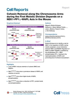
1. Report
Cohesin Removal along the Chromosome Arms
during the First Meiotic Division Depends on a
NEK1-PP1g-WAPL Axis in the Mouse
Graphical Abstract
Highlights
d Loss of NEK1 induces retention of cohesin SMC3 and
RAD21L
d Nek1kat2j/kat2j
mice show a premature release of WAPL and
retention of sororin
d Nek1kat2j/kat2j
mice show higher levels of WAPL
phosphorylation
d Nek1kat2j/kat2j
mice show a reduction of PP1g protein levels
and phosphorylation
Authors
Miguel A. Brien˜ o-Enrı´quez,
Stefannie L. Moak, Melissa Toledo, ...,
Jose´ L. Barbero, Paula E. Cohen,
J. Kim Holloway
Correspondence
paula.cohen@cornell.edu (P.E.C.),
jkh44@cornell.edu (J.K.H.)
In Brief
Brien˜ o-Enrı´quez et al. identify a role for
NEK1 in the regulation of WAPL during
meiotic prophase I, via an interaction
between NEK1 and PDS5B. Regulation of
WAPL by NEK1-PDS5B is mediated by
the phosphatase PP1g. NEK1
phosphorylates PP1g, leading to the
dephosphorylation of WAPL and
promoting loss of cohesion at prophase I.
Brien˜ o-Enrı´quez et al., 2016, Cell Reports 17, 977–986
October 18, 2016 ª 2016 The Author(s).
http://dx.doi.org/10.1016/j.celrep.2016.09.059
2. Cell Reports
Report
Cohesin Removal along the Chromosome Arms
during the First Meiotic Division Depends
on a NEK1-PP1g-WAPL Axis in the Mouse
Miguel A. Brien˜ o-Enrı´quez,1 Stefannie L. Moak,1 Melissa Toledo,1 Joshua J. Filter,2 Stephen Gray,1 Jose´ L. Barbero,3
Paula E. Cohen,1,4,* and J. Kim Holloway1,*
1Department of Biomedical Sciences and Center for Reproductive Genomics, Cornell University, Ithaca, NY 14853, USA
2Department of Molecular Biology and Genetics, Cornell University, Ithaca, NY 14853, USA
3Department of Cellular and Molecular Biology, Laboratory of Chromosomal Dynamics in Meiosis, Centro de Investigaciones Biolo´ gicas
(CSIC), Madrid 28040, Spain
4Lead Contact
*Correspondence: paula.cohen@cornell.edu (P.E.C.), jkh44@cornell.edu (J.K.H.)
http://dx.doi.org/10.1016/j.celrep.2016.09.059
SUMMARY
Mammalian NIMA-like kinase-1 (NEK1) is a dual-
specificity kinase highly expressed in mouse germ
cells during prophase I of meiosis. Loss of NEK1
induces retention of cohesin on chromosomes at
meiotic prophase I. Timely deposition and removal
of cohesin is essential for accurate chromosome
segregation. Two processes regulate cohesin
removal: a non-proteolytic mechanism involving
WAPL, sororin, and PDS5B and direct cleavage by
separase. Here, we demonstrate a role for NEK1 in
the regulation of WAPL loading during meiotic pro-
phase I, via an interaction between NEK1 and
PDS5B. This regulation of WAPL by NEK1-PDS5B
is mediated by protein phosphatase 1 gamma
(PP1g), which both interacts with and is a phos-
photarget of NEK1. Taken together, our results reveal
that NEK1 phosphorylates PP1g, leading to the
dephosphorylation of WAPL, which, in turn, re-
sults in its retention on chromosome cores to
promote loss of cohesion at the end of prophase I
in mammals.
INTRODUCTION
Meiosis is a specialized cell division characterized by a single
round of DNA replication followed by two rounds of chromosome
segregation, resulting in the formation of haploid gametes. In
order to achieve accurate segregation at both divisions, tension
must be established on the meiotic spindles, which is achieved
by the formation of crossovers between homologous chromo-
somes in prophase I of meiosis, and by cohesion between sister
chromatids in both meiosis I and meiosis II. Cohesion is estab-
lished by the cohesin complex and is an essential component
of prophase I events, along with the defining feature of prophase
I: the synaptonemal complex. Early in prophase I of meiosis,
a proteinaceous structure called the axial element (AE) begins
to form along replicated sister chromatids, consisting of proteins
such as synaptonemal complex proteins-2 and -3 (SYCP2 and
SYCP3), along with several cohesin components. Subsequently,
the AEs of homologous chromosomes become juxtaposed by
proteins of the central element of the synaptonemal complex
(SYCP1, TEX12, etc.). The paired AEs, now termed lateral
elements (LEs), are now joined by the transverse elements of
the central element, collectively forming the tripartite synap-
tonemal complex (SC) that connects all homologs at pachytene
(Llano et al., 2012).
Timely deposition and removal of cohesin is essential for SC
dynamics and for accurate chromosome segregation during
both mitosis and meiosis. Cohesin complexes differ in mitosis
compared to meiosis, consisting of SMC3, SMC1a, STAG1/2,
and RAD21 in the former (Haering and Jessberger, 2012; Hirano,
2015; Nasmyth and Haering, 2009) and consisting of REC8,
RAD21L, STAG3, SMC1a/b, and SMC3 in the latter (Haering
and Jessberger, 2012; McNicoll et al., 2013). Cohesin removal
during mitosis is a two-step process, beginning in prophase
(the ‘‘prophase pathway’’) and continuing with separase-medi-
ated proteolytic cleavage of centromeric cohesin during the
metaphase-anaphase transition. During mitosis, the prophase
pathway mediates cohesin removal via a non-enzymatic process
that involves the cohesin-associated proteins, WAPL (Wings
apart-like homolog), PDS5B (Sister chromatid cohesion protein
PDS5 homolog B), and sororin (Tedeschi et al., 2013). WAPL
facilitates the unloading of cohesin through an antagonistic
mechanism that involves competition with sororin for binding
to PDS5B (Nishiyama et al., 2010). In mitotic prophase, sororin
is phosphorylated by cyclin-dependent kinase (CDK) 1 (CDK1)
and Aurora kinase B (AURKB), which results in its release from
PDS5B, thus allowing the interaction of WAPL and PDS5B that
leads to cohesin release (Dreier et al., 2011; Nishiyama et al.,
2013). Thus, sororin-PDS5B interactions mediate cohesin
loading/stabilization, while WAPL-PDS5B interactions mediate
cohesin unloading.
WAPL is highly expressed in mouse testis and is localized to
meiotic chromosome cores during zygotene and pachytene of
Cell Reports 17, 977–986, October 18, 2016 ª 2016 The Author(s). 977
This is an open access article under the CC BY-NC-ND license (http://creativecommons.org/licenses/by-nc-nd/4.0/).
3. prophase I in the mouse (Kuroda et al., 2005). In mouse oocytes,
WAPL colocalizes with SYCP2 during pachytene (Zhang et al.,
2008). PDS5B is present in spermatogonia and spermatocytes,
and during meiotic prophase, it is associated with the AE inde-
pendently of the presence of synaptonemal complex proteins
(Fukuda and Hoog, 2010). Sororin is also localized within the
central element of the SC independently of the presence of co-
hesin (Go´ mez et al., 2016). However, to date, the function of
these proteins and the importance of the prophase pathway in
mammalian meiosis remain undescribed.
The never-in-mitosis-gene-A (NIMA)-related kinases (NEKs)
are a family of serine/threonine kinases involved in mitotic and
meiotic events (Fry et al., 2012; Meirelles et al., 2014), the found-
ing member of which is Aspergillus nidulans NIMA (Oakley and
Morris, 1983). In mammals, there are 11 orthologous Nek genes,
but Nek1 is a unique, dual-specificity kinase highly expressed in
mouse germ cells (Letwin et al., 1992; Upadhya et al., 2000). We
previously showed that NEK1 interacts with FKBP6, a compo-
nent of the mammalian synaptonemal complex, during prophase
I (Crackower et al., 2003; Holloway et al., 2011) Furthermore, our
studies of Nek1kat2j/kat2j
mice showed that the loss of NEK1 in-
duces retention of the cohesin component SMC3 during diplo-
tene (Holloway et al., 2011), leading us to propose a role for
NEK1 in the sequential removal of cohesin during meiosis.
Here, we demonstrate that the release of cohesin during
prophase I is dependent on NEK1 regulation of WAPL during
prophase I. This regulation of WAPL by NEK1 is mediated by
NEK1-PDS5B interaction and by protein phosphatase 1 gamma
(PP1g), which both interacts with NEK1 and is a phosphotarget
thereof. In support of this, loss of PP1g activity mimics the
meiosis I phenotype observed in Nek1kat2j/kat2j
mice, while
overexpression of PP1g results in retention of WAPL into diplo-
tene and premature release of cohesin subunits. Thus, we
have established a phosphorylation cascade regulating timely
cohesin removal that involves NEK1-mediated phosphorylation
of targets during prophase I. Importantly, this study establishes
a role for the prophase pathway in chromosome segregation
events during meiosis I.
RESULTS
Loss of NEK1 Induces Abnormal Phosphorylation of
WAPL and Premature Release from Chromosome Cores
Loss of NEK1 in mouse spermatocytes leads to abnormal reten-
tion of SMC3 on chromosome arms at the diplotene and diaki-
nesis (Figure S1) (Holloway et al., 2011). To identify candidate
targets of NEK1 that might affect the removal of cohesin during
prophase I, we compared the spectrum of phosphorylated pro-
teins in Nek1+/+
and Nek1kat2j/kat2j
littermate testes by mass
spectrometry (MS) (Figure 1A; Table S1). A total of 445 proteins
were found to have abnormal phosphorylation profiles in the
mutants compared to WT mice, with 105 proteins showing
increased phosphorylation in the mutant protein extracts and
340 showing a reduction in phosphorylation. Of particular rele-
vance to the persistent cohesin retention was the prophase
pathway component, WAPL, which showed an abnormal eleva-
tion in phosphorylation at the serine residue in position 226 (Fig-
ure 1A). In light of the well-documented role of WAPL in cohesin
release in mitotic cells, deregulation of WAPL during meiosis
would be well placed to be causative for the abnormal persis-
tence of cohesin during prophase I. Moreover, no direct post-
translational regulation of WAPL, such as we describe herein,
has been reported, either in mitosis or in meiosis. Confoundingly,
however, the fact that loss of NEK1 kinase activity leads to
increased phosphorylation of WAPL, rather than a predicted
decline in phosphorylation, led us to predict that WAPL was
not a direct target of NEK1 but might be phosphorylated or de-
phosphorylated by another protein that is, itself, a target of
NEK1. Therefore, we performed an immunoprecipitation (IP)
experiment with an antibody against NEK1 in both mutant and
WT mouse testis extracts followed by MS (proteins that appear
in both IP-MS results were considered as false positive and
eliminated) (Table S2). Interestingly, our results demonstrated
that PDS5B is a binding partner of NEK1, whereas WAPL is
not. The NEK1-PDS5B interaction and lack of interaction be-
tween NEK1 and WAPL were both confirmed by IP followed by
western blotting (WB) (Figure S2).
The proteomics data described earlier indicate a physical
interaction between PDS5B and NEK1, and at least a functional
(though indirect) interaction between WAPL and NEK1, leading
us to hypothesize that WAPL/PDS5B interactions with NEK1
might explain the retention of SMC3 during prophase I. To
examine this further, chromosome spreads were prepared
from spermatocytes of both Nek1kat2j/kat2j
mice and Nek1+/+
mice and were subjected to immunofluorescence (IF) using anti-
bodies against components of the prophase pathway (PDS5B,
WAPL, and sororin). The distribution of PDS5B observed in our
studies was similar to that described previously (Fukuda and
Hoog, 2010), with no difference being observed in the temporal
distribution or frequency of PDS5B loading on chromosome
cores in Nek1kat2j/kat2j
mice compared to Nek1+/+
mice (Fig-
ure 1A). The presence and dynamics of WAPL protein localiza-
tion were evaluated in both WT and mutant animals. In Nek1+/+
male spermatocytes, WAPL is observed from leptotene to
pachytene of prophase I, colocalizing with SYCP3 along the
chromosome cores independently of the synapsis. Finally, at
diplotene, WAPL is removed from chromosome cores (Fig-
ure 1C). However, in the absence of NEK1, WAPL shows prema-
ture release from the chromosome cores in early prophase I, as
early as late zygotene in Nek1kat2j/kat2j
males (Figure 1C).
Release of cohesion via the prophase pathway represents a
balance between WAPL and sororin (Nishiyama et al., 2010).
Given the premature release of WAPL from PDS5B in the
absence of NEK1, we evaluated the localization of sororin by
IF. Indeed, analysis of chromosome spreads shows the retention
of sororin along cores during pachytene in the Nek1kat2j/kat2j
mice
compared to WT littermates (Figure 1D). Our data are consistent
with the requirement for sororin removal in order to facilitate co-
hesin removal at the end of prophase I, with a stronger and more
persistent signal of sororin found in Nek1kat2j/kat2j
spermatocytes.
The changes we observed in the dynamics of WAPL and sororin
presence during late prophase I were analyzed by WB of protein
extracts from isolated pachytene spermatocytes, with WAPL
showing a decrease in protein abundance in Nek1kat2j/kat2j
mice
(Figure S2B). By contrast, sororin shows a subtle increase in pro-
tein abundance in extracts from Nek1kat2j/kat2j
males, while no
978 Cell Reports 17, 977–986, October 18, 2016
4. changes were observed in PDS5B protein extracts from
Nek1kat2j/kat2j
relative to that seen in WT extracts (Figure S2B).
Localization of all three components of the prophase pathway
(WAPL, PDS5B, and sororin) is coincident with the localization
of NEK1 described previously (on chromosome cores and
increased signal at the sex body) (Yeo et al., 2015).
Identification of PP1g as a Candidate Phosphatase that
Is a Target of NEK1
The aforementioned data demonstrate that loss of NEK1 results
in abnormal phosphorylation of WAPL. We hypothesize that this
aberrant phosphorylation leads to the premature release of
WAPL from chromosome cores during prophase I, allowing
for the retention of sororin, although certainly this is, by no
means, the only possible explanation for such an observation.
However, the abnormal phosphorylation of WAPL is consistent
with the idea that, under physiological conditions, NEK1 could
be phosphorylating a phosphatase that promotes the dephos-
phorylation of WAPL to aid its retention on chromosome cores.
To test this hypothesis, IP-MS was performed using an antibody
against WAPL (Table S3). Results obtained from WAPL IP-MS
(Table S3) were cross-referenced with MS results obtained
from the aforementioned phosphoproteomics of NEK1 targets
(Table S1) and with NEK1 IP-MS data (Table S2). A single candi-
date emerged that was present in all three sets of results: PP1g.
PP1g is reduced in the global MS data from Nek1kat2j/kat2j
males,
is found in the IP-MS data for NEK1 interactors, and shows a
reduction in phosphorylation at serine 129 in Nek1kat2j/kat2j
males
Figure 1. Loss of NEK1 Induces Changes in Phosphorylation Profile, Provoking a Premature Removal of WAPL from Chromosome Cores and
Retention of Sororin
(A) Distribution of phosphoproteins analyzed by mass spectrometry. Black dots indicate detected phosphopeptides without changes in Àlog10 of p value or fold
change (FC); red dots indicate p > 0.05 without change in log2 FC; orange dots show Àlog10 of p > 0.05 and log2 FC > 1. Green dots indicate Àlog10 of p > 0.05 and
log2 FC > 1 (n = 3).
(B) IF against WAPL (green) and SYCP3 (red) in spermatocyte spreads from Nek1+/+
and Nek1kat2j/kat2j
male mice.
(C) IF against PDS5B (green) and SYCP3 (red) in spermatocyte spreads from Nek1+/+
and Nek1kat2j/kat2j
male mice.
(D) IF against sororin (green) and SYCP3 (red) in spermatocyte spreads from Nek1+/+
and Nek1kat2j/kat2j
male mice.
See also Figures S1 and S4 and Table S1.
Cell Reports 17, 977–986, October 18, 2016 979
5. (Table S1). Thus, PP1g emerges as a strong candidate to interact
with, and become modified (directly or indirectly) by, NEK1 while
at the same time potentially phosphorylating WAPL.
Lack of NEK1 Disrupts PP1g Localization and Levels in
Spermatocytes
The interaction between WAPL and PP1g was confirmed by
IP-WB (Figure 2A). Evaluation of total protein level revealed a sig-
nificant reduction in PP1g in protein extracts from isolated
pachytene cells from Nek1kat2j/kat2j
males, as detected by WB
(Figure 2B). In addition, PP1g localization on chromosome
spreads from WT and NEK1-deficient males was demonstrated
by IF using antibodies against total PP1g and was found to be
loaded along the chromosome cores in WT males at late zygo-
tene, after which time, the protein signal diminishes and is
restricted to occasional ‘‘flares’’ that cross the chromosome
axes in a perpendicular direction. These flares are detectable
on chromosome spreads until pachytene (Figure 2C). By
contrast, chromosome spreads from Nek1kat2j/kat2j
males show
no detectable PP1g at zygotene, and few cells displaying limited
flare pattern at pachytene (Figure 2C).
Inhibition of PP1g Induces Phosphorylation of WAPL
The identification of PP1g as an interacting protein for both WAPL
and NEK1, its status as a potential phosphotarget for NEK1, and
the reduction of PP1g protein during pachytene in Nek1kat2j/kat2j
mutants all implicate PP1g as a candidate phosphatase that might
dephosphorylate WAPL during prophase I under normal condi-
tions. Thus, we hypothesized that the reduction in PP1g levels
and/or activity in NEK1-deficient males would result in the WAPL
phosphorylation observed during prophase I in these animals.
To test this directly, we used a pharmacological approach by
Figure 2. Loss of NEK1 Induces Low Protein Levels and Premature Removal of PP1g from Chromosome Cores
(A) Immunoprecipitation with WAPL, PP1g, and NEK1 antibodies, followed by WB.
(B) PP1g protein levels in isolated pachytene cells from Nek1+/+
and Nek1kat2j/kat2j
testes relative to actin (n = 3).
(C) IF against PP1g (green) and SYCP3 (red) in spermatoctyes from Nek1+/+
and Nek1kat2j/kat2j
male mice. Arrowheads indicate the flare pattern of PP1g along
chromosome cores from pachytene and diplotene spermatocytes.
See also Figure S2 and Tables S2 and S3.
980 Cell Reports 17, 977–986, October 18, 2016
6. inhibiting PP1g activity and assaying for the status of WAPL phos-
phorylation. Spermatocytes from Nek1+/+
males were cultured for
6 hr in the presence of the pan-phosphatase inhibitors, okadaic
acid (OA) and calyculin A (CLA). OA was used at doses that mainly
inhibit protein phosphatase 2 (1, 10, and 100 nM), while CLA was
usedatdosesthatinhibitPP1(2and 20nM)(Kitaetal.,2002;Swin-
gle et al., 2007). WB revealed that CLA significantly reduced the
amount of WAPL protein compared to those spermatocytes
cultured with vehicle alone (ethanol) (Figure 3A). By contrast, there
were no changes in WAPL protein when spermatocytes were
cultured in the presence of OA (Figure 3B). Interestingly, IP with
anti-phosphoserine antibody followed by WB with an antibody
against WAPL revealed that CLA, but not OA, induced increased
all the possible forms of phosphorylated WAPL (phospho-WAPL)
in short-term cultures of WT spermatocytes (Figure 3C). Thus,
pharmacological inhibition of PP1g using CLA in WT spermato-
cytes results in increased phospho-WAPL and subsequent desta-
bilization ofWAPL protein, supportingthe hypothesis that WAPLis
a direct target of PP1g activity.
PP1g Mutant Mice Showed a Phenotype that Mimics
Nek1 Mutant Mice
To confirm our in vitro inhibitor results, an in vivo genetics
approach was used with previously characterized PP1g
knockout mice (Ppp1ccÀ/À
) (Varmuza et al., 1999). Chromosome
spread analysis of WAPL localization in Ppp1ccÀ/À
mice phe-
nocopied the spermatocyte phenotypes observed in
Nek1kat2j/kat2j
mice, with premature release of WAPL from the
chromosome cores at late zygotene, leaving a faint residual
signal during pachytene in the Ppp1ccÀ/À
mice compared to
WT controls (Figure 3D). WB with an antibody against WAPL
on testis protein from Ppp1ccÀ/À
mice confirmed these results,
with a reduction in the WAPL protein level in the mutant testis
extracts compared to that observed in WT testis extracts (Fig-
ure 3E). IP with anti-phosphoserine antibody followed by WB
with an antibody against WAPL confirmed an increase in phos-
pho-WAPL in the spermatocytes of Ppp1ccÀ/À
mice (Figure 3F).
To test indirectly the activity of PP1g on WAPL and SMC3, we
performed IF in the previously described Ppp1cc overexpressing
PPP1CC2 mouse (Ppp1cc+/+Tg(Ppp1cc2/Ppp1cc2
) (Sinha et al.,
2013). Our results show that PP1g overexpression leads to the
retention of WAPL into diplotene and release of SMC3 (Fig-
ure S3). Thus, these data demonstrate that the absence of
NEK1, or of PP1g phosphatase activity, results in abnormal
phosphorylation of WAPL, leading to premature release of
WAPL from chromosome cores in late prophase I, disrupting
the prophase pathway in mouse meiosis.
DISCUSSION
During mitosis, WAPL is a constituent of the prophase pathway
(Gandhi et al., 2006; Huis in ’t Veld et al., 2014; Kueng et al.,
2006; Tedeschi et al., 2013), forming a complex with PDS5B to
facilitate the unloading of cohesin during prophase (Carretero
et al., 2013; Shintomi and Hirano, 2009). During S phase,
WAPL-PDS5B function is antagonized by sororin, which dis-
places WAPL from PDS5B, thereby promoting cohesin loading
and/or stabilization (Shintomi and Hirano, 2009; Zhang et al.,
2008). Recent studies in Caenorhabditis elegans, Saccharo-
myces cerevisiae, and Arabidopsis thaliana demonstrated a
role for WAPL in cohesin regulation during meiosis (Challa
et al., 2016; Crawley et al., 2016; De et al., 2014). In C. elegans
oogenesis, WAPL-1 antagonizes the binding of cohesin
containing the COH-3/4 kleisins, but not REC-8, and promotes
a WAPL-1-independent mechanism that removes cohesin
before metaphase I (Crawley et al., 2016). In S. cerevisiae, the or-
tholog of WAPL (Rad61/Wpl1) negatively regulates chromosome
axis compaction and is required for the efficient resolution of
telomere clustering during meiosis I (Challa et al., 2016). Muta-
tion of WAPL in A. thaliana blocks the removal of cohesin from
chromosomes during meiosis, resulting in chromosome bridges,
broken chromosomes, and uneven chromosome segregation
(De et al., 2014). However, studies in the mouse have been
limited only to a brief description of WAPL localization on chro-
mosome core (Kuroda et al., 2005; Zhang et al., 2008). Here,
we demonstrate that loss of NEK1 results in abnormal phosphor-
ylation of WAPL, which leads to premature loss of WAPL from the
chromosome cores and reduction of protein levels in prophase I
(Figure 1C). The premature release of WAPL in the absence of
NEK1 allows for the persistence of sororin (Figure 1D) and
subsequent retention of SMC3 during diplotene and diakinesis
(Figure S1). Our data also indicate that NEK1 mutant spermato-
cytes retain RAD21L on meiotic chromosome cores at the end of
prophase I (Figure S4), indicating that SMC3 is not the only
cohesin subunit to be disrupted. Post-translational modifications
of WAPL have been described in the phosphoproteomics
screening of polo-like kinase targets (Grosstessner-Hain et al.,
2011) and in PP1g mutant mice (MacLeod et al., 2014). However,
the role of such regulation of WAPL has not been described in
mammalian meiosis, nor have changes in the phosphoproteomic
profile of WAPL been associated with prophase I defects in the
mouse. Alignment of WAPL orthologs across species (Figure S5)
shows conservation of serine 226 in mouse and human but
within a larger domain of other conserved serines (for example,
serine 228 and serine 236). In all, at least five serine residues
in this region are conserved across mammals and in
D. melanogaster, providing the potential for this region to be
highly regulated by phosphorylation events.
Interaction between PDS5B and WAPL is mediated by HEAT
repeats (Losada et al., 2005; Nishiyama et al., 2010; Shintomi
and Hirano, 2009) and by a YSR motif (Ouyang et al., 2016) on
PDS5B. These domains interact with three FGF motifs of
WAPL, none of which include the serine 226 identified herein.
Thus, NEK1 activity through PP1g is not directed to the FGF re-
peats in WAPL. Instead, we propose that phosphorylation of
serine 226, or of any of the other highly conserved serines in
the vicinity of serine 226, could induce a conformational change
in the WAPL protein that destabilizes its interaction with PDS5B,
allowing PDS5B-sororin interactions to predominate and result-
ing in cohesin retention. Alternatively, WAPL phosphorylation
could act as a signal for WAPL degradation, resulting in its
premature removal from chromosome cores. Finally, the phos-
phorylation of WAPL at serine 226 may act to suppress key
phosphorylation events on other residues. Taken together, our
studies implicate a direct regulatory action on WAPL itself, lead-
ing to activation of the prophase pathway rather than an indirect
Cell Reports 17, 977–986, October 18, 2016 981
7. Figure 3. Increased WAPL Phosphorylation
Is Caused by the Lack of Phosphorylation of
PP1g by NEK1
(A) Quantitation of WAPL protein levels in extracts
from Nek1+/+
testes relative to GAPDH control in
cultured spermatocytes treated with calyculin A
(CLA; 2–20 nM) (n = 5).
(B) Quantitation of WAPL protein levels in extracts
from Nek1+/+
testes relative to GAPDH control in
cultured spermatocytes treated with okadaic acid
(n = 5).
(C) In vitro phosphorylation of WAPL in cultured
spermatocytes vehicle (ethanol), calyculin A (CLA)
(2 and 20 nM), and okadaic acid (OA) (1, 10, and
100 nM), analyzed by immunoprecipitation
pull-downs with antibody against phospho-serine
followed by WB. Input lane represents spe-
rmatocyte protein extracts without immuno-
precipitation.
(D) IF against WAPL (green) and SYCP3 (red) in
spermatocytes from Ppp1cc+/+
and Ppp1ccÀ/À
male mice.
(E) Quantitation of WAPL protein levels relative to
GAPDH control in extracts from Ppp1cc+/+
and
Ppp1ccÀ/À
testes using antibody against WAPL
(n = 3).
(F) In vivo phosphorylation of WAPL in Ppp1cc+/+
and Ppp1ccÀ/À
whole testis lysate using immu-
noprecipitation pull-downs with antibody against
phospho-serine followed by western blotting.
*Statistically significant differences (one-way
ANOVA followed by Dunnett’s multiple compari-
sons test, p < 0.05).
See also Figure S3.
982 Cell Reports 17, 977–986, October 18, 2016
8. Figure 4. Model the Role of NEK1 during Meiotic Prophase I
(A) Top panel represents the role of NEK1 during meiotic prophase I in WT mice. During prophase I, NEK1 binds to PDS5B at the cohesin ring. Additionally, NEK1
binds to PP1g and phosphorylates it; phosphorylation of PP1g subsequently inhibits WAPL phosphorylation and permits its activity during the prophase-
metaphase transition without changes in sororin.
(B) The bottom panel represents the effects of loss of NEK1, which results in retention of SMC3. The absence of NEK1 prevents PP1g phosphorylation which, in
turn, permits a hyper-phosphorylation of WAPL. Hyper-phosphorylation of WAPL affects its stability and/or binding to PDS5B, inducing a premature removal of
WAPL from the cohesin ring and persistence of sororin. Light gray text denotes events that require further confirmatory studies.
Cell Reports 17, 977–986, October 18, 2016 983
9. action through regulation of sororin, as has previously been
described in other cell systems (Nishiyama et al., 2010; Rankin
et al., 2005; Zhang and Pati, 2012).
The data presented herein demonstrate a role for NEK1 during
meiotic prophase I, regulating—directly or indirectly—the pro-
teins involved in the prophase pathway, specifically in regulation
of the PDS5B-WAPL complex. Importantly, NEK1 function on
the PDS5B-WAPL complex appears predicated on its interaction
with PDS5B, yet PDS5B itself is not a target of either of the two-
kinase activities of NEK1. Instead, our data indicate that this
function could be mediated through NEK1 interaction with
PP1g and through the altered phosphorylation status of PP1g.
During mitosis, PP1g phosphorylation is orchestrated preferen-
tially by CDKs (Dohadwala et al., 1994; Eto, 2009; Kim et al.,
2003; Li et al., 2007). However, our phosphoproteomics analysis
did not reveal any changes in the phosphorylation status of
CDKs in the absence of NEK1. Moreover, the NEK1 IP data failed
to identify a candidate CDK that could be a potential substrate
for NEK1, and/or kinase for PP1g. It is highly unlikely, therefore,
that PP1g is phosphorylated by a canonical CDK during meiotic
prophase. Instead, we propose that PP1g is a substrate of NEK1,
or that another kinase is acting as an intermediary between these
two proteins. Our analysis of the phosphoproteomics and NEK1-
IP data failed to reveal such a candidate kinase, but it is possible,
as with many kinase-substrate interactions, that NEK1-substrate
interactions may be too rapid/transient to permit their identifica-
tion by IP-MS. Another important factor is the regulation of PP1g
by its regulatory subunits and natural inhibitors. Our MS results
showed that there are changes in the protein levels of some of
these regulatory subunits and that these may, in turn, regulate
the activity or abundance of PP1g. Thus, analysis of NEK1/
PP1g/WAPL interactions during meiosis are made more com-
plex by involvement of a complex PP1 regulatory network, the
particularly large size of the protein constituents of this network,
and the potential for multiple serines on WAPL to be the targets
of PP1g activity.
In summary, we propose a model whereby NEK1 docks with
PDS5B to bring it into close proximity with the cohesin ring, where-
upon it phosphorylates a number of direct targets, altering the
phosphorylation status of PP1g and, hence, its localization and/or
activity. PP1g, in turn, dephosphorylates WAPL, securing its inter-
action with PDS5B and displacing sororin, resulting in accurate
and timely opening of the cohesin ring to facilitate loss of cohesion
along the chromosome arms at the end of meiosis I (Figure 4).
EXPERIMENTAL PROCEDURES
All mouse studies were conducted with the prior approval of the Cornell
Institutional Animal Care and Use Committee (protocol 2010-0054). The
Nek1kat2j/kat2j
mouse line was obtained originally from The Jackson
Laboratory and maintained on a C57Bl/6J line for >10 years at Cornell
University. At least three sets of 8-week-old homozygous mutant animals
(Nek1kat2J/kat2J
) were compared with WT (Nek1+/+
) littermates. Ppp1ccÀ/À
and
Pppc1+/+Tg(Ppp1cc2/Ppp1cc2)
mice were generously donated by Dr. Vijayaraghavan
(Department of Biomedical Sciences, Kent State University).
Chromosome Spread Preparations and IF Staining
Prophase I spermatocyte chromosome spreads were performed as
previously described (Sun et al., 2015). Primary antibodies included: rabbit
anti-Sororin (anti-Sororin serum C-106 was generated using a full-length re-
combinant mouse protein cloned in pET-12a vector and expressed in E. coli
[Carretero et al., 2013]), anti-mouse SYCP3 (ab97672), rabbit anti-PDS5B
(ab84918) (both from Abcam), rabbit anti-WAPL (16370-1-AP, from
Proteintech), rabbit anti-PP1g (PA5-21671, from Thermo Fisher), rabbit
anti-SMC3 (from J.L.B.), and rabbit anti-RAD21L (from Alberto Penda´ s).
Alexa Fluor secondary antibodies were used (Molecular Probes). Image
acquisition was performed using a Zeiss Imager Z1 microscope and
captured using a Zeiss charge-coupled device (CCD). Images were
processed using AxioVision (version 4.8, Zeiss).
Isolation of Pachytene Cells
Nek1+/+
and Nek1kat2j/kat2j
enrichment of specific spermatogenic cell types was
performed using the STA-PUT method based on separation by cell diameter/
density at unit gravity (Bellve´ , 1993; Romrell et al., 1976). Purity of resulting
fractions was determined by microscopy based on cell diameter and
morphology.
Culture of Spermatocytes
Culture was performed following the protocol described elsewhere (Wiltshire
et al., 1995), with some modifications. Briefly, we dissociated testes in 4 mL
of spermatocyte culture medium (SCM) (DMEM without red phenol [21063-
029], fetal calf serum [10082139], penicillin-streptomycin 1003 [15140-122],
all from GIBCO; lactic acid [L13750, NaHCO3 9s8761] and sodium pyruvate
1003 [11360-070], both from Sigma). Cells were placed in treated culture
dishes (#430167, Corning), washed five times with SCM, and centrifuged
1,000 3 g for 1 min to pellet cells. The pellet was resuspended in 600 mL
SCM. 100 mL cell suspension was placed into each 35-mm plate with and
without okadaic acid (135 mM, 50 mg/mL) (O9381, Sigma) or with and without
calyculin A (2–200 nM) (sc-24000, Santa Cruz Biotechnology). Cells were
cultured for 6 hr at 32
C before analysis.
Western Blot and IP
Proteins were extracted by sonication in RIPA buffer, denaturalized, and
then separated using 4%–15% Mini-PROTEAN TGX Precast Protein Gels
(#4561086, from Bio-Rad) and transferred onto nitrocellulose membrane.
Primary antibody incubation was performed for 12 hr at 4
C at 1:1,000
dilution. Secondary HRP (horseradish peroxidase)-conjugated antibodies
were obtained from Pierce and Life Technologies. Signal detection was
carried out using the SuperSignal substrate (Thermo Scientific). Images
were captured with Bio-Rad Image Lab 5.1 and analyzed by ImageJ
(http://rsbweb.nih.gov/ij). IP was performed using 1 mg of whole testis
protein. Incubation with the specific antibody was performed at 4
C over-
night. The protein lysate was then centrifuged, and the supernatant was
removed. The remaining beads were washed in fresh cold RIPA buffer
three times, and the final bead slurry was resuspended in 40 mL 2 3 SDS
protein loading dye and run in 4%–15% Mini-PROTEAN TGX Precast
Protein Gels.
Mass Spectrometry
MS was performed in the Cornell University Proteomics and Mass Spectrom-
etry Facility. Experiments were performed using three different sets of Nek1+/+
and Nek1kat2j/kat2j
mice. To obtain the specific postranslational modification of
the peptides, we performed TiO2 enrichment followed by the proteomics anal-
ysis. After nano-liquid chromatography-tandem MS (nano-LC-MS/MS), raw
data files were acquired using Orbitrap Elite (Thermo Scientific). We performed
a database search against the SwissProt mouse database from the UniProt
website (http://www.uniprot.org) using Mascot software version 2.3.02 (Matrix
Science) and MouseRefSeq (http://www.ncbi.nlm.nih.gov/refseq). The default
Mascot search settings were as follows:
1. One missed cleavage site by trypsin allowed with fixed MMTS
modification of cysteine; fixed 4-plex iTRAQ modifications on Lys and
N-terminal amines; and variable modifications of methionine oxidation,
deamidation of Asn and Gln residues, and 4-plex iTRAQ on Tyr for
iTRAQ 4-plex analysis.
2. One or two missed cleavage sites by trypsin allowed with fixed carbox-
amidomethyl modification of cysteine; fixed 6-plex TMT modifications
984 Cell Reports 17, 977–986, October 18, 2016
10. on Lys and N-terminal amines; and variable modifications of methionine
oxidation, deamidation of Asn and Gln residues, and 6-plex TMT on Tyr
for TMT 6-plex analysis.
3. Peptide mass tolerance was set at 0.01 Da, and fragment mass toler-
ance was set at 0.1 Da; decoy search: yes; ion score cutoff: 0.1.
4. The quantitative protein ratios were weighted and normalized by the
median ratio with outlier removal set automatically in Mascot for each
set of experiments.
We performed NEK1-IP on three different protein sets (from three distinct
mice) following the parameters described earlier. Quantification analysis of
NEK1-IP was performed following the label-free quantitation analysis using
MaxQuant software (Max Planck Institute of Biochemistry; http://www.
biochem.mpg.de/5111795/maxquant).
Statistics
Statistical analyses were performed using GraphPad Prism version 6.00 for
Macintosh (GraphPad Software; http://www.graphpad.com).
SUPPLEMENTAL INFORMATION
Supplemental Information includes five figures and three tables and can be
found with this article online at http://dx.doi.org/10.1016/j.celrep.2016.09.059.
AUTHOR CONTRIBUTIONS
M.A.B.-E., P.E.C., and J.K.H. designed experiments. M.A.B.-E., S.L.M., M.T.,
J.J.F., and S.G. carried out the experiments. M.A.B.-E., S.L.M., P.E.C., and
J.K.H. analyzed and interpreted data. J.L.B. provided critical antibodies.
M.A.B.-E. and P.E.C. wrote the manuscript.
ACKNOWLEDGMENTS
We thank Dr. Lee Zhou for the anti-NEK1 antibody, Dr. Ana Losada for the anti-
Sororin antibody, Dr. Alberto Penda´s for the anti-RAD21L antibody,
Dr. Srinivasan Vijayaraghavan for Ppp1ccÀ/À
and Ppp1cc+/+Tg(Ppp1cc2/Ppp1cc2)
mice, Dr. Michael Goldberg for advice and reagents pertaining to PP1g analysis,
and Dr. Sheng Zhang from the Cornell Proteomics and Mass Spectrometry Facil-
ity forhis adviceinthedevelopment ofMSexperiments.WethankMr.PeterBorst
for his help in the care and maintenance of experimental animals. This project is
funded by grants from the NICHD to J.K.H. (5R00HD065870) and from NIGMS
and March of Dimes to P.E.C. (1R01GM097263 and MOD2006-844). J.L.B. is
funded by a grant from MINECO (BFU2014-59307), Spain.
Received: April 29, 2016
Revised: July 25, 2016
Accepted: September 16, 2016
Published: October 18, 2016
REFERENCES
Bellve´ , A.R. (1993). Purification, culture, and fractionation of spermatogenic
cells. Methods Enzymol. 225, 84–113.
Carretero, M., Ruiz-Torres, M., Rodrı´guez-Corsino, M., Barthelemy, I., and
Losada, A. (2013). Pds5B is required for cohesion establishment and Aurora
B accumulation at centromeres. EMBO J. 32, 2938–2949.
Challa, K., Lee, M.S., Shinohara, M., Kim, K.P., and Shinohara, A. (2016).
Rad61/Wpl1 (Wapl), a cohesin regulator, controls chromosome compaction
during meiosis. Nucleic Acids Res. 44, 3190–3203.
Crackower, M.A., Kolas, N.K., Noguchi, J., Sarao, R., Kikuchi, K., Kaneko, H.,
Kobayashi, E., Kawai, Y., Kozieradzki, I., Landers, R., et al. (2003). Essential
role of Fkbp6 in male fertility and homologous chromosome pairing in meiosis.
Science 300, 1291–1295.
Crawley, O., Barroso, C., Testori, S., Ferrandiz, N., Silva, N., Castellano-Pozo,
M., Jaso-Tamame, A.L., and Martinez-Perez, E. (2016). Cohesin-interacting
protein WAPL-1 regulates meiotic chromosome structure and cohesion by
antagonizing specific cohesin complexes. eLife 5, e10851.
De, K., Sterle, L., Krueger, L., Yang, X., and Makaroff, C.A. (2014). Arabidopsis
thaliana WAPL is essential for the prophase removal of cohesin during meiosis.
PLoS Genet. 10, e1004497.
Dohadwala, M., da Cruz e Silva, E.F., Hall, F.L., Williams, R.T., Carbonaro-Hall,
D.A., Nairn, A.C., Greengard, P., and Berndt, N. (1994). Phosphorylation and
inactivation of protein phosphatase 1 by cyclin-dependent kinases. Proc.
Natl. Acad. Sci. USA 91, 6408–6412.
Dreier, M.R., Bekier, M.E., 2nd, and Taylor, W.R. (2011). Regulation of sororin
by Cdk1-mediated phosphorylation. J. Cell Sci. 124, 2976–2987.
Eto, M. (2009). Regulation of cellular protein phosphatase-1 (PP1) by phos-
phorylation of the CPI-17 family, C-kinase-activated PP1 inhibitors. J. Biol.
Chem. 284, 35273–35277.
Fry, A.M., O’Regan, L., Sabir, S.R., and Bayliss, R. (2012). Cell cycle regulation
by the NEK family of protein kinases. J. Cell Sci. 125, 4423–4433.
Fukuda, T., and Hoog, C. (2010). The mouse cohesin-associated protein
PDS5B is expressed in testicular cells and is associated with the meiotic chro-
mosome axes. Genes (Basel) 1, 484–494.
Gandhi, R., Gillespie, P.J., and Hirano, T. (2006). Human Wapl is a cohesin-
binding protein that promotes sister-chromatid resolution in mitotic prophase.
Curr. Biol. 16, 2406–2417.
Go´ mez, R., Felipe-Medina, N., Ruiz-Torres, M., Berenguer, I., Viera, A., Pe´ rez,
S., Barbero, J.L., Llano, E., Fukuda, T., Alsheimer, M., et al. (2016). Sororin
loads to the synaptonemal complex central region independently of meiotic
cohesin complexes. EMBO Rep. 17, 695–707.
Grosstessner-Hain, K., Hegemann, B., Novatchkova, M., Rameseder, J.,
Joughin, B.A., Hudecz, O., Roitinger, E., Pichler, P., Kraut, N., Yaffe, M.B.,
et al. (2011). Quantitative phospho-proteomics to investigate the polo-like
kinase 1-dependent phospho-proteome. Mol. Cell. Proteomics 10,
M111008540.
Haering, C.H., and Jessberger, R. (2012). Cohesin in determining chromosome
architecture. Exp. Cell Res. 318, 1386–1393.
Hirano, T. (2015). Chromosome dynamics during mitosis. Cold Spring Harb.
Perspect. Biol. 7, pii: a015792.
Holloway, K., Roberson, E.C., Corbett, K.L., Kolas, N.K., Nieves, E., and
Cohen, P.E. (2011). NEK1 facilitates cohesin removal during mammalian sper-
matogenesis. Genes (Basel) 2, 260–279.
Huis in ’t Veld, P.J., Herzog, F., Ladurner, R., Davidson, I.F., Piric, S., Kreidl, E.,
Bhaskara, V., Aebersold, R., and Peters, J.M. (2014). Characterization of a
DNA exit gate in the human cohesin ring. Science 346, 968–972.
Kim, Y.M., Watanabe, T., Allen, P.B., Kim, Y.M., Lee, S.J., Greengard, P.,
Nairn, A.C., and Kwon, Y.G. (2003). PNUTS, a protein phosphatase 1 (PP1)
nuclear targeting subunit. Characterization of its PP1- and RNA-binding do-
mains and regulation by phosphorylation. J. Biol. Chem. 278, 13819–13828.
Kita, A., Matsunaga, S., Takai, A., Kataiwa, H., Wakimoto, T., Fusetani, N.,
Isobe, M., and Miki, K. (2002). Crystal structure of the complex between caly-
culin A and the catalytic subunit of protein phosphatase 1. Structure 10,
715–724.
Kueng, S., Hegemann, B., Peters, B.H., Lipp, J.J., Schleiffer, A., Mechtler, K.,
and Peters, J.M. (2006). Wapl controls the dynamic association of cohesin with
chromatin. Cell 127, 955–967.
Kuroda, M., Oikawa, K., Ohbayashi, T., Yoshida, K., Yamada, K., Mimura, J.,
Matsuda, Y., Fujii-Kuriyama, Y., and Mukai, K. (2005). A dioxin sensitive
gene, mammalian WAPL, is implicated in spermatogenesis. FEBS Lett. 579,
167–172.
Letwin, K., Mizzen, L., Motro, B., Ben-David, Y., Bernstein, A., and Pawson, T.
(1992). A mammalian dual specificity protein kinase, Nek1, is related to the
NIMA cell cycle regulator and highly expressed in meiotic germ cells. EMBO
J. 11, 3521–3531.
Li, T., Chalifour, L.E., and Paudel, H.K. (2007). Phosphorylation of protein
phosphatase 1 by cyclin-dependent protein kinase 5 during nerve growth fac-
tor-induced PC12 cell differentiation. J. Biol. Chem. 282, 6619–6628.
Cell Reports 17, 977–986, October 18, 2016 985
11. Llano, E., Herra´ n, Y., Garcı´a-Tun˜ o´ n, I., Gutie´ rrez-Caballero, C., de A´ lava, E.,
Barbero, J.L., Schimenti, J., de Rooij, D.G., Sa´ nchez-Martı´n, M., and Penda´ s,
A.M. (2012). Meiotic cohesin complexes are essential for the formation of the
axial element in mice. J. Cell Biol. 197, 877–885.
Losada, A., Yokochi, T., and Hirano, T. (2005). Functional contribution of Pds5
to cohesin-mediated cohesion in human cells and Xenopus egg extracts.
J. Cell Sci. 118, 2133–2141.
MacLeod, G., Taylor, P., Mastropaolo, L., and Varmuzaa, S. (2014). Compar-
ative phosphoproteomic analysis of the mouse testis reveals changes in
phosphopeptide abundance in response to Ppp1cc deletion. EuPA Open Pro-
teom. 2, 1–16.
McNicoll, F., Stevense, M., and Jessberger, R. (2013). Cohesin in gametogen-
esis. Curr. Top. Dev. Biol. 102, 1–34.
Meirelles, G.V., Perez, A.M., de Souza, E.E., Basei, F.L., Papa, P.F., Melo Han-
chuk, T.D., Cardoso, V.B., and Kobarg, J. (2014). ‘‘Stop Ne(c)king around’’:
How interactomics contributes to functionally characterize Nek family kinases.
World J. Biol. Chem. 5, 141–160.
Nasmyth, K., and Haering, C.H. (2009). Cohesin: its roles and mechanisms.
Annu. Rev. Genet. 43, 525–558.
Nishiyama, T., Ladurner, R., Schmitz, J., Kreidl, E., Schleiffer, A., Bhaskara, V.,
Bando, M., Shirahige, K., Hyman, A.A., Mechtler, K., and Peters, J.M. (2010).
Sororin mediates sister chromatid cohesion by antagonizing Wapl. Cell 143,
737–749.
Nishiyama, T., Sykora, M.M., Huis in ’t Veld, P.J., Mechtler, K., and Peters, J.M.
(2013). Aurora B and Cdk1 mediate Wapl activation and release of acetylated
cohesin from chromosomes by phosphorylating Sororin. Proc. Natl. Acad. Sci.
USA 110, 13404–13409.
Oakley, B.R., and Morris, N.R. (1983). A mutation in Aspergillus nidulans that
blocks the transition from interphase to prophase. J. Cell Biol. 96, 1155–1158.
Ouyang, Z., Zheng, G., Tomchick, D.R., Luo, X., and Yu, H. (2016). Structural
basis and IP requirement for Pds5-dependent cohesin dynamics. Mol. Cell 62,
248–259.
Rankin, S., Ayad, N.G., and Kirschner, M.W. (2005). Sororin, a substrate of the
anaphase-promoting complex, is required for sister chromatid cohesion in ver-
tebrates. Mol. Cell 18, 185–200.
Romrell, L.J., Bellve´ , A.R., and Fawcett, D.W. (1976). Separation of mouse
spermatogenic cells by sedimentation velocity. A morphological characteriza-
tion. Dev. Biol. 49, 119–131.
Shintomi, K., and Hirano, T. (2009). Releasing cohesin from chromosome arms
in early mitosis: opposing actions of Wapl-Pds5 and Sgo1. Genes Dev. 23,
2224–2236.
Sinha, N., Puri, P., Nairn, A.C., and Vijayaraghavan, S. (2013). Selective abla-
tion of Ppp1cc gene in testicular germ cells causes oligo-teratozoospermia
and infertility in mice. Biol. Reprod. 89, 128.
Sun, X., Brieno-Enriquez, M.A., Cornelius, A., Modzelewski, A.J., Maley, T.T.,
Campbell-Peterson, K.M., Holloway, J.K., and Cohen, P.E. (2015). FancJ
(Brip1) loss-of-function allele results in spermatogonial cell depletion during
embryogenesis and altered processing of crossover sites during meiotic pro-
phase I in mice. Chromosoma 125, 237–252.
Swingle, M., Ni, L., and Honkanen, R.E. (2007). Small-molecule inhibitors of
ser/thr protein phosphatases: specificity, use and common forms of abuse.
Methods Mol. Biol. 365, 23–38.
Tedeschi, A., Wutz, G., Huet, S., Jaritz, M., Wuensche, A., Schirghuber, E.,
Davidson, I.F., Tang, W., Cisneros, D.A., Bhaskara, V., et al. (2013). Wapl is
an essential regulator of chromatin structure and chromosome segregation.
Nature 501, 564–568.
Upadhya, P., Birkenmeier, E.H., Birkenmeier, C.S., and Barker, J.E. (2000).
Mutations in a NIMA-related kinase gene, Nek1, cause pleiotropic effects
including a progressive polycystic kidney disease in mice. Proc. Natl. Acad.
Sci. USA 97, 217–221.
Varmuza, S., Jurisicova, A., Okano, K., Hudson, J., Boekelheide, K., and
Shipp, E.B. (1999). Spermiogenesis is impaired in mice bearing a targeted mu-
tation in the protein phosphatase 1cgamma gene. Dev. Biol. 205, 98–110.
Wiltshire, T., Park, C., Caldwell, K.A., and Handel, M.A. (1995). Induced prema-
ture G2/M-phase transition in pachytene spermatocytes includes events
unique to meiosis. Dev. Biol. 169, 557–567.
Yeo, A.J., Becherel, O.J., Luff, J.E., Graham, M.E., Richard, D., and Lavin, M.F.
(2015). Senataxin controls meiotic silencing through ATR activation and chro-
matin remodeling. Cell Discov 1, 15025.
Zhang, N., and Pati, D. (2012). Sororin is a master regulator of sister chromatid
cohesion and separation. Cell Cycle 11, 2073–2083.
Zhang, J., Hakansson, H., Kuroda, M., and Yuan, L. (2008). Wapl localization
on the synaptonemal complex, a meiosis-specific proteinaceous structure
that binds homologous chromosomes, in the female mouse. Reprod. Domest.
Anim. 43, 124–126.
986 Cell Reports 17, 977–986, October 18, 2016