
More Related Content
Similar to ivu.docx
Similar to ivu.docx (20)
More from KATHMANDUPHARMACY
Recently uploaded
Recently uploaded (20)
ivu.docx
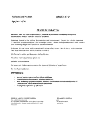
- 1. Name: Rekha Pradhan Date2075-07-24 Age/Sex: 36/M CT SCAN OF KUB/CT IVU Multislice plain and contrast enhanced CT scan of KUB performed followed by multiplanar reformations. Delayed scans are obtained for CT IVU. Rt Kidney: Normal in size, outline, density and cortical enhancement. There is tiny calculus measuring 2.5 mm seen in the midpole pole calyx of the right kidney. There is mild hydronephrosis is seen. There is mild thickening of right renal pelvis wall with enhancement. Lt Kidney: Normal in size, outline, density and cortical enhancement. No calculus or hydronephrosis. Two separate ureter seen uniting proximal to the VUJ. UB: Normal in outline and distension. No focal lesion. Visualized liver, GB, pancreas, spleen and Prostate is unremarkable. No bowel wall thickening or mass seen. No abnormal dilatation of bowel loops. No free fluid in abdomen. IMPRESSION: - Normal contrast excretion from bilateral kidneys. - Tiny right nephrolithiasis with mild hydronephrosis - Mild thickening of right renal pelvis wall with enhancement likely due to pyelitis/UTI. Correlate with clinical and laboratory parameter. - Incomplete duplication of left ureter PROF DR UMESH KUMAR SHARMA DR AABESH KOIRALA MD, RADIODIAGNOSIS (AIIMS) MBBS FELLOWSHIP IN INTERVENTIONAL RADIOLOGY (SOUTH KOREA) MD, RADIODIAGNOSIS NEURORADIOLOGY TRAINING IN UW MEDICAL CENTER (USA) NMC#12190 NMC#2023