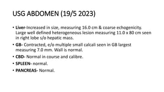
This case involves a 29-year-old female teacher presenting with right upper quadrant pain for 3 months and jaundice for 20 days. Investigations revealed elevated liver enzymes and bilirubin. Imaging showed a large hepatic mass and biopsies indicated metastatic adenocarcinoma. The second case is a 61-year-old female with recurrent abdominal pain and vomiting over 4 months. Imaging found an enlarged liver with a large heterogeneous mass. Biopsies were inconclusive but imaging suggested possible gallbladder carcinoma with metastases. Both patients underwent further endoscopic and imaging workup to characterize the liver lesions and evaluate for obstruction.
A 50-year-old female presented with a 10-day history of pain in the lower right abdomen. Examination revealed an 8x10cm mass in the right iliac fossa. Imaging showed an appendicular mass and abscess. The mass did not resolve with conservative treatment over 15 days. Exploratory laparotomy found a mucinous adenocarcinoma of the appendix that had spread. A right hemicolectomy was performed. Histopathology confirmed a well-differentiated mucin-producing adenocarcinoma with lymph node metastases. Appendiceal adenocarcinoma is a rare and often delayed diagnosis that requires surgical resection.
1) The 80-year-old male patient presented with 5 months of abdominal pain, weight loss, and recent vomiting. Imaging showed a pancreatic mass encasing the splenic artery and gastric outlet obstruction.
2) Laboratory tests showed elevated tumor markers consistent with a probable malignant pancreatic mass.
3) The pre-op diagnosis is a pancreatic mass likely malignant causing gastric outlet obstruction. The proposed surgical plan is a palliative double bypass surgery including gastrojejunostomy, jejunostomy, and cholecystojejunostomy.
This document discusses the case of a 55-year-old male patient who underwent surgery for small bowel stricture suspected to be lymphoma. The patient had a 10-year history of right lower abdominal pain and recent weight loss. Imaging and biopsy results were presented from the case. The surgical specimen showed diffuse ganglioneuromatosis of the intestine, a rare condition characterized by proliferation of nerve cells and fibers in the gastrointestinal tract.
This document discusses the role of imaging in evaluating patients presenting with an acute abdomen. It begins by defining an acute abdomen and describing the nonspecific clinical presentation. Potential causes are categorized as self-limiting, life-threatening, or surgical vs. nonsurgical. The role of imaging is to help determine if surgery is needed and to narrow the differential diagnosis. Imaging modalities discussed include plain radiography, ultrasound, CT scan, and others. The document then reviews how different modalities can help evaluate for specific common acute abdominal conditions like appendicitis, cholecystitis, and diverticulitis. It also describes signs to screen for on imaging like free air, free fluid, bowel wall thickening, and ileus.
The document discusses a case of a 40-year-old woman with peritoneal tuberculosis who presented with abdominal distension for 3 weeks and abdominal pain for 2 weeks. Her medical history included a previous diagnosis of pulmonary tuberculosis. The case presentation provides details of her symptoms, physical exam findings, lab and imaging results, and proposed treatment plan.
NAME- REG NO- STATE OF REGISTRATION: MOBILE NO- TRANSACTION ID- đJUST COPY &...AruneshVenkataraman
Â
1. Mr. Nagappan, a 42-year-old male, presented with abdominal pain and was found to have an infiltrating adenocarcinoma in his rectum.
2. Imaging found a large tumor involving the rectum and sigmoid colon. He underwent neoadjuvant chemoradiation.
3. Follow up imaging after treatment showed the tumor remained infiltrating the bowel wall and surrounding structures, with enlarged lymph nodes. Further PET scan was advised to evaluate for surgery.
This 55-year-old male presented with melena, weakness, weight loss, and anemia over the past year. Endoscopy revealed a polypoidal stomach mass, and biopsy showed a gastrointestinal stromal tumor (GIST). CT scan found a 5.7x6.8 cm stomach wall mass without metastases. He underwent a sleeve gastrectomy surgery to remove the tumor. Histopathology of the tumor was consistent with a GIST, and he was referred for adjuvant imatinib therapy due to risk of recurrence or metastases.
A 41-year-old woman presented with a large abdominal mass. She had a history of fibroid uterus and total abdominal hysterectomy 6 years prior. On examination, a huge solid mass measuring 30x40cm was found in the upper abdomen. Imaging studies showed a large pelvic mass likely originating from the ovaries, with metastases to the liver and lungs. Biopsy was non-diagnostic. She was planned for staging laparotomy to further evaluate the ovarian mass and metastases.
(1) The patient is a 75-year-old female who presented with left lower quadrant pain, decreased bowel movements, weight loss, and recent vomiting of coffee ground material. (2) Physical exam revealed abdominal tenderness and a palpable mass in the left lower quadrant. (3) Imaging showed features suggestive of a colorectal mass. (4) During surgery, a rectosigmoid mass was found involving nearby structures, consistent with stage IV rectal cancer. (5) The patient underwent a Hartmann's procedure and ileostomy for palliation of bowel obstruction from metastatic rectal cancer.
A 4-year-old male presented with a 2-month history of abdominal distension and pain. Physical examination revealed an ill-looking child with abdominal distension and fluid thrill. Ultrasound showed features of veno-occlusive disease including hepatomegaly and ascites. Further evaluation with CT scan was advised to assess for Budd-Chiari syndrome, which is hepatic venous outflow tract obstruction of the hepatic veins, inferior vena cava, or both.
1) The document discusses various imaging modalities used to diagnose conditions that present with acute abdomen such as abdominal pain, including plain radiography, ultrasound, CT, and MRI.
2) Common causes of acute abdomen discussed include appendicitis, diverticulitis, cholecystitis, small bowel obstruction, mesenteric lymphadenitis, epiploic appendagitis, urolithiasis, ruptured aneurysm, and acute pancreatitis.
3) Imaging findings for diagnosing these conditions are provided, with ultrasound and CT noted as important first-line tests to identify the cause of acute abdomen and exclude serious complications.
This document provides an overview of pancreatic pathology, focusing on pancreatitis. It describes the anatomy of the pancreas and imaging modalities used to evaluate pancreatic conditions. Acute and chronic pancreatitis are discussed in detail, including causes, classification, imaging features, and complications like fluid collections, pseudocysts, abscesses, necrosis, and hemorrhage. Other pancreatic conditions summarized include autoimmune pancreatitis and hereditary pancreatitis.
A 40-year-old male presented with abdominal pain and was found to have an epigastric mass. Differential diagnoses included pancreatic cancer, but imaging revealed a pancreatic pseudocyst. Pancreatic pseudocysts develop due to pancreatic duct disruption from acute or chronic pancreatitis. They can be managed conservatively but often require drainage if causing symptoms. The patient underwent cystogastrostomy to drain the pseudocyst.
This document presents an oncology audit from 2071-2072 at KMCTH. It summarizes key findings including patient demographics, prevalence of different cancer types, management approaches, and outcomes. The most common cancers were hepatobiliary, colorectal, pancreatic/periampullary, and stomach. Surgical management was provided for many cases. Mortality causes included postoperative complications, metastatic disease, and refusal of further recommended treatment. The audit aims to understand cancer trends and guide quality improvement efforts.
A 73-year-old male farmer presented with gross hematuria for 1 month and a history of bladder cancer 6 years ago. Physical examination and tests found an irregular mass in the right side of the bladder and a shrunken right kidney. The patient underwent surgery to remove the right kidney, ureter, and bladder along with the prostate and create a left ureterostomy. Post-operative recovery was uneventful. Final pathology revealed high-grade papillary urothelial carcinoma involving the right ureter and bladder, with one positive lymph node. The patient was referred to oncology for further management.
Acute pancreatitis is an inflammatory process of the pancreas with varying involvement of surrounding tissues. Ultrasound typically shows an enlarged, hypoechoic pancreas with blurred margins due to edema. CT shows pancreatic enlargement with heterogeneous enhancement and infiltration of surrounding fat planes. Complications include pancreatic pseudocysts and fluid collections, as well as vascular complications. The document provides details on the diagnostic evaluation, treatment, and prognosis of acute pancreatitis.
This document presents the case of a 58-year-old female who presented with rectal bleeding and altered bowel habits for 5 months. Examination revealed pallor and a rectal mass. Investigations confirmed moderately differentiated adenocarcinoma of the rectum. Imaging showed the primary tumor in the rectosigmoid junction and suspicious lymph nodes. The patient underwent a laparoscopic low anterior resection with diverting ileostomy.
Pancreatitis -a detailed study ( medical information )martinshaji
Â
Pancreatitis is the Inflammation of the pancreatic parenchyma. Acute condition of diffuse pancreatic inflammation & auto digestion, presents with abdominal pain, and is usually associated with raised pancreatic enzyme levels in the blood &urine. this is a detailed study pancreatitis describing factors such as definition , epidemiology , etiology , pathophysiology , treatment , prevention , imaging techniques , diagnosis , lab investigations , images , drugs , control etc
please comment
thank u
- A 13-year-old female presented with epigastric pain and vomiting for the past 2 years. Imaging showed a cystic area in the common bile duct. MRCP revealed a well-defined cystic lesion with calculi, suggestive of a choledochal cyst type 2.
- The patient underwent excision of the choledochal cyst with Roux-en-Y loop formation. Post-op recovery was uneventful.
- Choledochal cysts are rare congenital dilations of the biliary tree that are more common in females. Complete surgical excision is the recommended treatment to prevent complications like cholangiocarcinoma.
This tumor conference case discusses a 75-year-old female patient who presented with abdominal pain and fever. Imaging found a pancreatic tail mass, as well as lesions in both kidneys suspected to be metastases. She underwent a distal pancreatectomy with splenectomy. Pathology revealed a 6.1cm neuroendocrine tumor of the pancreas, grade 2. Staging was stage II. Surveillance with follow up CT was recommended for curative intent.
This tumor board conference discussed the case of a 70-year-old male who presented with gradual abdominal distention. Imaging and biopsy revealed a large intra-abdominal mass arising from appendiceal mucinous neoplasm. The conference objectives were to discuss the patient's presentation, workup, and treatment options for appendiceal mucinous neoplasm. Key points included the classification, clinical presentation and risk stratification of patients for cytoreductive surgery and hyperthermic intraperitoneal chemotherapy, which can provide benefits for select patients with appendiceal mucinous neoplasms and peritoneal surface malignancies.
Presentation1.pptx, ultrasound examination of the liver and gall bladder.Abdellah Nazeer
Â
This document provides an ultrasound protocol and guidelines for examining the liver and gallbladder. It begins with an overview of the role and technique of ultrasound for the liver, including scanning positions and images to capture. Common liver pathologies such as fatty liver, cirrhosis, cysts, hemangiomas, abscesses, and metastases are described. Guidelines are provided for gallbladder ultrasound including patient preparation, technique, and anatomy. Normal findings and pathologies like stones, acute cholecystitis, and emphysematous cholecystitis are outlined. The document concludes with potential vascular disorders of the liver involving blood flow.
This document summarizes the case of a 72-year-old female patient presenting with multiple abdominal swellings for 6 years. On examination, she was found to have incisional hernias at her hysterectomy scar site, with the largest hernia containing the caecum. She underwent an open incisional hernia repair with mesh placement. Post-operatively, her condition was stable and she was discharged on a diabetic diet.
The document provides a summary of the differential diagnosis and key details about various conditions that could explain the patient's liver mass. The main differentials discussed include hemangioma, hamartoma, focal nodular hyperplasia, hepatoblastoma, hepatocellular carcinoma, undifferentiated embryonal sarcoma, liver abscess (pyogenic and amebic), eosinophilic liver infiltrations like DRESS syndrome, hypereosinophilic syndrome, parasites such as echinococcosis, toxocaria and fascioliasis. Physical exam findings, investigations and imaging characteristics of each condition are outlined. Based on the information provided, the patient's case does not fully match the features of any one
How to Setup Default Value for a Field in Odoo 17Celine George
Â
In Odoo, we can set a default value for a field during the creation of a record for a model. We have many methods in odoo for setting a default value to the field.
A Free 200-Page eBook ~ Brain and Mind Exercise.pptxOH TEIK BIN
Â
(A Free eBook comprising 3 Sets of Presentation of a selection of Puzzles, Brain Teasers and Thinking Problems to exercise both the mind and the Right and Left Brain. To help keep the mind and brain fit and healthy. Good for both the young and old alike.
Answers are given for all the puzzles and problems.)
With Metta,
Bro. Oh Teik Bin đđ¤đ¤đĽ°
Gender and Mental Health - Counselling and Family Therapy Applications and In...PsychoTech Services
Â
A proprietary approach developed by bringing together the best of learning theories from Psychology, design principles from the world of visualization, and pedagogical methods from over a decade of training experience, that enables you to: Learn better, faster!
Level 3 NCEA - NZ: A Nation In the Making 1872 - 1900 SML.pptHenry Hollis
Â
The History of NZ 1870-1900.
Making of a Nation.
From the NZ Wars to Liberals,
Richard Seddon, George Grey,
Social Laboratory, New Zealand,
Confiscations, Kotahitanga, Kingitanga, Parliament, Suffrage, Repudiation, Economic Change, Agriculture, Gold Mining, Timber, Flax, Sheep, Dairying,
THE SACRIFICE HOW PRO-PALESTINE PROTESTS STUDENTS ARE SACRIFICING TO CHANGE T...indexPub
Â
The recent surge in pro-Palestine student activism has prompted significant responses from universities, ranging from negotiations and divestment commitments to increased transparency about investments in companies supporting the war on Gaza. This activism has led to the cessation of student encampments but also highlighted the substantial sacrifices made by students, including academic disruptions and personal risks. The primary drivers of these protests are poor university administration, lack of transparency, and inadequate communication between officials and students. This study examines the profound emotional, psychological, and professional impacts on students engaged in pro-Palestine protests, focusing on Generation Z's (Gen-Z) activism dynamics. This paper explores the significant sacrifices made by these students and even the professors supporting the pro-Palestine movement, with a focus on recent global movements. Through an in-depth analysis of printed and electronic media, the study examines the impacts of these sacrifices on the academic and personal lives of those involved. The paper highlights examples from various universities, demonstrating student activism's long-term and short-term effects, including disciplinary actions, social backlash, and career implications. The researchers also explore the broader implications of student sacrifices. The findings reveal that these sacrifices are driven by a profound commitment to justice and human rights, and are influenced by the increasing availability of information, peer interactions, and personal convictions. The study also discusses the broader implications of this activism, comparing it to historical precedents and assessing its potential to influence policy and public opinion. The emotional and psychological toll on student activists is significant, but their sense of purpose and community support mitigates some of these challenges. However, the researchers call for acknowledging the broader Impact of these sacrifices on the future global movement of FreePalestine.
CapTechTalks Webinar Slides June 2024 Donovan Wright.pptxCapitolTechU
Â
Slides from a Capitol Technology University webinar held June 20, 2024. The webinar featured Dr. Donovan Wright, presenting on the Department of Defense Digital Transformation.
Temple of Asclepius in Thrace. Excavation resultsKrassimira Luka
Â
The temple and the sanctuary around were dedicated to Asklepios Zmidrenus. This name has been known since 1875 when an inscription dedicated to him was discovered in Rome. The inscription is dated in 227 AD and was left by soldiers originating from the city of Philippopolis (modern Plovdiv).
3. CASE 1
⢠29/F GORAKHPUR
⢠No comorbidity ,
⢠Teacher
ďąPresentation :
⢠Right upper quadrant pain 3months
⢠Yellowish discolouration of eyes and urine
20days
⢠Clay coloured stool + Vomiting+ , loss of
appetite , LOW+
⢠No fever/past surgeries/high risk
behaviour/
⢠No family h/o similar complaints or
hepatic/biliary disease
ďąExamination
⢠HT 150 cm Wt 44 kg
⢠BMI 19.5kg/m2
⢠Icterus present
⢠Palpable mass RUQ
epigastrium 10x8cm
⢠Hepatomegaly+
⢠Other systems normal
4. Investigations
LFT 15/06/20
23
16/07/23 22/07/2023
T. Bil 2.8 16.4 10.4
D. Bil 0.3 11.5 6.1
T.Protein 7.27 7.1 7.2
Albumin 4.7 4.2 4.1
SGOT 195 200 69
SGPT/GGT 289/230 72 60
ALP 250 343 244
INR 1.1 1.0 1.1
Ca 19.9 >10000u/ml
CEA 939 ng/ml
Ca 125 30u/ml
HBsAg/HIV/AntiHC
V
Neg
AFP 2.4
CBC 16/7/23 22/07/23
HGB 8.3 8.2
TLC 39.2 31.2
PLT 195 264
RFT Normal
13. USG ABDOMEN
⢠Liver-is enlarged in size (16.2 cm) with e/o large heteroechoic
mass lesion seen, measuring approx. 12.8x9.8cm involving liver
parenchyma. The lesion shows vascularity on colour doppler.
Portal vein is poorly visualized.
⢠Gall bladder- fully distended. No calculus in lumen Wall thickness
is normal.
⢠CBD- normal.
⢠Pancreas in normal in thickness. Clearly defined margins are seen.
Pancreatic duct is not dilated.
⢠Spleen is normal in size (9.6 cm). No focal lesion is seen.
14. CRNO: 2023482396 29/F
⢠CHIEF COMPLAINS: Pain right upper abdomen and epigastrium- 3
months.
⢠Jaundice- 15 days
15. IgG 1620
ESR 108
FLUID GLUCOSE 116
BUN 41.40
S. Creatinine 2.26
S.Chloride 117
S. Phosphorous 4.78
16. HISTORY OF PRESENT ILLNESS
⢠Pain in RHC which was insidious in onset, dull aching in nature, mild
to moderate intensity, radiating to back, no aggravating factors,
releaved after taking oral analgesics.
⢠It was associated with nausea and vomitting occasional every 4-5 days
gastric content 50- 100 ml, h/o post prandial fullness.
⢠H/o jaundice since 15 days insidious onset ,progressively increasing in
intensity, associated with high colored urine and clay colored stool.
17. NEGATIVE HISTORY
⢠No H/O fever
⢠No H/o hematemesis , malena
⢠No h/o altered bowel habits
⢠No H/s/o fat soluble vitamin deficiency
⢠No h/s/o CLD or hypersplenism
21. EXAMINATION
⢠Patient is conscious / oriented
⢠ECOG - 0,METS > 4, KPS 90
⢠HT 150 cm Wt 44 kg. BMI 19.55 BHT 35sec
⢠Icterus present
⢠No pallor/ cyanosis/ pedal odema
⢠Afebrile.
⢠PR - 80/min.
⢠BP - 120/80mmHg.
22. ⢠Chest : BLAE present
⢠PA : Umbilicus central inverted, no scar/ sinus.
Tender RHC on deep palpation,10 x8 cm lump palpable in
epigastrium, moving with respiration, dull on percussion, palpable
hepatomegaly present.
23. USG ABDOMEN (19/5 2023)
⢠Liver-Increased in size, measuring 16.0 cm & coarse echogenicity.
Large well defined heterogeneous lesion measuring 11.0 x 80 cm seen
in right lobe s/o hepatic mass.
⢠GB- Contracted, e/o multiple small calcali seen in GB largest
measuring 7.0 mm. Wall is normal.
⢠CBD- Normal in course and calibre.
⢠SPLEEN- normal.
⢠PANCREAS- Normal.
24. USG ABDOMEN (21/5 /2023)
⢠Liver-is enlarged in size (16.2 cm) with e/o large hetroechoic mass lesion is
seen, measuring approx. 12.8x9.8cm involving liver parenchyma. The lesion
shows vascularity on colour doppler. Portal vein is poorly visualized.
⢠Gall bladder- fully distended. No calculus in lumen Wall thickness in
normal.
⢠CBD- normal.
⢠Pancreas in normal in thickness. Clearly defined margins are seen.
Pancreatic duct is not dilated.
⢠Spleen is normal in size (9.6 cm). No focal lesion is seen. Diaphragmatic
movements are within normal limits on both sides.
25. CECT ABDOMEN ( 20/05/23)
⢠Liver is enlarged in size (16.0 cm) and shows a large heterogeneous
lesion with predominant peripheral enhancement on portovenous
phase. Lesion in predominantly located in segment 4. and 8. It is
causing mass effect is form of posterior displacement and splaying of
main portal branches.
⢠A similar smaller lesion of size-32 x 27 mm is noted in porto-caval
region.
⢠Gall bladder- is contracted and shows subtle hyperdensity-
?Cholelithiasis. CBD is not dilated
⢠Pancreas - normal
26. CECT ABDOMEN ( 21 /5/23)
⢠Liver is enlarged in size with e/o large peripheral arterial enhancing
mass, measuring approx 14x10x8.5cm predominantly involving right
lobe of liver. The lesion show increased enhancement during
photovenous and delayed phase with suggestion of centripetal filling
however no obvious discontinuous peripheral enhancement can be
made out.
⢠Mass effect over portal with compression of effacement of right
branch of the portal vein. minimal fluid is seen in pelvic cavity.
⢠Small similar focal lesion are also noted in portocaval region
extending into caudate lobe giant hemangioma /?? FNH / ??
adenoma
27. TPCT Abdomen-7/7/23
⢠Relatively well defined large (- 18.8 x 14.9 x 16.4 cm) with irregular
heterogenously enhancing lesion is seen in both lobes of liver, causing mass
effect over hila with no separate visualization of the gall bladder. Mass is
involving CHD and causing upstream bilobar IHBRD. Primary confluence is
just formed. Mass effect is seen in the form of displacement of adjacent
bowel loops and abdominal viscera, with maintained fat planes.
⢠BD is not visualised and is compressed by necrotic enlarged nodes.
Multiple variable sized arterially enhancing lesion is seen in the body and
tail region of the pancreas ? metastasis ??Less likely neuro endocrine
tumour (~12X14mm). MPD is diffusely dilated ~ 3.4 mm .No obvious
peripancreatic fat stranding/collections noted. Multiple heterogenously
enhancing lymph nodes are seen in pre and paraaortic, aortocaval, &
paracaval and periportal region, largest SAD ~ 13 mm. Mild ascites is seen.
28. ⢠IMPRESSION:
⢠Large well defined heterogeneously enhancing liver lesion with no
separate visualization of gall bladder....? Primary gall bladder
carcinoma. Multiple pancreatic and nodal metastasis Well defined
cystic lesion in right adnexa showing dependent pre- contrast
hyperdensity (s/o hemorrhagic content) --- ? Hemorrhagic cyst (adv:
USG correlation).
29. UGIE-10/7/23
⢠Impression: Duodenal ulcer forrest class III with partial GOO
Erythematous mucosa seen in antrum
Multiple D1 and antral biopsies taken
30. ERCP-20/7/23
⢠SVE-Papilla normal. Selective CBD cannulation done. Cholangiogram
done - showed left IHBRD > right. The wire was manipulated to seg III
duct - dilated. Cholangiogram done for confirmation. 10F x 10cm
plastic stent placed in LHD-seg III duct. Drainage of contrast seen.
31. HPE REPORT-11/7/23
⢠DUODENAL BIOPSY: NO E/O villous atrophy or pasarite; gastric antral
biopsy: Helicobactor pylori associated active gastritis.