Download to read offline
![International Research Journal of Engineering and Technology (IRJET) e-ISSN: 2395-0056
Volume: 06 Issue: 03 | Mar 2019 www.irjet.net p-ISSN: 2395-0072
© 2019, IRJET | Impact Factor value: 7.211 | ISO 9001:2008 Certified Journal | Page 5248
• The main idea is to define k centres, one for each
cluster.
• These centers should be placed in a cunning way
because of different location causes different result.
• The next step is to take each point belonging to a
given data set and associate it to the nearest centre.
iii)SVM ALGORITHM:
•” Support Vector Machine” (SVM) is a supervised
machine learning algorithm.
• In this algorithm, we plot each data item as a point in
n-dimensional space.
• where n is number of features with the value of each
feature being the value of a particular coordinate
• Then, we perform classificationbyfindingthehyper-
plane that differentiate the two classes very well.
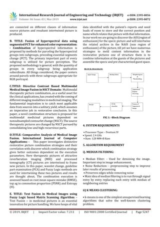
6. METHODOLOGY
• PREPROCESSING: -Preprocessing the input image
• IMAGE ENCHANCEMENT: -by denoising the image
using the algorithm called Median Filter.
•FEATURE EXTRACTION: -Extrac
ting the morphological features by using the k –means
clustering algorithm
• THRESHOLDING: -In addition to thresholding the
extracting image
• SEGMENTATION: -Then segmentation process will
be carried out for further performancetoidentifiedthe
tumor is benign and malignant
• SVM: -Now the Support Vector Machine (SVM)
classifier is used for classification as well as in
regression condition
• SVM classifies the tumor is benign or malignant
7. FUTURE ENHANCEMENT
In future work, it is fascinating to incorporate extra
component data. Other than the vitality, connection,
difference and homogeneity add more data to the
component extraction so as to make the framework
increasingly delicate; data from the surfaces or area. It
will intrigue keep growing increasingly versatile
models for different sorts of cerebrum tumors
followingasimilarprofessionintroducedhere.Another
future line would be the identification of little
threatening mind tumors. It ought to be evident that
numerous variables impact the presence of tumors on
pictures, and in spite of the fact that there are some
regular highlights of malignancies, there is likewise a
lot of variety that relies upon the tissue and the tumor
type. Trademark highlights are bound to be found in
expansive tumors. Little tumors might not have a
significant number of the highlights of danger andmay
even show themselves just by optional impacts, for
example, compositional mutilation.
8. CONCLUSION
Medical image fusion combines different modality of
medical pictures to deliver a high caliber combined
picture with spatial and ghastly data. Thus helps the
doctors and radiologist for cerebral tumor diagnosis.
9. REFERENCES
[1] T. Zaveri, and M. Zaveri, “A Novel Region Based
Multimodality Image Fusion Method”, Journal of
Pattern Recognition Research, vol. 2, pp. 140–153,
2011.
[2] V. Tsagaris, V. Anastassopoulos, and G.
Lampropoulos, “Fusion of hyperspectral data using
segmented PCT for enhanced color representation”,
IEEE Trans. Geosci. Remote Sens., vol. 43, no. 10, pp.
2365–2375, 2005.
[3] G. Bhatnagar, Q. M. J. Wu, and Z. Liu, “Directive
Contrast Based Multimodal Medical Image Fusion in
NSCT Domain. IEEE Transactions on Multimedia, vol.
15, no. 5, pp. 1014–1024, 2013. DOI:
10.1109/TMM.2013.2244870.
[4] A. Rana, and S. Arora, “Comparative Analysis of
Medical Image Fusion. International Journal of
Computer Applications,” vol. 73, no.9,pp.10–13,2013.
DOI: 10.5120/12768-9371.
[5] Pinki Jain, and Anu Aggarwal, “Text Fusion in
Medical Images using Fuzzy Logic based Matrix
Scanning Algorithm,” InternationalJournalofScientific
and Research Publications, vol. 2, no. 6, pp. 1-6, 2012.](https://image.slidesharecdn.com/irjet-v6i31350-191021073630/85/IRJET-Fusion-based-Brain-Tumor-Detection-3-320.jpg)

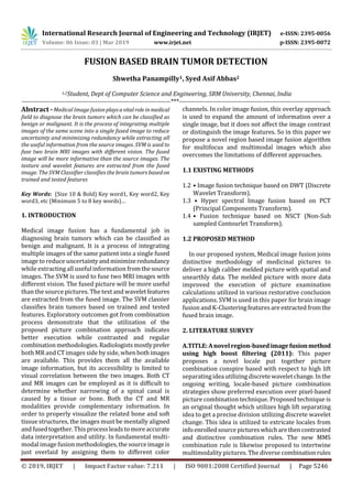
1. The document discusses a method for detecting brain tumors using medical image fusion and support vector machines (SVM). 2. It involves fusing two MRI images using SVM to create a single fused image with more information than the original images. Texture and wavelet features are then extracted from the fused image. 3. The SVM classifier classifies the brain tumors as benign or malignant based on the trained and tested features extracted from the fused image.