Reproductive Sequencing
•Download as PPTX, PDF•
1 like•568 views

The advances likes Next Generation Sequencing is more advanced than Microarray Compatability Genomic hybridization and it is 100% of sensitivity and specificity regarding aneuploidy sequencing from all biological samples.

Recommended
More Related Content
What's hot
What's hot (20)
Viewers also liked
Similar to Reproductive Sequencing
Similar to Reproductive Sequencing (20)
Recently uploaded
Recently uploaded (20)
Reproductive Sequencing
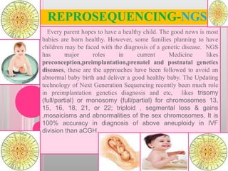
- 1. REPROSEQUENCING-NGS Every parent hopes to have a healthy child. The good news is most babies are born healthy. However, some families planning to have children may be faced with the diagnosis of a genetic disease. NGS has major roles in current Medicine likes preconception,preimplantation,prenatel and postnatal genetics diseases, these are the approaches have been followed to avoid an abnormal baby birth and deliver a good healthy baby. The Updating technology of Next Generation Sequencing recently been much role in preimplantation genetics diagnosis and etc, likes trisomy (full/partial) or monosomy (full/partial) for chromosomes 13, 15, 16, 18, 21, or 22; triploid , segmental loss & gains ,mosaicisms and abnormalities of the sex chromosomes. It is 100% accuracy in diagnosis of above aneuploidy in IVF division than aCGH.1
- 2. 2 Humans normally have 46 chromosomes in each cell, divided into 23 pairs. Two copies of chromosome 46, one copy inherited from each parent, form one of the pairs. X chromosomes has more than 150 millions base pairs with 1400 genes and Y chromosomes has 50millions base pairs with 200 genes ( 46XY). Sexual region of Y is responsible for testis determination. Nearly 30000 genes rolling for genes expression in a human being. FISH Normal Male Whole Genome Sequence
- 3. Normal Female Whole Genome Sequence 3 Humans normally have 46 chromosomes in each cell, divided into 23 pairs. Two copies of chromosome 46, one copy inherited from each parent, form one of the pairs. X chromosomes has more than 150 millions base pairs with 1400 genes and other X chromosomes has an inactivated while embryogenesis( 46XX). Most of genetics disorder from maternally an originated. Nearly 30000 genes rolling for genes expression in a cell. FISH
- 4. Chromosome Abnormalities In Human Pregnancies (IVF) 4 Ploidy: Normal chromosomes in a cell Euploidy: Embryo genetics normal Anuploidy: Embryo genetics abnormal. Ex: Trisomy, Tetrasomy, Monosomy, & Partial Trisomy-Monosomy. Segmental Gain & Loss. Polyploidy: Two more sets of chromosome than normal sets.
- 5. Cell division error on Genes 5 Meiosis is defined as process of producing haploid. Gametes are eggs and sperm. Meiosis cell division occur only in spermatogonia and Oogonia. Haploid is defined as single sets of chromosomes (1N) &diploid (2N). Cell division occur twice but with only one replication. Shuffling of genetics materials from both parents produce variety and diversity.
- 6. 6
- 7. Chromosome:1Trisomy-WGS 7 First largest auto chromosome in Humans. It has over 3000 genes for different genes coding with 249 millions base pairs(8% of total DNA of cell). Nearly 890 diseases and disorders are associated with chromosome 1 abnormality. These includes several cancers as well as brain degenerative disorders such as Parkinson's disease and Alzheimer's disease. Other conditions including glaucoma and deafness are also associated with chromosome 1 abnormalities. Trisomy of chromosome 1 is very rare. Source -htt://onlinelibrary.wiley.com/doi/10.1002/yea.955/pdf-web.udles www.ncbi.nlm.nih,gog
- 8. Chromosome:2 Trisomy-WGS 8 Chromosome 2 likely contains 2500 genes with 245 million base pairs that provide instructions for making proteins. These proteins perform a variety of different roles in the body. Changes in the structure or number of copies of a chromosome can also cause problems with health and development. The 1q37 deletion syndrome & Trisomy 2 found in myelodysplastic syndrome. Translocation between 2&3. ring chromosome 2, partial trisomy 2, partial monosomy 2 & etc. Source: National Human Genome research institute: Chromosome abnormalities
- 9. Chromosome:3 Trisomy-WGS 9 Chromosome 3 spans about 200 million base pairs with 1900 genes (the building blocks of DNA) and represents approximately 6.5 percent of the total DNA in cells. Many genetic conditions are related to changes in particular genes on chromosome 3. Source: National Human Genome research institute: Chromosome abnormalities
- 10. Chromosome:4 Tri &Monosomy-WGS 10 Chromosome 4 cover about 191 million DNA building blocks (base pairs) with 1600 genes and represents more than 6 percent of the total DNA in cells. Wolf-Hirschhorn syndrome is caused by a deletion of gene at the end of the short (p) arm of chromosome 4 at a position described as 4p16.3. PDGFRA-associated chronic eosinophilic leukemia is caused by genetic abnormalities that involve the PDGFRA gene on chromosome 4. Facioscapulohumeral muscular dystrophy is caused by genetic changes involving the long (q) arm of chromosome 4. Mosaic trisomy 4 is very rare; only a few cases have been reported. Trisomy 4 occurs when cells have three copies of chromosome 4 instead of the usual two copies. https://ghr.nlm.nih.gov/chromosome/4.
- 11. Chromosome:5 Trisomy-WGS 11 Chromosome 5 spans about 181 million DNA building blocks (base pairs) with 1700 genes and represents almost 6 percent of the total DNA in cells. Deletion of a region of DNA from the long (q) arm of chromosome 5 is involved in a condition called 5q minus (5q-) syndrome or myelodysplastic syndrome (MDS). Affected individuals also have an increased risk of developing a fast-growing blood cancer known as acute myeloid leukemia (AML). Cri-du-chat (cat's cry) syndrome is caused by a deletion of the end of the short (p) arm of chromosome 5p15.3. Several regions of chromosome 5 have been associated with the risk of developing Crohn disease((5q31). partial trisomy 5p or 5q and partial monosomy may causes different genetics disorder. continue
- 12. Cri Du Chat Syndrome 5P Del 12 Symptoms are includes small head, jaw, round face ,Eyes are very fare apart, folds of skin over their eyes and small nose bridge. The geneticist jerome legume identified cri-du-chart syndrome in 1963.He also discovered the genetics abnormality that causes down syndrome. In 80% cri-du-chart causes, the chromosomes caring the deletion come from father sperms rather than mother eggs. If pairs of chromosome don’t line up correctly during metaphase in meiosis the structure of a chromosome can be changed. When this happen With chromosome 5.it causes cri-du chart and it is commonest syndrome which is caused by deletion.
- 13. Chromosome:6 Trisomy-WGS 13 Chromosome 6 spans about 171 million DNA building blocks (base pairs) and represents between 5.5 and 6 percent of the total DNA in cells. Chromosome 6 likely contains 1,000 to 1,100 genes that provide instructions for making proteins. These proteins perform a variety of different roles in the body. The 6q24-related transient neonatal diabetes mellitus, a type of diabetes that occurs in infants, is caused by the over activity (over expression) of certain genes in a region of the long (q) arm of chromosome 6 called 6q24. Many genetic conditions are related to changes in particular genes on chromosome 6.
- 14. Chromosome:7 Trisomy & 12th Mosaicism 14 Chromosome 7 spans about 159 million DNA building blocks (base pairs) and represents more than 5 percent of the total DNA in cells. Chromosome 7 likely contains 900 to 1,000 genes that provide instructions for making proteins. These proteins perform a variety of different roles in the body. Changes in the number or structure of chromosome 7 occur frequently in human cancers. People inherit both copies of chromosome 7 from their mother instead of one copy from each parent. This phenomenon is called maternal uniparental disomy (UPD). Williams syndrome is caused by the deletion of genetic material from the long (q) arm of chromosome 7. Russell-Silver syndrome. continue
- 15. Williams Syndrome 7q del WS: is an autosomal dominant disorder caused by a deletion of about 26 genes from q arm of 7th chromosome. It is estimated about 1:7500-10000. Sign: developmental delay & highly verbal and social, tend to love music, mild to moderate mental retardation. Neonatal hypercalcimia and most of systems mildly disorder. 15
- 16. Chromosome:8 Trisomy-WGS 16 Chromosome 8 spans more than 146 million DNA building blocks (base pairs) contains about 700 genes and represents between 4.5 and 5 percent of the total DNA in cells. Translocations of genetic material between chromosome 8 and other chromosomes can cause 8p11 myelo- proliferative syndrome. The most common translocation involved in this condition, written as t(8;13)(p11;q12), fuses part of the FGFR1 gene on chromosome 8 with part of the ZMYM2 gene on chromosome 13. The translocations are found only in cancer cells. Trisomy 8 occurs when cells have three copies of chromosome 8 instead of the usual two copies. Langer-Giedion syndrome and recombinant 8 syndrome.
- 17. Chromosome:9 Trisomy-WGS 17 Chromosome 9 is made up of about 141 million DNA building blocks (base pairs) contains 800 to 900 genes and represents approximately 4.5 percent of the total DNA in cells. The Philadelphia chromosome has been identified in most cases of a slowly progressing form of blood cancer called chronic myeloid leukemia (CML). (partial trisomy &partial monosomy are not common. The 9q22.3 micro deletion, bladder cancer kleefstra syndrome are most offend disorder.
- 18. Chromosome:10 Trisomy-WGS 18 Chromosome 10 spans more than 135 million DNA building blocks (base pairs) contains 700 to 800 genes and represents between 4 and 4.5 percent of the total DNA in cells. Changes in the number and structure of chromosome 10 are associated with several types of cancer. Variations in a particular region of chromosome 10 have been associated with the risk of developing Crohn disease(10q21.1). A complex rearrangement (translocation) of genetic material between chromosomes 10 and 11 is associated with several types of blood cancer known as leukemia (MLL gene).
- 19. Chromosome:11 Monosomy-WGS 19 Chromosome 11 spans about 135 million DNA building blocks (base pairs) contains 1,300 to 1,400 genes and represents between 4 and 4.5 percent of the total DNA in cells. Beckwith-Wiedemann syndrome results from the abnormal regulation of genes on part of the short (p) arm of chromosome 11. The genes are located close together in a region designated 11p15.5 near one end of the chromosome. Emanuel syndrome is caused by the presence of extra genetic material from chromosome 11 and chromosome 22 in each cell. WAGR syndrome (11p13 del). Russell-Silver syndrome (11p15.5 region). Potocki-Shaffer syndrome (11p11.2). Jacobsen syndrome (11q terminal deletion disorder) are common.
- 20. Chromosome:12 Trisomy-WGS 20 Chromosome 12 spans almost 134 million DNA building blocks (base pairs) contains 1,100 to 1,200 genes and represents between 4 and 4.5 percent of the total DNA in cells. Changes in chromosome 12 have been identified in several types of cancer. Pallister-Killian mosaic syndrome is usually caused by the presence of an abnormal extra chromosome called an iso-chromosome 12p or I (12p). Translocations involving chromosome 12 are involved in a type of blood cell cancer called PDGFRB-associated chronic eosinophilic leukemia. Several different changes involving chromosome 12 have been reported.
- 21. Chromosome:13 Monosomy-WGS 21 Chromosome 13 is made up of about 115 million DNA building blocks (base pairs) contains 300 to 400 genes and represents between 3.5 and 4 percent of the total DNA in cells. The most of the abnormality have been observed in the 8p11myeloproliferative syndrome,retinoplastoma and trisomy 13. Patau syndrome Fusion of the eyes or a small or absent eye-Cleft lip and palate Extra fingers and toes Mental retardation
- 22. Chromosome:14 & 16,18,20&21Trisomy 22 Chromosome 14 spans more than 107 million DNA building blocks (base pairs) contains 800 to 900 genes and represents about 3.5 percent of the total DNA in cells. Rearrangements (translocations) of genetic material between chromosome 14 and other chromosomes have been associated with several types of cancer. The FOXGI syndrome and ring chromosome 14 were observed.
- 23. Chromosomal Translocations - Translocations In a reciprocal translocation two broken off chromosome pieces of non-homologous chromosomes are exchanged. This is a relatively frequent anomaly. One finds it with an incidence of 1:500 newborns. Reciprocal translocations are frequently balanced because the entire genetic material is present. Problems occur, though, in gamete formation. Another frequently observed anomaly (1:1'000 newborns) is the robertsonian translocation, which occurs between two acrocentric chromosomes of groups G and D. It is also referred to as the centric fusion of two acrocentric chromosomes. It is a special kind of translocation in that on the acrocentric chromosomes (most often chromosomes 14 and 21 or 22) the very short, satellite-bearing arm is lost and a centric fusion t(14q21q or 14q22q) of the two remainder chromosomes, i.e., the long arms of the two pieces, results.
- 24. Chromosome:15 ,5& 22Trisomy-WGS 24 Chromosome 15 spans more than 102 million DNA building blocks (base pairs) contains 600 to 700 genes and represents more than 3 percent of the total DNA in cells. The chromosomal changes are observed in the 15q13.3 &15q24microdeletion,acute promylocytic leukemia,angelman syndrome & Prader-willi syndrome.Sensorineural deafness and male infertility.
- 25. Chromosome:16& 22 Trisomy-16-22Monosomy 25 Chromosome 16 spans more than 90 million DNA building blocks (base pairs) contains 800 to 900 genes and represents almost 3 percent of the total DNA in cells. Alveolar capillary dysplasia with misalignment of pulmonary veins (ACD/MPV) is a disorder that affects the development of blood vessels in the lungs (16q24.1).Rubinstein-Taybi syndrome. Trisomy 16 occurs when cells have three copies of chromosome 16 instead of the usual two copies. The 16p11.2 deletion and duplication are observed.
- 26. Clinical details of 16th chromosomes 26
- 27. Chromosome:17 Monosomy-WGS 27 Chromosome 17 spans about 81 million DNA building blocks (base pairs) contains 1,200 to 1,300 genes and represents between 2.5 and 3 percent of the total DNA in cells. Changes in chromosome 17 have been identified in several additional types of human cancer.
- 28. Chromosome:18 Trisomy-WGS 28 Chromosome 18 spans about 78 million DNA building blocks (base pairs) contains 200 to 300 genes and represents approximately 2.5 percent of the total DNA in cell. Partial Trisomy & Monosomy of chromosome 18p (18p-q) & deletion and translocation are analyzed in different population. Edward Syndrome. Severe physical and mental disabilities/Development stops at the 6 month level/Oddly clenched fists-Liver and heart problems Low-set ears-Small mouth/Unusual or absent fingerprints
- 29. Chromosome:19 Trisomy-WGS 29 Chromosome 19 spans about 59 million base pairs (the building blocks of DNA) contains about 1,500 genes and represents almost 2 percent of the total DNA in cell. Source: National Human Genome Research Institute &Chromosomal Abnormalities.
- 30. Chromosome:20&21 Trisomy-WGS 30 Chromosome 20 spans about 63 million DNA building blocks (base pairs) contains 500 to 600 genes and represents approximately 2 percent of the total DNA in cell. Changes in chromosome 20 have been identified in several types of cancer. Ring chromosome 20 & alagille are related disorder.
- 31. Chromosome:21 Trisomy-WGS 31 Chromosome 21 is the smallest human chromosome, spanning about 48 million base pairs (the building blocks of DNA) contains 200 to 300 genes and representing 1.5 to 2 percent of the total DNA in cells. A genetic rearrangement (translocation) involving chromosome 21 is associated with a type of blood cancer known as core binding factor acute myeloid leukemia (CBF-AML).The feature of down syndrome. continue
- 32. 32
- 33. Chromosome:21& 22 Trisomy-WGS 33 Chromosome 22 is the second smallest human chromosome, spanning more than 51 million DNA building blocks (base pairs) contains 500 to 600 genes and representing between 1.5 and 2 percent of the total DNA in cells. Several types of blood cancer known as leukemia are associated with a translocation of genetic material between chromosomes 9 and 22. The Philadelphia chromosome has been identified in most cases of a slowly progressing form of blood cancer called chronic myeloid leukemia (CML). The 22q11.2 deletion syndrome,22q11.2 duplication,22q13.3 deletion syndrome, dermato- fibrosarcoma pro-tuberans, Emanuel syndrome, Ewing sarcoma and Optiz G/BBB syndrome have observed commonly.
- 34. 12th Chromosome Mosaicism-WGS 34 The extra copy is due to an intra chromosomal duplication or an extra copy on a derivative chromosome. Patients with tetrasomy 12p, or Pallister-Killian syndrome (PKS), additionally present with sparse temporal hair, eyebrows, and eyelashes, prominent forehead, a cupid- bow shaped mouth, and large ears. Only about 24 cases with mosaicism for a structural abnormality of an auto some have been reported in the literature.
- 35. Segmental Loss(4th) & Gain(7th)-WGS 35 Wolf-Hirschhorn syndrome is caused by a deletion of genetic material near the end of the short (p) arm of chromosome 4 at a position described as 4p16.3-A specific translocation involving chr4&14 t(4;14)(p16;q32), is found in multiple myeloma(bone cancer). Greig cephalopolysyndactyly syndrome results from a rearrangement (translocation) of genetic material between chromosome 7 and another chromosome.
- 36. Segmental Loss(18q) &Gain(16p)-WGS 36 The 18q deletion syndrome is caused by a deletion of genetic material from the long (q) arm of chromosome 18. This chromosomal change is written as 18q-. The signs and symptoms of 18q deletion syndrome are probably related to the loss of multiple genes in this region. Two types are distal 18q deletion syndrome & proximal 18q deletion syndrome with different clinical features. A 16p11.2 duplication is an extra copy of the same 600 kb segment of chromosome 16 that is missing in 16p11.2 deletion syndrome . The 600 kb region contains more than 25 genes, and in many cases little is known about their function.
- 37. Segmental Gain(13p&19thp)-WGS 37 Translocation of genetic material involving chromosome 13 has been identified in most people with a rare blood cancer called 8p11 myelo-proliferative syndrome(myelo-proliferative disorder) and the development of lymphoma, a blood-related cancer that causes tumor formation in the lymph nodes( t(8;13)(p11;q12). The 19th chromosomes losses might have different clinical disorder.
- 38. Deletion (18q del)-WGS 38 The 8q deletion syndrome is caused by a deletion of genetic material from the long (q) arm of chromosome 18. The18q deletion syndrome is often categorized into two types: individuals with deletions near the end of the long arm of chromosome 18 are said to have distal 18q deletion syndrome, and those with deletions in the part of the long arm near the center of chromosome 18 are said to have proximal 18q deletion syndrome. The sign and symptoms of both deletion: hearing loss and heart abnormalities are more common in people with distal 18q deletion syndrome, while seizures occur more often in people with proximal 18q deletion syndrome.
- 39. Deletion (3p gain-del- +9p-9q)-WGS 39 The 9q22.3 micro-deletion is a chromosomal change in which a small piece of the long (q) arm of chromosome 9 is deleted in each cell. Affected individuals are missing at least 352,000 base pairs, also written as 352 kb, in the q22.3 region of chromosome ,the largest reported deletion included 20.5 million base pairs (20.5 Mb).
- 40. Deletion 7th q & 8th q del-WGS 40 Williams syndrome is caused by the deletion of genetic material from a portion of the long (q) arm of chromosome 7. The deleted region, which is located at position 11.23 (written as 7q11.23), is designated the Williams- Beuren region. This region includes 26 to 28 genes, and researchers believe that the characteristic features of Williams syndrome are probably related to the loss of several of these genes. Langer-Giedion syndrome is caused by a deletion or mutation in several genes on the long (q) arm of chromosome 8 at a position described as 8q24.1.The signs and symptoms of this condition are related to the deletion or mutation in at least two genes from this part of the chromosome.
- 42. Sex Chromosome:45 X Turner Syndrome-WGS 42 continue
- 43. Turner Syndrome 45x Sex chromosomal abnormality Delayed puberty 99% are not born Infertility if born a female baby X is depart because of Mosaicism 43
- 44. Klinefelter Syndrome 46+XXY-Male Sex-chromosome disorder in Male Sexually oriented system underdeveloped (Small testes, sparse facial and pubic hair) Long arms and legs and big feet and hands May develop breast tissue Often infertile A male with Klinefelter has one extra copy of the X chromosome (XXY), but is fully male because of the presence of the Y chromosome. 44
- 45. Triplo-X (46+XXX) Female Tall stature Menstrual abnormalities All but 1 X is inactivated 45
- 46. 46 Causes: Females XXX syndrome have an extra X chromosome. This condition is not usually inherited. This mistake in egg or sperm cell division is called nondisjunction.Triple x is also known as trisomy X or Triplo-X. Symptoms: Tall, low weight compared to height, mental retardation ovarian failure, menstrual irregularities and increased width between eyes. Complications a girl can sometime face with having this condition is mental or developmental challenges. This could later lead to academic disabilities, trouble socializing and stress and infertilities. The extra chromosome cant eliminate but if you are expose to good environment it can good life. 1.every 1000 newborn females suffer from XXX syndrome. It is not specific to any one ethnic group or race. Since many girl with XXX syndrome are healthy and have normal life. Mother can go to genetics tests when they are pregnant
- 47. 47
- 48. 46+XXYY Syndrome (Male) Slightly delayed childhood development Behavioral problems ADD, OCD, and learning disabilities Leg ulcers due to poor circulation Sexual development is delayed Testes do not descend Infertile Abnormal (YY) sperm & abnormal (XX) egg 48
- 49. Jacobs Syndrome 46+XYY Sex-chromosome abnormality-Male 1/1000 males has an extra Y 96% of XYY males are normal Tall height and acne Criminals with chromosomal abnormalities tend to have XYY 49