This document summarizes a presentation on obstructive sleep apnea (OSA) given by Dr. Jean-Marc Retrouvey. The presentation defines OSA, discusses its manifestations, describes common patient types, recognizes the difference between OSA and snoring, and suggests treatment approaches. It provides details on quantifying OSA severity using the apnea-hypopnea index and discusses common contributing factors like obesity, allergies, and genetics. The role of orthodontics in treating OSA and potential craniofacial impacts of untreated OSA are also summarized.
The Indian Dental Academy is the Leader in continuing dental education , training dentists in all aspects of dentistry and
offering a wide range of dental certified courses in different formats.for more details please visit
www.indiandentalacademy.com
The Indian Dental Academy is the Leader in continuing dental education , training dentists in all aspects of dentistry and
offering a wide range of dental certified courses in different formats.for more details please visit
www.indiandentalacademy.com
This document provides an overview of rapid maxillary expansion (RME) in orthodontics. It discusses the history of RME dating back to 1860. It also covers anatomy related to RME, including the midpalatal suture. Key topics covered include indications and contraindications for RME, types of expansion screws used, jackscrew turn schedules, and different types of RME appliances such as the Haas expander and Hyrax expander. The document is an educational resource on the clinical use and mechanics of RME.
The document provides information on the history and development of the Andresen activator. It describes:
1. How the activator was originally developed in 1909 by Viggo Andresen to treat his daughter's Class II malocclusion.
2. How the activator works to correct malocclusions through guiding the mandible into a forward position using interocclusal acrylic planes and muscular forces.
3. The controversies around the different construction bite concepts and theories on how the activator stimulates skeletal adaptation.
4. The guidelines for taking the construction bite and fabricating the activator, including registering the bite, articulating models, wiring, and trimming.
orthodontic Myofunctional appliances /certified fixed orthodontic courses by...Indian dental academy
The Indian Dental Academy is the Leader in continuing dental education , training dentists in all aspects of dentistry and offering a wide range of dental certified courses in different formats.
Finishing and retention in Begg appliance / fixed orthodontics coursesIndian dental academy
The Indian Dental Academy is the Leader in continuing dental education , training dentists in all aspects of dentistry and
offering a wide range of dental certified courses in different formats.for more details please visit
www.indiandentalacademy.com
This document discusses the classification, causes, and treatment options for Class II and Class III malocclusions. It begins with an overview of Class II malocclusions, including the dental and skeletal classifications. Common causes are discussed, such as heredity and environmental factors. Treatment options for Class II malocclusions include camouflage with orthodontics alone, extraction of premolars with orthodontics, and distalization of maxillary molars with appliances or temporary skeletal anchorage. Class III malocclusions are also briefly covered, discussing etiology, classification, and treatment including camouflage or surgical options.
Clinical digital photography in orthodonticsFaizan Ali
This document discusses photographic records for orthodontic treatment. It provides information on the types of photographs needed, including extra-oral and intra-oral views. Extra-oral photos should include frontal at rest, frontal smiling, right profile at rest, and oblique smiling views. Intra-oral views include frontal occlusion, bilateral buccal occlusion, and upper and lower occlusal views using mirrors. Digital photography provides advantages over film such as immediate viewing and editing. Proper equipment, techniques and minimum of 9 photos are recommended for comprehensive records.
The Indian Dental Academy is the Leader in continuing dental education , training dentists in all aspects of dentistry and
offering a wide range of dental certified courses in different formats.for more details please visit
www.indiandentalacademy.com
The Indian Dental Academy is the Leader in continuing dental education , training dentists in all aspects of dentistry and
offering a wide range of dental certified courses in different formats.for more details please visit
www.indiandentalacademy.com
This document provides an overview of rapid maxillary expansion (RME) in orthodontics. It discusses the history of RME dating back to 1860. It also covers anatomy related to RME, including the midpalatal suture. Key topics covered include indications and contraindications for RME, types of expansion screws used, jackscrew turn schedules, and different types of RME appliances such as the Haas expander and Hyrax expander. The document is an educational resource on the clinical use and mechanics of RME.
The document provides information on the history and development of the Andresen activator. It describes:
1. How the activator was originally developed in 1909 by Viggo Andresen to treat his daughter's Class II malocclusion.
2. How the activator works to correct malocclusions through guiding the mandible into a forward position using interocclusal acrylic planes and muscular forces.
3. The controversies around the different construction bite concepts and theories on how the activator stimulates skeletal adaptation.
4. The guidelines for taking the construction bite and fabricating the activator, including registering the bite, articulating models, wiring, and trimming.
orthodontic Myofunctional appliances /certified fixed orthodontic courses by...Indian dental academy
The Indian Dental Academy is the Leader in continuing dental education , training dentists in all aspects of dentistry and offering a wide range of dental certified courses in different formats.
Finishing and retention in Begg appliance / fixed orthodontics coursesIndian dental academy
The Indian Dental Academy is the Leader in continuing dental education , training dentists in all aspects of dentistry and
offering a wide range of dental certified courses in different formats.for more details please visit
www.indiandentalacademy.com
This document discusses the classification, causes, and treatment options for Class II and Class III malocclusions. It begins with an overview of Class II malocclusions, including the dental and skeletal classifications. Common causes are discussed, such as heredity and environmental factors. Treatment options for Class II malocclusions include camouflage with orthodontics alone, extraction of premolars with orthodontics, and distalization of maxillary molars with appliances or temporary skeletal anchorage. Class III malocclusions are also briefly covered, discussing etiology, classification, and treatment including camouflage or surgical options.
Clinical digital photography in orthodonticsFaizan Ali
This document discusses photographic records for orthodontic treatment. It provides information on the types of photographs needed, including extra-oral and intra-oral views. Extra-oral photos should include frontal at rest, frontal smiling, right profile at rest, and oblique smiling views. Intra-oral views include frontal occlusion, bilateral buccal occlusion, and upper and lower occlusal views using mirrors. Digital photography provides advantages over film such as immediate viewing and editing. Proper equipment, techniques and minimum of 9 photos are recommended for comprehensive records.
The document discusses various skeletal maturity indicators used to assess skeletal maturity, including hand-wrist radiographs, cervical vertebrae, and dental indicators. It provides details on the anatomy of the hand and wrist bones and stages of ossification visible in hand-wrist radiographs according to different methods. It also describes the six stages of cervical vertebral maturation as seen on lateral cephalograms according to Lamparski. Comparing the stages of ossification seen in the middle phalanx of the third finger (MP3) to the cervical vertebral maturation stages shows similarities between the MP3-F stage and initiation stage, MP3-FG stage and acceleration stage, and MP3-G stage and transition stage.
This document discusses the management of impacted canines. It begins with definitions and the most commonly impacted teeth. It then covers the incidence, classification, etiology, theories, localization, and prognosis of canine impactions. Regarding management, it discusses interceptive treatment, surgical exposure techniques for labial and palatal impactions, methods of applying orthodontic traction, and retention considerations. Radiographic diagnosis methods including periapical films, occlusal views, parallax technique, and CT are also summarized.
The document discusses the Bionator, a functional appliance used to treat Class II malocclusions. It begins with an introduction and overview, then covers the history, principles, types, construction, clinical management, and modifications of the Bionator. Studies have found the Bionator is effective at restricting maxillary growth, increasing mandibular length, improving molar relationships, and correcting Class II malocclusions through modulating muscle activity and jaw positions. The summary provides a high-level overview of the key points covered.
This document discusses anchorage in orthodontics. It defines anchorage as the resistance to unwanted tooth movement. It classifies anchorage according to site (intraoral vs extraoral), number of units (simple vs compound), and arch form (Moyers and Burstone classifications). Biological factors like tooth morphology and muscles affect anchorage. Mechanically, friction influences anchorage. The document reviews anchorage considerations for removable and fixed appliances historically used like edgewise and Begg appliances. It also discusses anchorage preparation and loss.
“The study of orthodontia is indissolubly connected with that of art as related to the human face.” -Dr. Edward H. Angle.
Each person shares with the rest of the population a great many characteristics, but there are enough differences to make each human being a unique individual. Such limitless variation in the size, shape and relationship of the dental, skeletal and soft tissue facial structures are important in providing each individual with his or her own identity.
Face – Difficult object to measure accurately because of
– complex morphology
– sensitivity to eyes
– its soft nature.
One of goals of orthodontic treatment is creating a balanced & harmonious facial appearance. Craniofacial symmetry is one of the aspect of this harmony. Subject of symmetry or lack of symmetry of human face has been of considerable interest, particularly in the field of Orthodontics. Minor variation is a desirable variation of craniofacial structure which is perceived as esthetically pleasing and has no esthetic or functional significance. Asymmetry becomes important when it affects the function or esthetics of the person.
The Indian Dental Academy is the Leader in continuing dental education , training dentists in all aspects of dentistry and
offering a wide range of dental certified courses in different formats.for more details please visit
www.indiandentalacademy.com
Muscle function in orthodontics /certified fixed orthodontic courses by Indi...Indian dental academy
The Indian Dental Academy is the Leader in continuing dental education , training dentists in all aspects of dentistry and offering a wide range of dental certified courses in different formats.
Indian dental academy provides dental crown & Bridge,rotary endodontics,fixed orthodontics,
Dental implants courses.for details pls visit www.indiandentalacademy.com ,or call
0091-9248678078
The document provides a history and overview of rapid maxillary expansion (RME). It discusses:
- The early history of RME dating back to the 1800s and its reintroduction by Haas in the 1960s.
- Classification of RME based on factors like rate of expansion, direction, and type of appliance.
- Indications for RME including dental issues like posterior crossbites and medical issues like poor nasal breathing.
- Contraindications such as single tooth crossbites or severe anteroposterior skeletal discrepancies.
- Examples of RME appliances including the Hyrax expander and bonded expanders, discussing their advantages.
The Indian Dental Academy is the Leader in continuing dental education , training dentists in all aspects of dentistry and
offering a wide range of dental certified courses in different formats.for more details please visit
www.indiandentalacademy.com
Introduction
Clinical application of images
+ Case documentation
+ Laboratory communication
+ Patient education
+ Dental education
Dental photography
Preoperative photography
+ Extraoral photography
# Full face- front and profile
# Dentofacial smile
# Close-up lip photographs
Intraoral photography
# Occlusal photographs
# Full dentition retracted view (Front & Lateral)
- Teeth slightly open & in occlusion
# Maxillary Anterior teeth
Postoperative photography
Features of camera
+ Lenses
# Close-Up photography lenses (Macro lenses)
# Alternatives to Macro lenses
+ Focal length
+ Working distance
+ Magnification ratio
Principle-based photography concepts
+ Exposure control
# Factors affecting exposure control
# Exposure modes
+ White balance
+ Depth of field
Camera system equipment
+ Classification of camera based on a recording of the image
+ Most common types of camera currently available
# Digital Single-lens reflex camera (DSLR)
# Pocket-sized cameras
# Portable electronic devices
How to select a camera
Important features in choosing an SLR camera
+ Lenses
+ Number of pixels
+ Functions
+ Available cameras for dental photography
Understanding lighting
+ Flashes
# Ring flash versus Point source flash
+ Accessories for Smartphone
+ How to modify the light system?
+ F-Stop and Flash setting
Accessories for Intraoral photography
+ Retractors
+ Photographic mirror
+ Contrasters
Basic general photographic technique
Technical problems area
+ Camera
+ Film
+ Developing
Image management
+ File format and compression
Summery
Conclusion
References
Corrective orthodontics- deep bite & open biteDrSusmita Shah
Management of deep bite and open bite (anterior, posterior) has been covered in this presentation. Removable as well as fixed corrective orthodontic treatment options have been mentioned.
Overview of Diagnostic Aids
Case History and Clinical Examination- General examination
Extra-oral examination
Functional examination
Photographic Analysis
This document provides an overview of the Twin Block appliance. It discusses the history, components, phases of treatment, modifications, advantages/disadvantages, and comparisons to other appliances. The Twin Block was developed in 1977 to correct Class II malocclusions by posturing the mandible forward full-time. It consists of upper and lower bite blocks connected by inclined planes. Treatment involves an active phase to correct the bite, followed by a support phase as teeth settle into occlusion. Modifications include adding screws, magnets, or facemasks for different applications. The Twin Block is effective at correcting malocclusions with minimal patient compliance issues.
The document discusses the classification, etiology, diagnosis and management of open bite malocclusions. Open bite can be classified based on the region involved, etiological factors, molar relationship and degree of clinical involvement. Common etiologies include thumb/digit sucking habits, tongue thrusting and mouth breathing. Management involves correcting habits, guiding growth in mixed dentition, and fixed appliances with or without surgery in permanent dentition depending on the skeletal versus dental components. Treatment aims to close the bite through mechanics like elastics, activators or myofunctional appliances combined with orthodontics or orthognathic surgery if needed.
This document discusses interceptive orthodontics and serial extraction procedures. It defines interceptive orthodontics as recognizing and eliminating potential irregularities in the developing dentofacial complex. Serial extraction involves removing primary and permanent teeth in a planned sequence to correct crowding and guide teeth into improved positions. The document describes several popular serial extraction methods, including Dewel's three-stage method and Tweed's method of extracting primary molars and canines. Factors such as a tooth-size discrepancy or premature tooth loss help determine if serial extraction is appropriate for correcting a developing malocclusion.
principles, applications, advantages, disadvantages, guidelines, uses of cone beam computed tomography in the field of orthodontics and dentistry in general
It include proximal stripping, Diagnostic aids, advantages, disadvantages, periodontal consideration, procedure for proximal stripping. Expansion, extraction, Distalization in detail as method of gaining space, Extra-oral, Intra-oral method for gaining space. uprighting, derotation of posterior teeth. proclination of anterior teeth.
This document provides an overview of twin block therapy. It discusses the history, philosophy, design, construction, and stages of treatment using twin blocks. Twin blocks are functional appliances composed of separate upper and lower bite blocks. They are worn 24 hours a day to achieve rapid correction of malocclusions through forces transmitted to inclined planes covering the posterior teeth. Treatment typically involves an initial active phase followed by a support phase to stabilize results.
I. The orthodontic examination involves gathering data about a patient's case to determine the presence or absence of any dentofacial abnormalities. This includes collecting a patient history and performing a clinical examination.
II. The clinical examination consists of extra-oral and intra-oral assessments to evaluate the patient's soft tissues, teeth, dental arch relationships, and functional behaviors. Malocclusions are classified based on anteroposterior, vertical, and transverse dental arch relationships.
III. The orthodontic examination thoroughly documents the patient's chief complaint, medical history, dental history, facial aesthetics, dental occlusion, and oral functions to establish the nature of any dentofacial deformities.
Obstructive sleep apnea (OSA) in children refers to the spectrum of repetitive complete or partial obstructions of the airway during sleep that disrupt breathing and sleep architecture. OSA is common in young children ages 2 to 8 years old, affecting approximately 2-3% of children. It is diagnosed if there is a greater than 90% drop in airflow for at least two missed breaths during sleep studies. Treatment involves weight reduction, avoiding sedatives, tonsillectomy and adenoidectomy which is usually first line treatment, and CPAP if symptoms persist after surgery or it is not performed. Children with Down syndrome are particularly at high risk for OSA due to anatomical factors. [/SUMMARY]
This document discusses considerations for anesthesia in children with obstructive sleep apnea (OSA). It provides background on OSA, including definitions and classifications. It notes that children with OSA are at risk of postoperative complications like hypertension and pulmonary edema. The document recommends careful preoperative evaluation in high risk children. It suggests avoiding sedative premedication and using techniques like jaw thrust and CPAP at induction to relieve airway obstruction. Maintenance should use low doses of opioids and extubation when fully awake. Postoperative monitoring on a high dependency unit is important due to risk of reobstruction.
Obstructive sleep apnea (OSA) is a common breathing disorder in children characterized by pauses in breathing during sleep. The document summarizes epidemiology, pathophysiology, clinical features, diagnostic tests, and treatment options for paediatric OSA. The largest risk group is children with adenotonsillar hypertrophy. Polysomnography is the gold standard diagnostic test. Treatment includes weight loss, nasal steroids, adenotonsillectomy, CPAP, and jaw surgery. Complications of adenotonsillectomy include bleeding and respiratory issues in high risk children. Residual OSA occurs in some children following surgery.
This document discusses sleep apnea and its diagnosis and treatment. It begins with an overview of sleep physiology and the different stages of sleep. It then discusses the different types of sleep apnea, most notably obstructive sleep apnea (OSA). The risk factors, symptoms, diagnostic tools and treatments for OSA are explained in detail. Key points include that OSA is caused by upper airway obstruction during sleep, and is diagnosed using polysomnography and treated initially through weight loss, sleep position changes, and CPAP therapy. Surgery may be considered if other treatments are unsuccessful.
1) Multiple studies have found anatomical factors such as a narrow maxilla, retrognathic mandible, enlarged tonsils, and increased neck circumference are risk factors for obstructive sleep apnea.
2) Craniofacial morphology plays an important role in determining airway size, with a constricted maxilla placing the upper pharynx at greater risk of collapse during sleep. Maxillary deficiency is a key predictor of sleep apnea severity.
3) Chronic mouth breathing in children is associated with narrower pharyngeal airway space and forward head posture, which can negatively impact craniofacial growth patterns and increase risk of sleep disordered breathing.
The document summarizes guidelines from the American Association of Orthodontists on the role of orthodontists in screening for and managing obstructive sleep apnea. It discusses how orthodontists can screen patients for risk factors and signs of sleep apnea. It also outlines how orthodontic treatments like oral appliances and orthognathic surgery can be used to treat sleep apnea in collaboration with physicians. The document separates discussions on adult and pediatric sleep apnea, covering etiology, risk factors, symptoms, diagnosis, and treatment approaches for each.
"Breath Easy: The Role of Orthodontics in Managing Obstructive Sleep Apnea"safabasiouny1
obstructive sleep apnea and orthodontics including diagnosis and treatment
Sleep disruption caused by breathing disorders are potentially life-threatening and therefore an important global health issue.
Sleep disorders, particularly untreated obstructive sleep apnea (OSA) has been known as a risk and possible causative factor in
1.
development of systemic hypertension,
2.
depression,
3.
stroke, angina
4.
cardiac dysrhythmias.
5.
can be associated with motor vehicle accidents,
6.
poor work performance and therefore, also makes a person prone to occupational accidents and reduced quality of life.
7.
adversely affects patients on their personal, social and professional levels.
Obstructive sleep apnea (OSA)
Definition: cessation of airflow for more than 10 seconds and hypopnoea is 50% reduction in air flow
It is Classified as central, obstructive and mixed and can be graded as mild, moderate and severe
This document provides an overview of obstructive sleep apnea (OSA), including its diagnosis and treatment. It discusses the orthodontist's role in diagnosing OSA using tools like Mallampati scoring and lateral cephalograms. Treatment options covered include rapid maxillary expansion, distraction osteogenesis, hypoglossal nerve stimulation, and pranayama breathing exercises. The document concludes that a multidisciplinary approach including orthodontic, orthopedic, and surgical craniofacial treatments throughout life can effectively manage sleep-disordered breathing with craniofacial risk factors.
Obstructive Sleep Apnea a type of sleep disorderGunalan M.M
OSA, or Obstructive Sleep Apnea, is a sleep disorder characterized by repeated interruptions in breathing during sleep due to blocked or narrowed airways. This obstruction can lead to fragmented sleep, daytime fatigue, and other health complications if left untreated. Treatment options include lifestyle changes, CPAP therapy, and in some cases, surgery.
1. Sleep Apnea Prepared by: dr. Mohamad Ghazi 1
2. Outline: • Sleep Apnea definition • Epidemiology • Types of Sleep Apnea • Risk factors for Obstructive sleep apnea • Diagnosis • OSA can increase the risk of ? • Treatment Options for Sleep Apnea • Conclusion 2
3. Sleep Apnea is defined as the stopping of airflow during sleep and preventing air from entering the lungs caused by an obstruction.(1) What is Sleep Apnea? 1.British Snoring & Sleep Apnoea Association . 2. Orthodontics - Current Principles and Techniques - Graber 5th edition - 2011 Just as allergic disease significantly affects quality of life, obstructive sleep apnea, if it is untreated, may affect adversely the ability of adults and children to function adequately at work and at school.(2) 3
4. 4
5. Is Sleep Apnea Significant Health Issue ? 22 million Americans suffer from sleep apnea, with 80 percent of the cases of moderate and severe obstructive sleep apnea undiagnosed. (3) 3.American Sleep Apnea Association 4.Young et al 1993 5.Young et al 2002 15. Marcus CL, Brooks LJ, Draper KA, Gozal D, Halbower AC, Jones J, et al. Diagnosis and management of childhood obstructive sleep apnea syndrome. Pediatrics. 2012; 130: 576-84. EPIDEMIOLOGY: OSA present in 25-58% of Male and 10-37% of Female. (4)(5) According to a report by American Academy of Pediatrics, depends on the population studied, the prevalence of OSAS is in the range of 1% to 5% (15) 5
6. Types of Sleep Apnea: Obstructive sleep apnea is the most common type of sleep apnea. It occurs when the soft tissue in the back of your throat relaxes during sleep and blocks the airway, often causing you to snore loudly. 6
7. Central sleep apnea is a much less common type of sleep apnea that involves the central nervous system, occurring when the brain fails to signal the muscles that control breathing. People with central sleep apnea seldom snore. Complex sleep apnea is a combination of obstructive sleep apnea and central sleep apnea. A) Obstructive sleep apnea. Note continued chest and abdominal effort in the absence of airflow. B) Central sleep apnea. Note absence of chest and abdominal effort, as well as absence of airflow 7
8. You have a higher risk for obstructive sleep apnea if you are: Overweight ??? (Most Important Factor) 5.Young et al 2002 6.National Institutes of Health 14.Malhotra et al 2002 • About 70% of those with OSA are obese (14) • Higher BMI associated with higher prevalence – BMI>30: 26% with AHI>15, 60% with AHI>5 – BMI>40: 33% with AHI>15, 98% with AHI>5 Obese people have extrinsic narrowing of the area surrounding collapsible region of the pharynx and regional soft tissue enlargement. Increased fat deposits posteriolateral to oropharyngeal airspace at level of soft palate, in the soft palate, and in submental area. Risk factors for Obstructive sleep apnea(6) 8
9. • Sex : Male are more likely than Female to have sleep apnea. • Age : the risk increases as you get older. • A family history of sleep apnea.
Obstructive sleep apnea (OSA) is a common sleep disorder where the airway becomes blocked during sleep, interrupting breathing. It is characterized by loud snoring, breathing pauses, and fatigue. Risk factors include obesity, large neck size, and family history. OSA can be classified by the site of airway obstruction and is diagnosed using tests like polysomnography. Untreated OSA has health consequences and increased mortality. Dentofacial features associated with OSA include a narrow upper airway and retrognathic mandible.
Obstructive sleep apnea (OSA) is a common sleep disorder where the airway becomes blocked during sleep, interrupting breathing. It is characterized by loud snoring, breathing pauses, and fatigue. Risk factors include obesity, large neck size, and family history. OSA can be classified by the site of obstruction and severity. Symptoms include excessive daytime sleepiness, impaired cognition, and increased health risks if left untreated. Dentofacial features associated with OSA include a narrow upper airway and retrognathic mandible.
This document discusses obstructive sleep apnea (OSA) in children. It defines OSA and other sleep-disordered breathing conditions. It describes the etiology of OSA, including anatomical abnormalities, reduced muscle tone, obesity, and other risk factors. Symptoms discussed include loud snoring, breathing pauses, restless sleep, daytime sleepiness, and neurobehavioral issues. The gold standard for diagnosis is an overnight polysomnogram. Treatment options include adenotonsillectomy, weight loss, positional therapy, continuous positive airway pressure, and addressing underlying risk factors.
Obstructive sleep apnea (OSA) is a common sleep disorder where the muscles in the back of the throat relax too much during sleep, blocking the airway and disrupting breathing. Left untreated, OSA can increase the risk of heart disease and diabetes. The document discusses risk factors for OSA like obesity, large neck size, and family history. It also describes evaluating patients for OSA through questionnaires, medical history, physical exam, and polysomnography sleep study. Treatment focuses on reducing airway obstruction through lifestyle changes and oral appliances.
Osa in children by DR shashidhar tatavarthySHASHIDHAR T B
Management of OSA in children. evaluation tools, contraversies , surgeries and challenges in OSA made by Dr Shashidhar Tatavarthy. head of ENT at artemis hospitals
This document discusses apnea of prematurity, which is more common in preterm infants and involves cessation of breathing. It defines apnea and describes the different types - obstructive, central, and mixed. Potential causes are discussed along with clinical manifestations. Treatment typically involves cardiorespiratory monitoring, stimulation, caffeine/theophylline, CPAP, or doxapram. Prognosis is generally good unless apnea is severe and refractory. The document also discusses SIDS and notes that while preterm infants are at higher risk, apnea of prematurity itself is not a risk factor.
This document provides an overview of obstructive sleep apnea (OSA) and its implications for anesthesia. It defines OSA and discusses its causes, diagnosis, and physiological effects. It outlines risks of anesthesia for those with OSA, including difficult intubation and postoperative respiratory depression. It recommends preoperative screening and treatment with CPAP or weight loss. Intraoperatively, it advises securing the airway and avoiding sedatives that could cause collapse. Postoperatively, supplemental oxygen is important due to risk of apnea and respiratory depression upon waking.
1. The document discusses office-based ENT practices for children, covering diagnostic tools, common conditions, and treatment approaches. Polysomnography is the gold standard for diagnosing sleep disorders, while adenotonsillectomy is often the primary treatment for sleep-disordered breathing.
2. Flexible laryngoscopy and other scoping procedures are important for evaluating the airway and detecting issues like laryngomalacia. Swallowing disorders are common in children with neurological conditions and may require videofluoroscopy.
3. Hemangiomas are a frequent congenital issue but often resolve spontaneously. Propranolol is now used to treat hemangiomas in addition to older surgical approaches.
Similar to Introduction to Sleep apnea for Orthodontists (20)
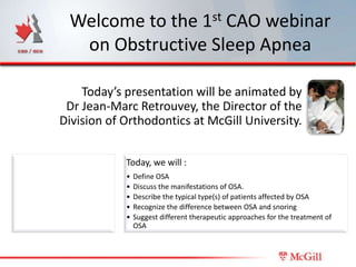
1. Welcome to the 1st CAO webinar
on Obstructive Sleep Apnea
Today’s presentation will be animated by
Dr Jean-Marc Retrouvey, the Director of the
Division of Orthodontics at McGill University.
Today, we will :
• Define OSA
• Discuss the manifestations of OSA.
• Describe the typical type(s) of patients affected by OSA
• Recognize the difference between OSA and snoring
• Suggest different therapeutic approaches for the treatment of
OSA
2. Obstructive Sleep Apnea
The Role of the Orthodontist:
The role of orthodontics in improving breathing in children, teenagers
and adults who suffer from sleep apnea
Dr Jean Marc Retrouvey
Director of Orthodontics
McGill University
3. Objectives
Define OSA
Discuss the manifestations of OSA.
Describe the typical type(s) of patients
affected by OSA
Recognize the difference between OSA
and snoring
Suggest different therapeutic approaches
for the treatment of OSA
4. Apnea–hypopnea index
WIKIPEDIA
• The apnea–hypopnea index (AHI) is an
index of sleep apnea severity that
combines apneas and hypopneas.
• AHI values are typically categorized as 5–
15/hr = mild;
• 15–30/hr = moderate;
• > 30/h = severe.)
5. Apnea–hypopnea index
WIKIPEDIA
• The apnea–hypopnea index (AHI) is an index of
sleep apnea severity that combines apneas and
hypopneas.
AHI values are typically
categorized as:
• 5–15/hr = mild
• 15–30/hr = moderate
• > 30/h = severe
6. Obstructive Sleep Apnea
27 % of
Snoring patients may
exhibit snoring
Upper Airway
UARS Resistance
Syndrome
4%
Obstructive
OSA Sleep Apnea
2-3%
Snoring and obstructive sleep apnea
By David N. F. Fairbanks, Samuel A. Mickelson, B. Tucker Woodson, p 243
7. Snoring: Benign condition
(annoying but not
dangerous)
UARS: Sleep disturbances
but no severe oxygen
desaturation (No cardiac
sequellae)
OSA: Oxygen desaturation
and sleep disturbances
(Cardiac disturbances:
Strokes, hypertension
arrhythmias)
Collpo N. Semin Respir Crit Care Med 2005; 26(1): 13-24
Pediatric Care Med 2005; 26(1): 13-24
8. 1. Excessive daytime
somnolence
Daytime
symptoms in 2. Abnormal daytime
children with behavior
obstructive
sleep apnea 3. Learning problems
4. Bizarre behavior
5. Morning headaches
6. Failure to thrive or
obesity
7. Repetitive upper
airway infections
8. Acute cardiac
failure Guilleminault C,
Korobkin R, and R
Winkle. A Review of
9. Cor pulmonale 50 Children with
Obstructive Sleep
Apnea
Syndrome. Lung
10. Hypertension 1981.
9. Most common
contributing factors
Obesity Allergies
and Combinations
Genetics (ex: Skeletal
malocclusions)
10. 1. Obesity
A fairly direct correlation has been established between obesity
and OSA in children1 and adolescents2
Apnea Hypoxia Index (AHI) scores are higher in obese than in
normal-weight children with OSA3
1 - The Correlation Among Obesity, Apnea-Hypopnea Index, and Tonsil Size in Children
Yuen-yu Lam, FHKAM(Paed), et al. Chest 2006: 1751-1756
2 - Obesity increases the risk for persisting obstructive sleep apnea after treatment in children
Louise M. O’Brien et al . International Journal of Pediatric Otorhinolaryngology (2006) 70, 1555—1560
3 - Outcome of adenotonsillectomy for obstructive sleep apnea in obese and normal-weight children
Ron B. Mitchell, MD, and James Kelly, PhD, St Louis, MO; Albuquerque, NM . Otolaryngology–Head and
Neck Surgery (2007) 137, 43-48
11. 1. Obesity
What about treating OSA in obese kids?
Both groups show a dramatic improvement in AHI
after adenotonsillectomy, but persistent OSA is more
common in obese children.
Outcome of adenotonsillectomy for obstructive sleep apnea in obese and normal-weight children
Ron B. Mitchell, MD, and James Kelly, PhD, St Louis, MO; Albuquerque, NM . Otolaryngology–
Head and Neck Surgery (2007) 137, 43-48
12. 1. Obesity
With treatment, improvement in OSA but…..
Weight gain!
Recommendation : Lose weight and improve physical
condition before starting OSA treatment.
Soultan, Z., et al., Effect of treating obstructive sleep apnea by tonsillectomy and/or adenoidectomy on
obesity in children. Archives of Pediatrics and Adolescent Medicine, 1999. 153(1): p. 33.
13. Treatment of OSA or UARS in
non-obese children
Impact of Orthodontic treatment
14. Common Contributing
Observations
Severely enlarged
tonsils and adenoids in
the young patient
presenting either
UARS or OSA
http://kidshealth.org.nz/index.php/ps_pagename/content
page/pi_id/303
15. Consequence of
Enlarged Tonsils and Adenoids
Dr Harvold, from the University of Toronto, performed studies on Monkeys which showed
that:
If you block nasal respiration, mouth breathing follows and
a severe malocclusion is observed (variable response)
Harvold EP et al. Primate experiments on oral respiration. Am J Orthod 79(4):359-72, 1981
Harvold EP et al. Experiments on the development of dental malocclusion. Am J Orthod 61:38-44, 1972.
16. Recognize early!
OSA will have an impact on normal growth
and development (early treatment must be
seriously considered)
• Growth hormone is mainly released during the
stage 3 of NREM sleep.
• http://youtu.be/HiNaJhO2Ht4
17. Importance of Early Detection
and Treatment
Such changes are also influenced by genetic factors. Facial growth is nearly complete between the ages
of 15
and 16 years in girls and between 18 and 19 years in boys, but the largest increments of growth occur
during the earliest years of life: By the age of 4 years, the craniofacial
skeleton has attained 60% of adult size, and by the age of 12
years it is 90% of adult size. Thus both genetic and environmental factors play a role in
teenage facial determination.
Our findings suggest that specific morphometric features may have been present in certain children ot
tonsilectomy and adenoectomy, some aspect of facial growth may even
have been modified by the early airway obstruction.
Morphometric facial changes and obstructive sleep apnea in adolescents
Christian Guilleminault, MD, Markku Partinen, MD, Jean Paul Praud, MD,
Maria-Antonia Quera-Salva, MD, Nelson Powell, MD, and
Robert Riley, DDS, MD
From Stanford University Medical Center, Stanford, California, Ullanlinnan Sleep Disorders
Clinic, Helsinki, Finland, Laboratoire d'Explorations Fonctionelles, Hopital Antoine Beclere,
Clamart, France, and Hopital Raymond Poincarré, Garches, France A, Jand ournal of Pediatrics 1989
18. Examination of a Patient Suffering
from OSA or UARS
1 • Reference to pneumologist for polysomnography
2 • Extra oral findings
3 • Intra oral findings
4 • Cephalometric or Cone Beam assessment
5 • Final diagnosis
6 • Treatment options
19. Examination of a Patient Suffering
from OSA or UARS
2 Extra oral findings
• Facial features
• ―Pockets‖ under the eyes
• Evidence of mouth
breathing
• Retrusive mandible (Cl II
malocclusion)
• Retrusive maxilla?
20. Examination of a Patient
Suffering
from OSA or UARS
3 Intra oral findings
• Openbite
• Narrow palate
• Curve of Spee
• Lower arch form
• Severe malocclusion
• Usually Cl II
21. Examination of a Patient
Suffering
from OSA or UARS
3 Intra oral findings
Compared with 48 asymptomatic children from the
same cohort, the obstructed children had a
narrower maxilla, a deeper palatal height, and a
shorter lower dental arch. In addition, the
prevalence of lateral crossbite was significantly
higher among the obstructed children.
Breathing obstruction in relation to craniofacial and dental arch morphology in 4-year-old
children
B Löfstrand-Tideström European Journal of Orthodontics
Volume 21, Issue 4 , 1999 Pp. 323-332
22. Examination of a Patient Suffering
from OSA or UARS
4 Cephalometric or Cone Beam assessment
• Consistent for a large number of OSA pediatric
patients
23. • Retrognathic mandible
• Steep mandibular plane angle
• Long anterior face height
• Short posterior face height
24. Examination of a Patient Suffering
from OSA or UARS
6
5 Treatment options
1. Tonsillectomy
2. Rapid Palatal Expansion
3. Mandibular Advancement
25. 1. Tonsillectomy?
Children, who were tonsillectomized because of sleep apnea
were examined with respect to facial growth and dental arch
morphology.
The findings were compared to data from children without
tonsillary obstruction. A higher proportion of malocclusion
than normal, especially openbite and crossbite, was noticed
before surgery.
Two years after surgery, 77% of the open bites were normalized
and 50-65% of the buccal and anterior crossbites. The best
results were seen in children operated before the age of 6.
E. Hultcrantz E., Larson M. , Hellquist R. , Ahlquist-Rastad J. , Svanholm H. and Jakobsson
O.P. : The influence of tonsillar obstruction and tonsillectomy on facial growth and dental arch
morphology
International Journal of Pediatric Otorhinolaryngology 22,2: 125-134 1991
26. 2. Rapid Palatal expansion
• Multiple articles point
towards an improvement
in the sleep apnea
condition.
• Expansion is done via
RPE and averages
4.5mm to 6 mm at the
palatal suture.
• On sleep apnea
patients, the earlier the
better.
27. Selection Criteria for RPE patients
• High narrow palate
• Deep bite
• Retrusive mandible
Villa, M.P., et al., Rapid maxillary
expansion in children with obstructive
sleep apnea syndrome: 12-month
follow-up. Sleep medicine, 2007. 8(2):
p. 128-134.
28. 3. Mandibular advancement
Has the same effect in growing children as
rapid palatal expansion
Randomized Controlled Study of an Oral Jaw-Positioning Appliance for the Treatment of Obstructive Sleep Apnea in
Children with Malocclusion. MARIA P. Villa, edoardo bernkopf, jacopo pagani, vanna broia, Marilisa montesano and
roberto ronchetti.
29. Impact of Orthodontics on Pediatric
OSA Management
Treatment will depend on the severity of the OSA, its
influence on the degree of malocclusion and the age of
the patient.
Take Home Message :
Early recognition (before age 7)
• Educate parents and dentists
Constant collaboration with the treating physician
(Respirologist, Plastics, ENT), the Orthodontist and
the Dentist.
Treat early and aggressively
• Through RPE; Mandibulat advancement and Maxillary
Vertical Control
30. OSA Treatment in the adult
Role of the orthodontist?
Therapy Provider
CPAP Pneumologist or
Sleep center
Soft tissue surgery ENT
MADs Sleep center
Dentist – TMJ
specialist
Orthodontist?
MMA surgery OMFS
SARPE Orthodontist
31. Mandibular advancement
devices
• May be efficient for moderate OSA
• Do not replace the CPAP in severe cases
33. What about SARPE?
Dr Fiore (Fiore et al., U de Montreal, 2012)
testing 9 patients treated with Sarpe and
comprehensive orthodontics.
Showed a small but not significant reduction
in respiratoy index.
Significant change in snoring index.
35. 43 yr male with snoring and
witnessed apneas.
• Sleep study
– RDI 67/hr, LSAT 83%
• Sleep study with CPAP
– RDI 15/hr, LSAT 86%
• Does not tolerate CPAP
39. Results
Pre- operative sleep study:
- RDI 67/hr
6 month post- operative sleep study
– RDI 9/hr, (was down to 15 with CPAP)
RDI : Respiratory Disturbance Index
LSAT: Saturation in oxygen
40. Long Term Follow up of a
TMJ- OSA Patient
Patient presenting with Long face
syndrome :
– Narrow palate
– Retrusive mandible
– Anterior tongue posture
– Severe to moderate crowding of dental arches
– Painful bilateral TMJ clicks
– Moderate OSA ( No C Pap used)
41. Treatments
1. Maxillary expansion at 8 years old (failed)
2. Dental alignment (camouflage failed)
3. Extractions were contemplated by
previous orthodontist (failed to recognize
OSA)
4. Mandibular protraction appliance contra-
indicated (High MP angle)
42. Long term Follow up of TMJ and
OSA Patient
In 2004, after
first rapid palatal
expansion
attempt
43. 2009: Ready for Ortho-Surgery
Orthodontics: 3 piece maxilla preparation
Uprighting of lower arch
46. Results:
TMJ pain is resolved ( no
splint worn)
Snoring and symptoms of
OSA have subsided
Patient is satisfied with
aesthetic result.
47. Conclusions
OSA is a medical condition and may be
potentially lethal
A positive diagnosis of OSA should be
obtained before starting any treatment
The dental profession has an important role
in screening young patients
Orthodontists have a greater role to play
(back to the future: treat early and
aggressively)
48. Conclusions
Tonsillectomy is making a comeback in preventive
therapy for this type of patients (OSA-UARS)
CPAP machine is still standard of care in adults
Growth modification may play an important aspect
of OSA treatment
Maxillary expansion
Mandibular protraction seem to have a positive effect
on OSA
Must start as early as possible ( do not allow upper
molars descent)