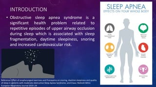
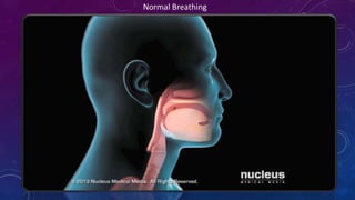
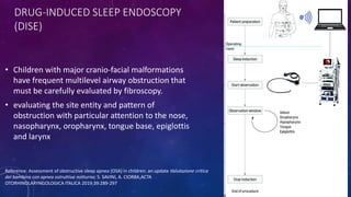
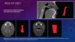
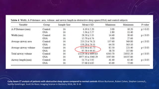
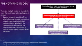
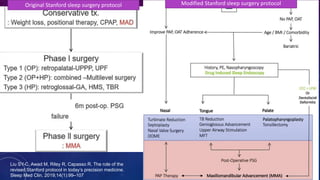
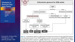
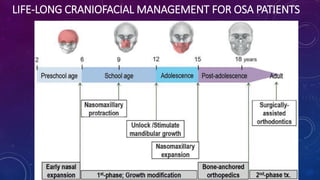
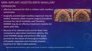
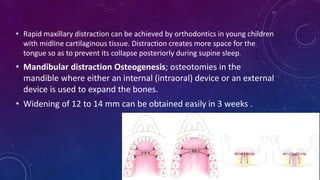
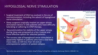
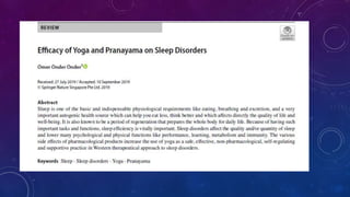
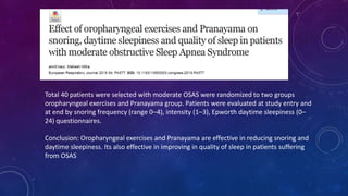
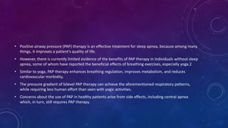
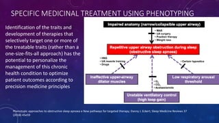
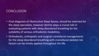
OBSTRUCTIVE SLEEP APNEA
ADVANCES IN DIAGNOSIS AND MANAGEMENT
GUIDED BY: DR. SUNITA SHRIVASTAV
PRESENTED BY: DR. SHRIYA MURARKA
CONTENTS
• Overview of Obstructive Sleep Apnea (OSA)
• Orthodontist’s perspective of Diagnosis
• Phenotyping in OSA
• Treatment protocols
• Role of RME and Distraction Osteogenesis in OSA
• Hypoglossal Nerve Stimulation
• Role of pranayama in treatment of OSA
• Conclusion
• References
INTRODUCTION
• Obstructive sleep apnea syndrome is a
significant health problem related to
repetitive episodes of upper airway occlusion
during sleep which is associated with sleep
fragmentation, daytime sleepiness, snoring
and increased cardiovascular risk.
Reference: Effect of oropharyngeal exercises and Pranayama on snoring, daytime sleepiness and quality
of sleep in patients with moderate obstructive Sleep Apnea Syndrome; amrit kaur, Mahesh Mitra
European Respiratory Journal 2019 ;54
NORMAL BREATHING
Normal Breathing
PATHOPHYSIOLOGY OF OSA
THEORIES OF SNORING
• Obstacle theory- (1990)- An increased negative pressure during inspiration
retracts the structures of the pharynx and makes them vibrate in the airflow to
produce snore and possible obstruction in OSA.
• Bernoulli theory (1738)- The velocity of streaming air is higher and the pressure
lower at a constriction of a tube compared with a larger part. This may cause
inward suction of the pharyngeal structures in a constricted area and causes
snoring by the vibration of wall structures.
Reference: Engelke W, Repetto G, Mendoza – Gaertner M, Knoesel M. Functional treatment of snoring using oral shields in conjunction with the
tongue repositioning manoevre. Int J Odontostomat. 2007;1(2):133–139.
CRANIOFACIAL ABNORMALITIES CAUSING OSA
• Retroposition of the
mandible
• Reduced cranial base flexure
measured at the nasion–
sella– basion angle
• Lower position with
displacement of the hyoid
bone.
ORTHODONTIST’S ROLE IN DIAGNOSING OSA
• Evaluation for tongue size and air space between soft palate and
tongue using Malampati Scores
• Tonsillar grading
• Lateral Cephalogram Analysis
O.P. Kharbanda; Orthodontics Diagnosis and management of malocclusion and dentofacial deformities; second edition.
Original Mallampati Scoring:
• Class 1: Faucial pillars, soft palate and uvula could be
visualized.
• Class 2: Faucial pillars and soft palate could be visualized,
but uvula was masked by the base of the tongue.
• Class 3: Only soft palate visualized.
Modified Mallampati Scoring:
• Class I: Soft palate, uvula, fauces, pillars visible.
• Class II: Soft palate, major part of uvula, fauces visible.
• Class III: Soft palate, base of uvula visible.
• Class IV: Only hard palate visible.
Reference: MaFreiberger, D; Liu, PL (Jul 1985). "A clinical sign to predict difficult tracheal intubation: a prospective study". Canadian Anaesthetists' Society Journal. 32 (4): 429–34.
llampati, SR; Gatt, SP; Gugino, LD; Desai, SP; Waraksa, B
Samsoon, GL; Young, JR (May 1987). "Difficult tracheal intubation: a retrospective study". Anaesthesia. 42 (5): 487–90.
TONSILS GRADING SCALE
In Grade III and IV the airway is
severely compromised.
• 0 — Tonsils are entirely within the tonsillar
pillar, or previously removed by surgery.
• 1+ — Tonsils occupy less than 25 % of the
lateral dimension of the oropharynx, as
measured between the anterior tonsillar
pillars (solid yellow arrow).
• 2+ — Tonsils occupy 26 to 50 % of the lateral
dimension of the oropharynx.
• 3+ — Tonsils occupy 51 to 75 % of the lateral
dimension of the oropharynx.
• 4+ — Tonsils occupy more than 75 % of the
lateral dimension of the oropharynx. (kissing
tonsils).
Reference: Brodsky L. Modern assessment of tonsils and adenoids. Pediatr Clin North Am 1989; 36:1551.
LATERAL CEPHALOGRAM ANALYSIS
ACOUSTIC RHINOMETRY
• Acoustic rhinometry (AR) is a simple, fast, and noninvasive diagnostic tool measuring nasal
cavity geometry and nasal airway change through acoustic reflection.
• The size and the pattern of the reflected sound waves provide information on the structure
and dimensions of anterior and middle parts of nasal cavity including nasal valve area, which
shows the greatest nasal airflow resistance
Acoustic pulse from a
speaker
Reflected back to a
microphone
Amplifier
Computer
DRUG-INDUCED SLEEP ENDOSCOPY
(DISE)
• Children with major cranio-facial malformations
have frequent multilevel airway obstruction that
must be carefully evaluated by fibroscopy.
• evaluating the site entity and pattern of
obstruction with particular attention to the nose,
nasopharynx, oropharynx, tongue base, epiglottis
and larynx
Reference: Assessment of obstructive sleep apnea (OSA) in children: an update Valutazione critica
del bambino con apnea ostruttiva notturna; S. SAVINI, A. CIORBA,ACTA
OTORHINOLARYNGOLOGICA ITALICA 2019;39:289-297
ROLE OF CBCT
• Cone-beam CT analysis of patients
with obstructive sleep apnea
Cone-beam CT analysis of patients with obstructive sleep apnea compared to normal controls Allison Buchanan, Ruben Cohen, Stephen Looney3,,
Sajitha Kalathingal, Scott De Rossi; Imaging Science in Dentistry 2016; 46: 9-16
Cone-beam CT analysis of patients with obstructive sleep apnea compared to normal controls Allison Buchanan, Ruben Cohen, Stephen Looney3,,
Sajitha Kalathingal, Scott De Rossi; Imaging Science in Dentistry 2016; 46: 9-16
DYNAMIC SLEEP MRI
• Dynamic sleep MRI has
advantages of dynamic nature,
the ability to evaluate the
airway in a multiplane fashion,
and more realistic information
obtained in the sleeping state
or a simulated sleep state.
PHENOTYPING IN OSA
There are multiple causes or phenotypic
traits that contribute to the pathogenesis
of OSA.
• Current emphasis is on identifying
which of several possible mechanisms
is (are) responsible in individual
patients (phenotyping) with the
therapeutic aim of targeting the
specific offending mechanisms by
nonmechanical means (individualized
medicine).
Phenotypic approaches to obstructive sleep apnoea e New pathways for targeted therapy
Danny J. Eckert; Sleep Medicine Reviews 37 (2018) 45
TREATMENT PROTOCOL
Original Stanford sleep surgery protocol Modified Stanford sleep surgery protocol
Liu SY-C, Awad M, Riley R, Capasso R. The role of the
revised Stanford protocol in today’s precision medicine.
Sleep Med Clin. 2019;14(1):99–107
Su-Jung Kim, Ki Beom KIM;
Orthodontics in OSA;
patients; Springer; 2020
LIFE-LONG CRANIOFACIAL MANAGEMENT FOR OSA PATIENTS
MINI-IMPLANT ASSISTED RAPID MAXILLARY
EXPANSION
• effective treatment for OSA in children with maxillary
constriction.
• Surgically assisted rapid maxillary expansion may be
helpful. However, these invasive surgical procedures
increase the risk of morbidity and Therefore,
MARME may be an effective treatment modality for
adults with OSA.
• Because of its reduced costs and far fewer risks
compared to alternative treatment options, this
novel MARME design and protocol offer great
promise for the future of nonsurgical maxillary
orthopedic expansion and minimally invasive
treatment for OSA in adult patients.
Obstructive sleep apnea treatment in adults, Hong-Po Chang, Yu-Feng Chen, Je-Kang Du, Kaohsiung J Med Sci. 2020;36:7–12.
• Rapid maxillary distraction can be achieved by orthodontics in young children
with midline cartilaginous tissue. Distraction creates more space for the
tongue so as to prevent its collapse posteriorly during supine sleep.
• Mandibular distraction Osteogenesis; osteotomies in the
mandible where either an internal (intraoral) device or an external
device is used to expand the bones.
• Widening of 12 to 14 mm can be obtained easily in 3 weeks .
HYPOGLOSSAL NERVE STIMULATION
• Surgical treatment of OSA has evolved in the era of
neurostimulation, including the advent of hypoglossal
nerve stimulation.
• Sleep surgeons surgically implant an upper-airway
stimulation device in OSA patients who have difficulty
tolerating or adhering to CPAP therapy.
• Neurostimulation for stability of the upper airway
during sleep was proposed as a less invasive and
more effective option for selected patients.
• The success rate of hypoglossal nerve stimulation is
apparently highest in patients who have a low body
mass index, an AHI less than 50 and an
anteroposterior pattern of palatal collapse.
Obstructive sleep apnea treatment in adults, Hong-Po Chang, Yu-Feng Chen, Je-Kang Du, Kaohsiung J Med Sci. 2020;36:7–12.
ROLE OF PRANAYAMA IN TREATMENT OF OSA
Bhujangasana (Cobra Pose)
• Opens up the chest and clears the lungs.
• Helps improve the flow of oxygen and blood circulation
•Helps regulate breathing.
•Opens up the chest muscles to allow deeper
inhalations and exhalations.
Dhanurasana (Bow Pose)
Bhramari Pranayama (Bee Breath)
• Helps increase concentration.
• Relieves anger and tension.
• Helps reduce blood pressure.
Ujjayi Pranayama (Victory Breath Technique)
• Helps strengthen the muscles of the throat and face.
• Helps regulate sleep patterns.
• Calms the mind.
Nadi Shodhan Pranayama (Alternate Nostril Breathing Technique)
• Helps to cleanse the circulation channels.
• Helps to alleviate throat infections.
• Helps regulate snoring and sleep apnea problems.
Kapal Bharti Pranayama (Skull Shining Breathing Technique)
• Helps cleanse the sinuses.
• Helps improve sleep.
Simha Garjasana (Roaring Lion Pose)
• Exercises the tongue.
• Relaxes the neck muscles.
• Helps prevent a sore throat.
• Stimulates the platysma, i.e., the muscles in front of the
throat.
• It is hypothesized that stress induces the autonomic imbalance by decreased
parasympathetic, increased sympathetic activity, gamma amino-butyric acid
(GABA) underactivity and increased allostatic load.
• The importance of the key neurotransmitter GABA which plays role in various
health conditions including insomnia has been recognized in recent years.
• Natural calming and anxiety reducing effect of GABA is obtained through
decreased neuronal excitability. Yogic practices are known to correct
underactivity of the parasympathetic nervous system and GABA through
stimulation of the vagus nerve and reduce allostatic load.
Total 40 patients were selected with moderate OSAS were randomized to two groups
oropharyngeal exercises and Pranayama group. Patients were evaluated at study entry and
at end by snoring frequency (range 0–4), intensity (1–3), Epworth daytime sleepiness (0–
24) questionnaires.
Conclusion: Oropharyngeal exercises and Pranayama are effective in reducing snoring and
daytime sleepiness. Its also effective in improving in quality of sleep in patients suffering
from OSAS
• Positive airway pressure (PAP) therapy is an effective treatment for sleep apnea, because among many
things, it improves a patient’s quality of life.
• However, there is currently limited evidence of the benefits of PAP therapy in individuals without sleep
apnea, some of whom have reported the beneficial effects of breathing exercises, especially yoga.2
• Similar to yoga, PAP therapy enhances breathing regulation, improves metabolism, and reduces
cardiovascular morbidity.
• The pressure gradient of bilevel PAP therapy can achieve the aforementioned respiratory patterns,
while requiring less human effort than seen with yogic activities.
• Concerns about the use of PAP in healthy patients arise from side effects, including central apnea
which, in turn, still requires PAP therapy.
SPECIFIC MEDICINAL TREATMENT USING PHENOTYPING
Identification of the traits and
development of therapies that
selectively target one or more of
the treatable traits (rather than a
one-size-fits-all approach) has the
potential to personalize the
management of this chronic
health condition to optimize
patient outcomes according to
precision medicine principles
Phenotypic approaches to obstructive sleep apnoea e New pathways for targeted therapy; Danny J. Eckert; Sleep Medicine Reviews 37
(2018) 45e59
CONCLUSION
• Final diagnosis of Obstructive Sleep Apnea, should be reserved for
the sleep specialists, however dentist plays a crucial role in
evaluating patients with sleep disordered breathing for the
suitability of various orthodontic modalities.
• Orthodontic, orthopedic and surgical craniofacial managements
for the sleep disordered breathing with craniofacial skeletal risk
factors can be timely applied throughout the life.
REFERENCES
• Effect of oropharyngeal exercises and Pranayama on snoring, daytime sleepiness and quality of sleep in
patients with moderate obstructive Sleep Apnea Syndrome; amrit kaur, Mahesh Mitra European
Respiratory Journal 2019 ;54
• Engelke W, Repetto G, Mendoza – Gaertner M, Knoesel M. Functional treatment of snoring using oral
shields in conjunction with the tongue repositioning manoevre. Int J Odontostomat. 2007;1(2):133–139
• O.P. Kharbanda; Orthodontics Diagnosis and management of malocclusion and dentofacial deformities;
second edition.
• Obstructive sleep apnea treatment in adults, Hong-Po Chang, Yu-Feng Chen, Je-Kang Du, Kaohsiung J
Med Sci. 2020;36:7–12.
• Phenotypic approaches to obstructive sleep apnoea e New pathways for targeted therapy; Danny J.
Eckert; Sleep Medicine Reviews 37 (2018) 45e59
THANKYOU

Obstructive sleep apnea- Orthodontist's perspective

  • 1.
    OBSTRUCTIVE SLEEP APNEA ADVANCESIN DIAGNOSIS AND MANAGEMENT GUIDED BY: DR. SUNITA SHRIVASTAV PRESENTED BY: DR. SHRIYA MURARKA
  • 2.
    CONTENTS • Overview ofObstructive Sleep Apnea (OSA) • Orthodontist’s perspective of Diagnosis • Phenotyping in OSA • Treatment protocols • Role of RME and Distraction Osteogenesis in OSA • Hypoglossal Nerve Stimulation • Role of pranayama in treatment of OSA • Conclusion • References
  • 3.
    INTRODUCTION • Obstructive sleepapnea syndrome is a significant health problem related to repetitive episodes of upper airway occlusion during sleep which is associated with sleep fragmentation, daytime sleepiness, snoring and increased cardiovascular risk. Reference: Effect of oropharyngeal exercises and Pranayama on snoring, daytime sleepiness and quality of sleep in patients with moderate obstructive Sleep Apnea Syndrome; amrit kaur, Mahesh Mitra European Respiratory Journal 2019 ;54
  • 4.
  • 5.
  • 6.
    THEORIES OF SNORING •Obstacle theory- (1990)- An increased negative pressure during inspiration retracts the structures of the pharynx and makes them vibrate in the airflow to produce snore and possible obstruction in OSA. • Bernoulli theory (1738)- The velocity of streaming air is higher and the pressure lower at a constriction of a tube compared with a larger part. This may cause inward suction of the pharyngeal structures in a constricted area and causes snoring by the vibration of wall structures. Reference: Engelke W, Repetto G, Mendoza – Gaertner M, Knoesel M. Functional treatment of snoring using oral shields in conjunction with the tongue repositioning manoevre. Int J Odontostomat. 2007;1(2):133–139.
  • 7.
    CRANIOFACIAL ABNORMALITIES CAUSINGOSA • Retroposition of the mandible • Reduced cranial base flexure measured at the nasion– sella– basion angle • Lower position with displacement of the hyoid bone.
  • 8.
    ORTHODONTIST’S ROLE INDIAGNOSING OSA • Evaluation for tongue size and air space between soft palate and tongue using Malampati Scores • Tonsillar grading • Lateral Cephalogram Analysis O.P. Kharbanda; Orthodontics Diagnosis and management of malocclusion and dentofacial deformities; second edition.
  • 9.
    Original Mallampati Scoring: •Class 1: Faucial pillars, soft palate and uvula could be visualized. • Class 2: Faucial pillars and soft palate could be visualized, but uvula was masked by the base of the tongue. • Class 3: Only soft palate visualized. Modified Mallampati Scoring: • Class I: Soft palate, uvula, fauces, pillars visible. • Class II: Soft palate, major part of uvula, fauces visible. • Class III: Soft palate, base of uvula visible. • Class IV: Only hard palate visible. Reference: MaFreiberger, D; Liu, PL (Jul 1985). "A clinical sign to predict difficult tracheal intubation: a prospective study". Canadian Anaesthetists' Society Journal. 32 (4): 429–34. llampati, SR; Gatt, SP; Gugino, LD; Desai, SP; Waraksa, B Samsoon, GL; Young, JR (May 1987). "Difficult tracheal intubation: a retrospective study". Anaesthesia. 42 (5): 487–90.
  • 10.
    TONSILS GRADING SCALE InGrade III and IV the airway is severely compromised. • 0 — Tonsils are entirely within the tonsillar pillar, or previously removed by surgery. • 1+ — Tonsils occupy less than 25 % of the lateral dimension of the oropharynx, as measured between the anterior tonsillar pillars (solid yellow arrow). • 2+ — Tonsils occupy 26 to 50 % of the lateral dimension of the oropharynx. • 3+ — Tonsils occupy 51 to 75 % of the lateral dimension of the oropharynx. • 4+ — Tonsils occupy more than 75 % of the lateral dimension of the oropharynx. (kissing tonsils). Reference: Brodsky L. Modern assessment of tonsils and adenoids. Pediatr Clin North Am 1989; 36:1551.
  • 11.
  • 12.
    ACOUSTIC RHINOMETRY • Acousticrhinometry (AR) is a simple, fast, and noninvasive diagnostic tool measuring nasal cavity geometry and nasal airway change through acoustic reflection. • The size and the pattern of the reflected sound waves provide information on the structure and dimensions of anterior and middle parts of nasal cavity including nasal valve area, which shows the greatest nasal airflow resistance Acoustic pulse from a speaker Reflected back to a microphone Amplifier Computer
  • 13.
    DRUG-INDUCED SLEEP ENDOSCOPY (DISE) •Children with major cranio-facial malformations have frequent multilevel airway obstruction that must be carefully evaluated by fibroscopy. • evaluating the site entity and pattern of obstruction with particular attention to the nose, nasopharynx, oropharynx, tongue base, epiglottis and larynx Reference: Assessment of obstructive sleep apnea (OSA) in children: an update Valutazione critica del bambino con apnea ostruttiva notturna; S. SAVINI, A. CIORBA,ACTA OTORHINOLARYNGOLOGICA ITALICA 2019;39:289-297
  • 14.
    ROLE OF CBCT •Cone-beam CT analysis of patients with obstructive sleep apnea Cone-beam CT analysis of patients with obstructive sleep apnea compared to normal controls Allison Buchanan, Ruben Cohen, Stephen Looney3,, Sajitha Kalathingal, Scott De Rossi; Imaging Science in Dentistry 2016; 46: 9-16
  • 15.
    Cone-beam CT analysisof patients with obstructive sleep apnea compared to normal controls Allison Buchanan, Ruben Cohen, Stephen Looney3,, Sajitha Kalathingal, Scott De Rossi; Imaging Science in Dentistry 2016; 46: 9-16
  • 16.
    DYNAMIC SLEEP MRI •Dynamic sleep MRI has advantages of dynamic nature, the ability to evaluate the airway in a multiplane fashion, and more realistic information obtained in the sleeping state or a simulated sleep state.
  • 17.
    PHENOTYPING IN OSA Thereare multiple causes or phenotypic traits that contribute to the pathogenesis of OSA. • Current emphasis is on identifying which of several possible mechanisms is (are) responsible in individual patients (phenotyping) with the therapeutic aim of targeting the specific offending mechanisms by nonmechanical means (individualized medicine). Phenotypic approaches to obstructive sleep apnoea e New pathways for targeted therapy Danny J. Eckert; Sleep Medicine Reviews 37 (2018) 45
  • 18.
  • 19.
    Original Stanford sleepsurgery protocol Modified Stanford sleep surgery protocol Liu SY-C, Awad M, Riley R, Capasso R. The role of the revised Stanford protocol in today’s precision medicine. Sleep Med Clin. 2019;14(1):99–107
  • 20.
    Su-Jung Kim, KiBeom KIM; Orthodontics in OSA; patients; Springer; 2020
  • 21.
  • 22.
    MINI-IMPLANT ASSISTED RAPIDMAXILLARY EXPANSION • effective treatment for OSA in children with maxillary constriction. • Surgically assisted rapid maxillary expansion may be helpful. However, these invasive surgical procedures increase the risk of morbidity and Therefore, MARME may be an effective treatment modality for adults with OSA. • Because of its reduced costs and far fewer risks compared to alternative treatment options, this novel MARME design and protocol offer great promise for the future of nonsurgical maxillary orthopedic expansion and minimally invasive treatment for OSA in adult patients. Obstructive sleep apnea treatment in adults, Hong-Po Chang, Yu-Feng Chen, Je-Kang Du, Kaohsiung J Med Sci. 2020;36:7–12.
  • 23.
    • Rapid maxillarydistraction can be achieved by orthodontics in young children with midline cartilaginous tissue. Distraction creates more space for the tongue so as to prevent its collapse posteriorly during supine sleep. • Mandibular distraction Osteogenesis; osteotomies in the mandible where either an internal (intraoral) device or an external device is used to expand the bones. • Widening of 12 to 14 mm can be obtained easily in 3 weeks .
  • 24.
    HYPOGLOSSAL NERVE STIMULATION •Surgical treatment of OSA has evolved in the era of neurostimulation, including the advent of hypoglossal nerve stimulation. • Sleep surgeons surgically implant an upper-airway stimulation device in OSA patients who have difficulty tolerating or adhering to CPAP therapy. • Neurostimulation for stability of the upper airway during sleep was proposed as a less invasive and more effective option for selected patients. • The success rate of hypoglossal nerve stimulation is apparently highest in patients who have a low body mass index, an AHI less than 50 and an anteroposterior pattern of palatal collapse. Obstructive sleep apnea treatment in adults, Hong-Po Chang, Yu-Feng Chen, Je-Kang Du, Kaohsiung J Med Sci. 2020;36:7–12.
  • 25.
    ROLE OF PRANAYAMAIN TREATMENT OF OSA Bhujangasana (Cobra Pose) • Opens up the chest and clears the lungs. • Helps improve the flow of oxygen and blood circulation
  • 26.
    •Helps regulate breathing. •Opensup the chest muscles to allow deeper inhalations and exhalations. Dhanurasana (Bow Pose)
  • 27.
    Bhramari Pranayama (BeeBreath) • Helps increase concentration. • Relieves anger and tension. • Helps reduce blood pressure.
  • 28.
    Ujjayi Pranayama (VictoryBreath Technique) • Helps strengthen the muscles of the throat and face. • Helps regulate sleep patterns. • Calms the mind.
  • 29.
    Nadi Shodhan Pranayama(Alternate Nostril Breathing Technique) • Helps to cleanse the circulation channels. • Helps to alleviate throat infections. • Helps regulate snoring and sleep apnea problems.
  • 30.
    Kapal Bharti Pranayama(Skull Shining Breathing Technique) • Helps cleanse the sinuses. • Helps improve sleep.
  • 31.
    Simha Garjasana (RoaringLion Pose) • Exercises the tongue. • Relaxes the neck muscles. • Helps prevent a sore throat. • Stimulates the platysma, i.e., the muscles in front of the throat.
  • 33.
    • It ishypothesized that stress induces the autonomic imbalance by decreased parasympathetic, increased sympathetic activity, gamma amino-butyric acid (GABA) underactivity and increased allostatic load. • The importance of the key neurotransmitter GABA which plays role in various health conditions including insomnia has been recognized in recent years. • Natural calming and anxiety reducing effect of GABA is obtained through decreased neuronal excitability. Yogic practices are known to correct underactivity of the parasympathetic nervous system and GABA through stimulation of the vagus nerve and reduce allostatic load.
  • 34.
    Total 40 patientswere selected with moderate OSAS were randomized to two groups oropharyngeal exercises and Pranayama group. Patients were evaluated at study entry and at end by snoring frequency (range 0–4), intensity (1–3), Epworth daytime sleepiness (0– 24) questionnaires. Conclusion: Oropharyngeal exercises and Pranayama are effective in reducing snoring and daytime sleepiness. Its also effective in improving in quality of sleep in patients suffering from OSAS
  • 36.
    • Positive airwaypressure (PAP) therapy is an effective treatment for sleep apnea, because among many things, it improves a patient’s quality of life. • However, there is currently limited evidence of the benefits of PAP therapy in individuals without sleep apnea, some of whom have reported the beneficial effects of breathing exercises, especially yoga.2 • Similar to yoga, PAP therapy enhances breathing regulation, improves metabolism, and reduces cardiovascular morbidity. • The pressure gradient of bilevel PAP therapy can achieve the aforementioned respiratory patterns, while requiring less human effort than seen with yogic activities. • Concerns about the use of PAP in healthy patients arise from side effects, including central apnea which, in turn, still requires PAP therapy.
  • 37.
    SPECIFIC MEDICINAL TREATMENTUSING PHENOTYPING Identification of the traits and development of therapies that selectively target one or more of the treatable traits (rather than a one-size-fits-all approach) has the potential to personalize the management of this chronic health condition to optimize patient outcomes according to precision medicine principles Phenotypic approaches to obstructive sleep apnoea e New pathways for targeted therapy; Danny J. Eckert; Sleep Medicine Reviews 37 (2018) 45e59
  • 38.
    CONCLUSION • Final diagnosisof Obstructive Sleep Apnea, should be reserved for the sleep specialists, however dentist plays a crucial role in evaluating patients with sleep disordered breathing for the suitability of various orthodontic modalities. • Orthodontic, orthopedic and surgical craniofacial managements for the sleep disordered breathing with craniofacial skeletal risk factors can be timely applied throughout the life.
  • 39.
    REFERENCES • Effect oforopharyngeal exercises and Pranayama on snoring, daytime sleepiness and quality of sleep in patients with moderate obstructive Sleep Apnea Syndrome; amrit kaur, Mahesh Mitra European Respiratory Journal 2019 ;54 • Engelke W, Repetto G, Mendoza – Gaertner M, Knoesel M. Functional treatment of snoring using oral shields in conjunction with the tongue repositioning manoevre. Int J Odontostomat. 2007;1(2):133–139 • O.P. Kharbanda; Orthodontics Diagnosis and management of malocclusion and dentofacial deformities; second edition. • Obstructive sleep apnea treatment in adults, Hong-Po Chang, Yu-Feng Chen, Je-Kang Du, Kaohsiung J Med Sci. 2020;36:7–12. • Phenotypic approaches to obstructive sleep apnoea e New pathways for targeted therapy; Danny J. Eckert; Sleep Medicine Reviews 37 (2018) 45e59
  • 40.

Editor's Notes

  • #4 Human beings spend approximately one-third of their lives sleeping and this is recognised as having an important physiological role. Sleep disorders of the upper airway result from any condition or disease that causes partial or complete obstruction of airway when the patient assumes a supine position and goes to sleep.
  • #5 This is the video demonstrating normal sleep . There is a patent airway and air flow reaches to the wind pipe effectively. Spreading through the bronchioles in to the lungs. Each time we breath the negative suction pulls the soft tissue structures of the mouth, inwards. It is normal for our muscles to relax slightly when we sleep but not completely that it can block the airway.
  • #6 The ventilatory activity of respiratory muscles including the musculature of the upper airway is reduced in sleep. This results in a fall in ventilation and increase in resistance of upper airway leading to increasing CO2 saturation. Ventilatory activity in sleep is stimulated by chemical drive from hypoxia and hypercapnia. The resistance of the system is increased, and at the same time, both chemical and mechanical sensors are depressed.
  • #7 Frequently osa patients demonstrates snoring. So what causes snoring.
  • #8 Removal of obstructions to facilitate mouth breathing and orthodontic treatment to allow normal growth of the mandible facilitate patency of the airway and therefore are likely to reduce the possibility of OSA developing in future.
  • #9 Periodontal and soft tissue examination, TMJ evaluation, arch size, maximum protrusive movement and clearance between central incisors at full mouth opening
  • #10 The assessment is made by asking the patient to open the mouth with tongue protruding fully: Score I: soft palate, uvula and the pharyngeal wall is fully visible. Score II: the pharyngeal wall is slightly visible, and space between uvula and tongue is reduced. Score III: The uvula is partially obscured by the tongue and pharyngeal wall not visible at all. Score IV: uvula and part of soft palate obscured and pharyngeal wall not visible at all. Mallampati score is a means of determining airway patency. The score is a predictor for determining the severity of sleep apnoea. A patient with a score of III or IV is at a higher risk of developing OSA.
  • #11 Given by Brodsky in 1989
  • #13 Acoustic rhinometry uses a reflected sound signal to measure the cross-sectional area and volume of the nasal passage. Acoustic rhinometry gives an anatomic description of a nasal passage, whereas rhinomanometry gives a functional measure of the pressure-flow relationships during the respiratory cycle. The test is carried out by generating an acoustic pulse from a speaker or spark source and this sound pulse is transmitted to the nose along a tube. This sound pulse is reflected back to a microphone and this reflection is generated according to the changes in the local acoustic impedance related to cross-sectional area of the nasal cavity. The microphone transmits the sound signal to an amplifier and to a PC which processes it an area distance graph.
  • #14 Endoscopic assessment. Endoscopy with flexible optic fibres allows evaluating patency of nasal cavities (i.e. hypertrophy of the inferior turbinates, presence of septal deviations or choanal atresia, adenoid hypertrophy), tongue base tropism or the possible presence of laryngomalacia. In selected patients, DISE can be indicated. In children, DISE assesses the residual OSA after adenotonsillectomy
  • #17 Dynamic MR imaging can accurately diagnose the cause and level of upper airway narrowing in patients with OSA. It can characterize and anatomically classify the level of narrowing for planning reparative surgery..
  • #20 Nonsurgical conservative treatment was firstly considered, and phase I surgical procedures were applied according to the main obstruction sites. Only when phase I alone was not successful in achieving surgical success, the sleep surgeon could proceed to phase II surgical protocol encompassing MMA. This approach has been advocated to minimize morbidity while maximizing opportunity for successful outcomes. Here, MMA could not be considered as a primary option regardless of severe craniofacial abnormality. This revised protocol in 2019 is defined by precision in patient selection, procedural selection, and procedural accuracy. Here, MMA surgery can be considered as a primary option for OSA patient with definite dentofacial deformity and/or with complete concentric collapse of lateral pharyngeal wall in DISE, as well as secondary option for the patients who failed to other therapies. In addition, they introduced another orthodontic option of surgical maxillary expansion, called distraction osteogenesis maxillary osteotomy (DOME), for the patients with nasal obstruction. With increasing demand of orthodontist’s roles, we need our precision protocol not only to decide primary intervention but to participate in multiprofessional team better.
  • #23 he maxillary skeletal expander (MSE) can enlarge the size of the nasal cavity and substantially increase the airflow through the nasal airway. Although RME can produce some maxillary skeletal expansion, it often produces large unwanted tooth movements, especially in mature patients increase treatment costs for the patients. For mature patients, mini-implant assisted rapid maxillary expansion (MARME) is now a common approach because it reduces or eliminates adverse dental side effects. Recent studies suggest that nonsurgical MARME is achievable and predictable in young adults. In addition to providing an effective solution for maxillary transverse deficiency in numerous patients, MARME can apparently reduce upper airway resistance. Compared to RME, a MARME procedure has larger skeletal effects, which enables a larger midpalatal suture separation and a larger nasal cavity volume increase. An MSE is a particular MARME appliance characterized by four mini-implants positioned in the posterior part of the palate with bicortical engagements of the palatal and nasal cortical bone
  • #24 Rapid maxillary distraction needs to occur before the cartilage becomes bone, and is therefore most often performed between the ages of 5 and 16 years. The concept of mandibular distraction Osteogenesis is very similar to that of rapid maxillary distraction, except that the surgeon uses a saw to create osteotomies in the mandible where either an internal (intraoral) device or an external device is used to expand the bones. Patients accept internal devices better than external ones, but the former are difficult to use in patients with small bones. External devices offer flexibility and vector selection, but often cause scarring of the skin. Widening of 12 to 14 mm can be obtained easily in 3 weeks .This approach is normally postponed until 10 to 13 years of age, and by 12 to 13 years of age both rapid maxillary distraction and mandibular distraction osteogenesis can be performed simultaneously.
  • #38 Improved phenotyping approaches are an important step toward the goal of personalized medicine for OSA patients. Recent work focused on pathophysiologic risk factors for OSA (eg, arousal threshold, craniofacial morphology, chemoreflex sensitivity) appears to capture heterogeneity in OSA, but requires clinical validation.