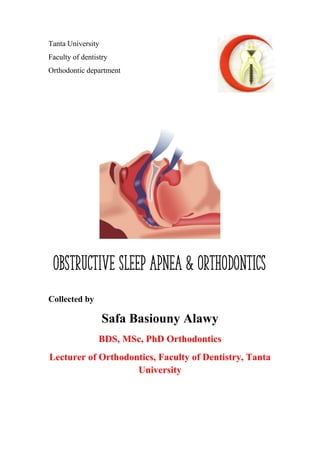
SlideShare a Scribd company logo
1 of 21
Download to read offline
Tanta University
Faculty of dentistry
Orthodontic department
Collected by
Safa Basiouny Alawy
BDS, MSc, PhD Orthodontics
Lecturer of Orthodontics, Faculty of Dentistry, Tanta
University
Obstructive sleep apnea & Orthodontics
Safa Basiouny Obstructive Sleep Apnea
1
Contents
• Introduction
• Classification of sleep disorders
• Epidemiology
• Causes
• Symptoms
• Diagnosis
• Polysomnogram
• MRI/ CT/ Tompgraphy
• Lateral cephalometric
• Acoustic reflection test
• Other examinations
• Craniofacial anatomy in patients with upper airway sleep disorders
• complications
• Treatment
• Team
• Non dental treatment
• Dental and orthodontic treatment
Oral appliances
• Philosophy
• Mandibular repositioning appliances
• Tongue retaining device
Surgical orthodontics
• Advancement genioplasty
• Maxillomandibular advancement
• Maxillomandibular transverse distraction
• Other surgeries
Treatment in children
Safa Basiouny Obstructive Sleep Apnea
2
• Introduction
Sleep disruption caused by breathing disorders are potentially life-threatening and
therefore an important global health issue.
Sleep disorders, particularly untreated obstructive sleep apnea (OSA) has been
known as a risk and possible causative factor in
1. development of systemic hypertension,
2. depression,
3. stroke, angina
4. cardiac dysrhythmias.
5. can be associated with motor vehicle accidents,
6. poor work performance and therefore, also makes a person prone to
occupational accidents and reduced quality of life.
7. adversely affects patients on their personal, social and professional levels.
• Classification of sleep disorders
Sleep disorders commonly considered are:
• Snoring
• Upper airway resistance syndrome (UARS)
• Obstructive sleep apnea (OSA)
• Sleep bruxism.
• Snoring:
It occurs as a result of the base of the tongue compromising the upper airway. The
obstruction happens when a patient falls asleep in the supine position.
This results in decreased air flow and the patient subsequently attempts to increase
the speed of air flow to maintain the required oxygen saturation. The increased air
flow velocity results in vibration of the soft tissues which causes the sound of
snoring.
Safa Basiouny Obstructive Sleep Apnea
3
• Upper airway resistance syndrome (UARS):
-Some authors believe that UARS is condition between primary snoring and OSAS ,
whereas others believe that it is a distinct syndrome from OSAS.
-Some authors support that both UARS and OSAS have the same symptoms and as
their pathophysiology do not significantly differ from each other.
-Currently, UARS is subsumed under the diagnosis of Obstructive Sleep Apnea
Syndrome (OSAS) by the American Academy of Sleep Medicine (AASM)
• Sleep bruxism:
Sleep bruxism (SB) is an oral parafunctional activity that occurs when the individual
is asleep. the condition does not affect sleep and awake state. The pathophysiology
of this condition is not clear. It has been classified as primary (idiopathic) and
secondary (iatrogenic forms). The secondary forms are associated with neurological,
psychiatric, OSA or with administration and withdrawal of drugs. Management
includes behavioural and stress management, lifestyle changes and oral hard acrylic
splints to protect the teeth from grinding.
• Obstructive sleep apnea (OSA)
Definition: cessation of airflow for more than 10 seconds and hypopnoea is 50%
reduction in air flow
It is Classified as central, obstructive and mixed and can be graded as mild,
moderate and severe
 Central sleep apnea: the respiratory muscles make no attempt to breathe as a
result of a central nervous system disorder lead to diaphragmatic excursions.
Orthodontists have no role in these cases
 Obstructive sleep apnea (OSA) syndrome: occurrence of at least 5 apneas or
hypoapneas per hour (AHI > 5 hr), resulting in sleep fragmentation and decreased
oxygen saturation. These apneic/hypoapneic spells last for 10-30 seconds.
Orthodontists have a major role in these cases
Safa Basiouny Obstructive Sleep Apnea
4
Apnea is defined as cessation of air flow during sleep, which lasts for at least 10
seconds with oxygen desaturation of more than 3% and/or associated with arousal.
Hypoapnoea is defined as reduction in amplitude of air flow of greater than 50% of
baseline measurement, for at least 10 seconds with accompanying oxygen
desaturation of at least 3% and/or associated with arousal.
 Mixed combination of central and obstructive sleep apnea. Oral appliances alone
cannot address mixed apnea effectively.
• Epidemiology
o Men ˃ women
o all ages can be affected but OSA prevalence increases steadily with age
throughout midlife with a 2- to 3-fold higher prevalence in persons above 65
years of age compared with those between 30-64 years of age. After 65 years it
seems to plateau. This is due to
o Dimensional changes in the airways related to age,
o increased size of the soft palate,
o Posterior positioned tongue
o the inferior repositioning of the hyoid bone,
o moderately to severely overweight have the highest prevalence.
o 1 to 3 % of the general pediatric population.
o 22 to 65 % of children with cleft lip and/or palate.
o 40 to 68 % of children with Apert, Crouzon, and craniosynostosis syndromes
o 85 % of infants with Pierre Robin sequence
o Risk factors: smoking, alcohol intake, sedatives, hypothyroidism, hormonal
(increase testosterone, decrease progesterone or menopause, hypothyroidism,
Down's)
• Anatomical basis of OSA
Upper airway is a non-rigid structure which includes hypopharynx, oropharynx and
nasopharynx. During inspiration, air pressure in the upper airway space becomes
subatmospheric caused by diaphragm attempting to pull air through the airway and
Safa Basiouny Obstructive Sleep Apnea
5
the walls of the airway resisting this air flow. The negative pressure tends to cause
a change in shape of the airway which is resisted by the activity of tensor veli
palatine and the genioglossus muscles.
• In OSA patients, there is a reduction
in the activity of these muscles that
result in decreased airway space.
• Causes
1. CNS disorder affecting respiratory
muscles
2. Mandibular deficiency or functional retrusion… the tongue is placed posterior
resulting in obstruction
3. Obesity can also narrow the upper airway.
4. Maxillary deficiency can cause approximation of the soft palate with the
posterior pharyngeal wall, thus reducing the airway.
5. Adenoids and enlarged tonsils.
6. Posterior and inferior placement of hyoid bone
7. Syndromes that affect craniofacial morphology (Pierre Robin, Treacher Collins)
• Symptoms
1. loud snoring,
2. excessive day time sleepiness,
3. feeling of choking or gasping, restless sleep,
4. nocturia, nocturnal sweating, nocturnal cough
5. drooling and xerostomia
6. Less common symptoms are morning headaches,
7. Impaired concentration
8. Depression and irritability
Safa Basiouny Obstructive Sleep Apnea
6
9. Potential fatal illnesses associated with this disorder include hypertension, heart
failure, nocturnal cardiac dysrhythmia, myocardial infarction and ischaemic
stroke
• Diagnosis
1. Polysomnogram (PSG):
PSG is the gold standard test for diagnosis of OSA. The
test involves overnight recording of:
• Sleep time,
• Sleep stages,
• Respiratory effort,
• Airflow,
• Cardiac rhythm,
• Oximetry,
• Limb movements
• Body position
• PSG provides the Apnea-hypopnea index (AHI) scores which are an estimation
of apnoeic-hypopnoeic episodes per hour of sleep. Based on these scores, OSA
grouped into three categories:
• Mild OSA (5-15 AHI)
• Moderate OSA (16-30 AHI)
• Severe OSA (30 and above AHI).
2. Dynamic MRI and CT scans of the air way are useful imaging aids for snoring
and sleep apnea patients
3. Lateral cephalometric
• useful in examination of upper airway, craniofacial and soft tissue analysis.
• The lateral cephalogram should be standardized and recorded at end expiration and
not at deglutition because upper airway caliber is affected by respiratory cycle.
• The most important cephalometric measurements include:
Safa Basiouny Obstructive Sleep Apnea
7
SNA Relationship of maxilla to cranial base 82
SNB Relationship of mandible to cranial base 79
ANB Relationship of maxilla to mandible 3
PAS Posterior airway space: distance from posterior tongue
margin to posterior pharyngeal wall measured on line
from point B to gonion (Go)
11mm
PNS-P Length of soft palate (PNS to tip of soft palate) 35mm
MPH Distance of hyoid measure perpendicular to mandibular
plane to superior most point on the hyoid bone
15mm
MAS
or MPAS
Minimum anteroposterior airway space. Shortest
linear distance between anterior and posterior pharyngeal
wall
Upper pharyngeal space = 15-20 mm
Lower pharyngeal space = 11-14 mm
G Thickness of the soft palate 8mm
Cephalometric features:
o Retruded mandible
o Retruded maxilla
o Posterior vertical maxillary deficiency
o Retropositioned tongue
o High occlusal plane
o High mandibular plane angle
o Short chin neck line
o Narrow airway
Safa Basiouny Obstructive Sleep Apnea
8
4. Acoustic reflection test
• Special clinical test can be done in an orthodontic
clinic.
• can be used to determine the airway obstruction and
also the corresponding effect of mandibular
advancement and protrusion on upper airway.
• Technique: sound wave is projected into the airway
and is reflected back into the tube to a computer
which creates an image that determines the location of obstruction.
5. Other examinations including
• ENT visual examination and assessment.
• laryngoscopy,
• endoscopy during wakefulness
• Oropharyngeal size, viewed through the mouth (Mallampati
classification). Modified Mallampati Scoring:
I. Class I: Soft palate, uvula, fauces, pillars visible.
II. Class II: Soft palate, uvula, fauces visible.
III. Class III: Soft palate, base of uvula visible.
IV. Class IV: Only hard palate visible.
• Craniofacial anatomy in patients with upper
airway sleep disorders
1. Patients with long face syndrome Increased lower anterior
face height, steep mandibular plane angle, are found to be more susceptible to
OSA. In dolichocephalism, there is a tendency towards mandibular retrusion and a
convex profile
2. mandibular deficiency/ retrusion.
3. may be associated with craniofacial syndromes like Pierre Robin, Treacher-
Collins, and mandibular deficiencies associated with TMJ ankylosis.
4. maxillary deficiency, retropalatal space is decreased.
5. narrow posterior airway space,
6. enlarged tongue and soft palate,
7. inferior positioned hyoid bone
8. high arch palate and narrow maxilla
Safa Basiouny Obstructive Sleep Apnea
9
• complications
1. negative influence on physical and mental growth
2. drowsiness during day
3. pulmonary hypertension and diabetes
4. heart failure
5. sudden death
• Treatment protocol
A. Team
Comprehensive management of upper airway sleep disorder requires an
interdisciplinary approach. The management team comprises of:
• Sleep physician or pulmonologist
• Otolaryngologist
• Orthodontist
• Maxillofacial surgeon
• Prosthodontist
• Radiologist
• Sleep and dental lab technician.
Orthodontist can play the role of secondary care provider by helping the sleep
physician to analyze craniofacial morphology by cephalometric analysis of
upper airway, design and fabrication of oral appliances for mandibular
advancement, orthodontic treatment during maxillomandibular advancement by
orthognathic surgery or distraction osteogenesis, use of functional appliances in
children to address mandibular deficiency
B. Non dental treatment
✓ Behavioral modifications:
-sleep position changes (Patients may be asked to lie on their side and place a
pillow behind them so that they cannot role on to their back to a supine position)
-weight control ( Increase in weight results in loss in diameter of the upper
airway because fat deposits accumulate in the walls around pharynx.
Safa Basiouny Obstructive Sleep Apnea
10
-stopping sedatives and alcohol (CNS depressant)
✓ continuous positive air pressure (CPAP):
This involves continuously pumping room air under
pressure through a sealed gauge or nose mask which
passes through the upper airway to the lungs.
Disadvantage: poor patient compliances because of
portability problems, pump noise, dryness of airway and mask discomfort
✓ Adenoidectomy and tonsillectomy
C. Dental and orthodontic treatment
(Oral appliances)
• Philosophy
Oral appliances are designed to maintain the mandible in a protruded position,
oral appliances are worn only during sleep and thus increasing the space between
post-pharyngeal wall and tongue.
The posterior movement of the tongue is minimized or prevented by use of
mandibular advancement device (MAD) or tongue retaining device
A. Mandibular repositioning appliances or Mandibular advancement device
(MAD)
-It was first described by Robin in 1934
-MADs are of two types, one with fixed mandibular advancement and other
titratable where mandibular advancement can be adjusted.
- Whether fixed or adjustable, the sagittal advancement of the appliance should not
exceed 70-75% of maximum protrusion
-Offered a viable alternative to patients with mild to moderate OSA, intolerant to
CPAP
Safa Basiouny Obstructive Sleep Apnea
11
Classification of MAD
The currently available appliances could be broadly classified into three
types, based on a succession of design modifications, which importantly
permit incremental advancement of the mandible:
A. First generation. (non-adjustable) These were primarily
one-piece in design, with no ability to incrementally advance
the mandible.
B. Second generation. (semi-adjustable) This type of appliance
was principally two-piece in design and offered the potential
for incremental advancement. However, this would often
necessitate laboratory support and potentially were more time-
consuming at the chair side.
C. Third generation. (fully-adjustable) These appliances may be
regarded as the ‘gold standard’ in design. They not only permit
incremental advancement, which is self-adjustable, but also
lateral movement of the mandible, and ensure the mandible is
retained in its postured state during sleep
MADs with fixed/recorded mandibular advancement include:
1. A simple mandibular advancement splint
It is a simple maxillomandibular splint which helps in keeping the mandible
in a pre-recorded protrusive position
2. Bionator
Safa Basiouny Obstructive Sleep Apnea
12
3. Removable Herbst appliance (Telescopic Herbst):
Serves as mandibular forward positioner during the night
time wear. The positioner is made in laboratory on upper
and lower study models articulated on a recorded wax
bite, recorded in postural forward position of the
mandible
4. Karwetzky activator:
-It is the most widely used appliance in OSA.
-Karwetzky activator is a tooth and tissue-borne
activator which is split along the occlusal plane
and joined by two U loops in the lingual acrylic
area of first molars. -This design permits lateral
and vertical jaw movements during sleep.
Appliance fabrication:
1. upper and lower dental impressions are obtained.
2. Range of motion is measured, including maximum opening, left and right lateral
excursion, and maximum protrusion.
3. The appliance is constructed using a position approximately one half to two thirds
the patient's maximum protrusion and several millimeters open (7-8mm).
4. Bite recording can also be done conveniently with a device called George bite
gauge which allows indexing of anterior teeth and uses bite fork along with a
scale to determine the amount of vertical opening and advancement
Safa Basiouny Obstructive Sleep Apnea
13
5. The impressions and bite registrations can then be sent to a commercial
laboratory for appliance fabrication, or an appliance may be made on site.
Adjustable/titratable mandibular advancement devices
(MADs)
-They are preferred for their inbuilt system by which mandibular protraction
can be titrated or sequentially advanced in the sagittal plane until an acceptable
level of subjective improvement occurs.
-Most modern labs make appliances with thermoplastic materials which are more
comfortable to patients.
-Titratable mandibular advancement device helps in slowly moving the mandible
either anteriorly or posteriorly using the adjustable mechanism until successful
results are achieved with minimum possible protrusive position.
-Titanium Halstrom hinges and modified unidirectional expansion screws are used
for incremental advancement of mandible.
- Following completion of titration, screw is replaced with sealing plates which
help to keep the mouth closed.
-example: Thornton-Adjustable-Positioner
https://www.youtube.com/watch?v=s6H57rYEzAI
-advantage:
1. simpler in design.
2. enables limited lateral excursions of the jaw during sleep
3. Comfortable, custom fit
Safa Basiouny Obstructive Sleep Apnea
14
4. Durable construction
5. Easy to use
6. Adjustable while in the mouth
7. Patient can adjust at home and achieve maximum treatment results night
to night
8. Smaller and less bulky than other oral appliances
9. Convenient for travel
10.No masks or straps involved
B. Tongue retaining device
TRD is a tooth-tissue-borne appliance. The appliance
consists of hollow bulb attached to plates that fit over
maxillary or mandibular teeth or edentulous ridges. The
patient projects the tip of the tongue into the hollow bulb
and the appliances are retained by suction.
Available in 3 sizes: small, medium, large. If the tongue
size is between two sizes, the manufacture recommends the bigger size.
Disadvantage: not used widely because most patient found it uncomfortable.
Note: in rare cases the tongue can be surgically tethered to the genial tubercle
creating tongue tie. Because of the associated speech defect, it is not usually
performed.
Cochrane review: by Carvalho 2008 for apnoea in children. It found that at present
there is no sufficient evidence to state that oral appliances or functional orthopaedic
appliances are effective in the treatment of OSAS in children. Another recent
Cochrane review evaluated randomised trials in adults with OSA (Lim 2004). The
review found that oral appliances were less effective than nasal CPAP and the use
of oral appliances should be restricted to OSA subjects unwilling or unable to cope
with nasal CPAP.
Safa Basiouny Obstructive Sleep Apnea
15
(Surgical orthodontics)
Although dental appliances often work well in the patient with mild to moderate
OSA, these devices are not universally effective and may not be appropriate in more
severe cases.
• Advancement genioplasty:
-The best candidates have a functional occlusion with good maxillomandibular
skeletal positioning but have deficient chin projection called retrogenia or micro-
genia. Retrogenia must be differentiated from retrognathia. With retrognathia the
mandible is small and in a poor sagittal position, but the bony chin button may be
adequate.
- In some patients a standard genioplasty is performed, taking care to have the
genial tubercles in the segment that is advanced.
-Other osteotomy designs include advancing a full-thickness area of the
mandibular symphysis containing the genial tubercles….genioglossus and
geniohyoid muscles are stretched… increase muscle tone…decrease tongue falling
against the back of throat… decrease airway obstruction.
-If only partial resolution of the symptoms results from genioplasty alone, a second
surgical phase of treatment that advances both the maxilla and the mandible can be
performed.
Safa Basiouny Obstructive Sleep Apnea
16
• Maxillomandibular advancement
-Some OSA patients will elect to pursue maxillomandibular advancement (MMA)
quickly because they want their symptoms resolved as soon as possible. Because this
does not allow presurgical orthodontic treatment, the published risks associated with
MMA include postoperative malocclusion.
-The preferred approach should address the
pretreatment malocclusion using presurgical
orthodontic therapy to prepare the dental arches.
-As with all surgical orthodontic patients,
presurgical orthodontic therapy for MMA should
focus on all three planes of space. The transverse,
sagittal, and vertical relationship of the teeth and
jaws should be assessed. The treatment plan must include where the teeth will be
positioned in each jaw and where each jaw will be positioned relative to the cranial
base. Special consideration is given to arch width, arch form, leveling, and arch
length deficiency.
-After MMS, a mean reduction in AHI of 87% has been reported and there is general
consensus that this represents the most effective surgical approach after
tracheotomy.
-The treatment should be reserved for selected patients when all other approaches and
first level surgery have failed or patients with established craniofacial malformations.
Because it is extremely invasive treatment often associated with complications and
aesthetic sequelae.
• Maxillomandibular transverse distraction
-The magnitude of the transverse deficiency varies, with some patients exhibiting
extreme narrowness in both jaws. Some pediatric or adolescent reports theorize that
expansion of the denial arches with rapid maxillary expansion can alleviate OSA in
children.
Safa Basiouny Obstructive Sleep Apnea
17
-Before the early 1990s, distraction osteogenesis as developed by Ilizarce was
confined to the long hones. One of the earliest reports of craniofacial distraction used
mandibular symphyseal transverse distraction osteogenesis to widen the mandible
concurrent with rapid maxillary expansion.
-Before the development of mandibular transverse distraction osteogenesis,
maxillary expansion was limited by the size, shape, and position of the mandible.
If a crossbite was not present, the maxilla could not be expanded because there was
no stable way to expand the mandible.
-Because of the encouraging results from rapid maxillary expansion in OSA children,
maxillary and mandibular transverse distraction osteogenesis is anticipated to
provide similar results in the adult population.
-When considering bimaxillary transverse distraction osteogenesis, the clinician first
must determine how narrow the jaws are and how much they can be expanded. A
useful clinical guideline is that the mandible cannot be expanded more than about
10 mm. As a result, if the patient presents with a narrow maxilla and narrow
mandible but no crossbite, no more than 10 mm of expansion in both arches should
be planned.
-If the maxilla and mandible are narrow, however, and a crossbite exists, the
mandible can be expanded 10 mm and the maxilla a greater amount. This can be
assessed using diagnostic models, occlusogram, and posteroanterior (PA)
cephalogram
-Custom-made rapid maxillary and mandibular expansion appliances are constructed.
In the mandible the expansion screw body should be placed lingual to the
mandibular incisors and oriented approximately 45 degrees to the occlusal
plane. This allows for easier activation of the appliance.
-If adequate interdental space is present between the maxillary and mandibular
central incisors, limited pre-distraction movement will be required. Even if not
needed to diverge the roots, it is helpful to have brackets on as many teeth as possible
so that the central incisors can be held in place during the distraction and early
Safa Basiouny Obstructive Sleep Apnea
18
consolidation periods. Movement too early into the distraction gap may lead to loss
of periodontal attachment. Later in the consolidation period, the brackets can allow
placement of an orthodontic archwire to serve as a track for the medial tooth
movement.
-The increase in transverse dimension will provide additional arch perimeter, which
can assist in initial alignment. The same space can also increase the ability to perform
non extraction orthodontic treatment where appropriate.
• Other surgeries
1. Uvulo palate pharyngeoplasty (UPPP)
This technique consists of the resection of uvula, part of the soft palate and
tissue excess in the oropharynx, and is usually performed with simultaneous
tonsillectomy.
Complications:
o velopharyngeal insufficiency (up to one-third of patients),
velopharyngeal insufficiency, might preclude the tolerability and the
response to a putative future treatment with CPAP; in fact, in many
patients treated with UPPP higher pressure will be necessary to
compensate air leakage
o Dry throat
o Swallowing difficulty
2. Radiofrequency ablation of the palate (RFA):
less invasive alternative to UPPP, consisting of submucosal scarring of the soft
palate in order to produce its stiffening
3. Hyoid suspension
4. Tongue based suspension sutures creating tongue tie
5. Midline glossectomy
6. Tracheotomy
Safa Basiouny Obstructive Sleep Apnea
19
is the most effective surgical treatment for OSA and must be reserved
exclusively for patients with severe OSA whose life is at risk and for whom
all other treatment approaches have failed
7. Bariatric surgery …. aid in weight loss
8. Septoplasty
9. Turbinate surgery
Treatment in children
Patient with this condition exhibit loud snoring.
 Non orthodontic treatment include:
• Tonsillectomy/Adenoidectomy: the first-line treatment recommended for
most children by the American Academy of Pediatrics
• CPAP but poor compliance
• Nutritional counselling
 Orthodontic treatment include:
• Rapid maxillary expansion:
-Increase airway space … increase air flow
-Provide additional room for the tongue
-Improve superior pharyngeal constrictor muscle and other orofacial muscle
tone
• Functional appliances (monoblock, herbest) for mandibular advancement
• Mandibular advancement with distraction osteogenesis effective in children
with mandibular retrognathia syndromes.
• Children with significant impairment of maxillary growth may also benefit
from facial skeletal surgery. Before skeletal maturation, Le Fort III distraction
osteogenesis may be safely performed; however, Le Fort I advancement is
risky because of potential disruption of maxillary teeth as they develop and
migrate. Transsutural distraction osteogenesis has been described as an
alternative to maxillary osteotomy in the skeletally immature patient and may
significantly improve midface hypoplasia.102 Once the child is skeletally
Safa Basiouny Obstructive Sleep Apnea
20
mature, revision distraction or definitive orthognathic surgery may be
performed
Distraction osteogenesis for craniofacial
advancement and airway expansion. (Above)
Midface distraction osteogenesis. A Le Fort
III distraction, which advances the malar,
orbital, maxillary, and nasal bones, is
represented. (Below) Mandibular distraction
osteogenesis. This procedure advances the
dental arch with its attached tongue base,
thereby expanding the retrolingual airway.
References:
1. Om Prakash Kharbanda. Orthodontics Diagnosis and Management of Malocclusion
and Dentofacial Deformities. First Edition 2009
2. Ravindra Nanda_ Sunil Kapila - Current therapy in orthodontics-Mosby Elsevier
(2010) page 251
3. Spicuzza et al. Obstructive sleep apnoea syndrome and its management. Therapeutic
Advances in Chronic Disease. 2015;6:273-85
4. Garg RK, Afifi AM, Garland CB, Sanchez R, Mount DL. Pediatric Obstructive Sleep
Apnea: Consensus, Controversy, and Craniofacial Considerations. Plast Reconstr
Surg. 2017 Nov;140:987-997.
5. Kumar N et al. Obstructive Sleep Apnea -An Orthodontic review. IOSR Journal of
Dental and Medical Sciences. 2013;6: 68-72

More Related Content

Similar to "Breath Easy: The Role of Orthodontics in Managing Obstructive Sleep Apnea"

Osa topic presentation
Osa topic presentationOsa topic presentation
Osa topic presentation
Sai Sai
 
Obstructive Sleep Apnea pathophysiology
Obstructive Sleep Apnea pathophysiology Obstructive Sleep Apnea pathophysiology
Obstructive Sleep Apnea pathophysiology
Ashraf ElAdawy
 
Sleep apnea in dentistry and Orthodontics
Sleep apnea in dentistry and Orthodontics Sleep apnea in dentistry and Orthodontics
Sleep apnea in dentistry and Orthodontics
Dr.Mohamad Ghazi
 

Similar to "Breath Easy: The Role of Orthodontics in Managing Obstructive Sleep Apnea" (20)

sleep apneas
sleep apneas sleep apneas
sleep apneas
 
Osa tongue-based- Surgical Procedures
Osa tongue-based- Surgical ProceduresOsa tongue-based- Surgical Procedures
Osa tongue-based- Surgical Procedures
 
Obstructive sleep Apnea
Obstructive sleep ApneaObstructive sleep Apnea
Obstructive sleep Apnea
 
OSA.pptx
OSA.pptxOSA.pptx
OSA.pptx
 
Osa topic presentation
Osa topic presentationOsa topic presentation
Osa topic presentation
 
Obstructive sleep apnea
Obstructive sleep apneaObstructive sleep apnea
Obstructive sleep apnea
 
OBSTRUCTIVE SLEEP APNEA.ppt
OBSTRUCTIVE  SLEEP APNEA.pptOBSTRUCTIVE  SLEEP APNEA.ppt
OBSTRUCTIVE SLEEP APNEA.ppt
 
Obstructive sleep apnea
Obstructive sleep apneaObstructive sleep apnea
Obstructive sleep apnea
 
Sleep apnoea in pcos by dr alka mukherjee nagpur m.s. india
Sleep apnoea in pcos by dr alka mukherjee nagpur m.s. indiaSleep apnoea in pcos by dr alka mukherjee nagpur m.s. india
Sleep apnoea in pcos by dr alka mukherjee nagpur m.s. india
 
Etiology Of Obstructive sleep apnea
Etiology Of Obstructive sleep apneaEtiology Of Obstructive sleep apnea
Etiology Of Obstructive sleep apnea
 
Etiology Of Obstructive sleep apnea
Etiology Of Obstructive sleep apneaEtiology Of Obstructive sleep apnea
Etiology Of Obstructive sleep apnea
 
Obstructive Sleep Apnoea
Obstructive Sleep ApnoeaObstructive Sleep Apnoea
Obstructive Sleep Apnoea
 
Obstructive sleep apnea
Obstructive sleep apneaObstructive sleep apnea
Obstructive sleep apnea
 
Obstructive sleep apnoea
Obstructive sleep apnoeaObstructive sleep apnoea
Obstructive sleep apnoea
 
Obstructive Sleep Apnea pathophysiology
Obstructive Sleep Apnea pathophysiology Obstructive Sleep Apnea pathophysiology
Obstructive Sleep Apnea pathophysiology
 
Osas iran
Osas iranOsas iran
Osas iran
 
Sleep apnea in dentistry and Orthodontics
Sleep apnea in dentistry and Orthodontics Sleep apnea in dentistry and Orthodontics
Sleep apnea in dentistry and Orthodontics
 
Dental management of sleep apnea
Dental management of sleep apneaDental management of sleep apnea
Dental management of sleep apnea
 
Obstructive sleep apnea- Orthodontist's perspective
Obstructive sleep apnea- Orthodontist's perspectiveObstructive sleep apnea- Orthodontist's perspective
Obstructive sleep apnea- Orthodontist's perspective
 
OSA (Obstructive Sleep Apnea)
OSA (Obstructive Sleep Apnea)OSA (Obstructive Sleep Apnea)
OSA (Obstructive Sleep Apnea)
 

More from safabasiouny1

Exploring Materials for Orthodontic Mini-Implants: A Comprehensive Overview.pdf
Exploring Materials for Orthodontic Mini-Implants: A Comprehensive Overview.pdfExploring Materials for Orthodontic Mini-Implants: A Comprehensive Overview.pdf
Exploring Materials for Orthodontic Mini-Implants: A Comprehensive Overview.pdf
safabasiouny1
 
Strategies for Managing White Spot Lesions in Orthodontic Patients and A Sugg...
Strategies for Managing White Spot Lesions in Orthodontic Patients and A Sugg...Strategies for Managing White Spot Lesions in Orthodontic Patients and A Sugg...
Strategies for Managing White Spot Lesions in Orthodontic Patients and A Sugg...
safabasiouny1
 

More from safabasiouny1 (12)

Exploring Materials for Orthodontic Mini-Implants: A Comprehensive Overview.pdf
Exploring Materials for Orthodontic Mini-Implants: A Comprehensive Overview.pdfExploring Materials for Orthodontic Mini-Implants: A Comprehensive Overview.pdf
Exploring Materials for Orthodontic Mini-Implants: A Comprehensive Overview.pdf
 
Strategies for Managing White Spot Lesions in Orthodontic Patients and A Sugg...
Strategies for Managing White Spot Lesions in Orthodontic Patients and A Sugg...Strategies for Managing White Spot Lesions in Orthodontic Patients and A Sugg...
Strategies for Managing White Spot Lesions in Orthodontic Patients and A Sugg...
 
delayed eruption in dentistry.pdf
delayed eruption in dentistry.pdfdelayed eruption in dentistry.pdf
delayed eruption in dentistry.pdf
 
Damon system in orthodontics.pdf
Damon system in orthodontics.pdfDamon system in orthodontics.pdf
Damon system in orthodontics.pdf
 
removable functional appliances in orthodontics.pdf
removable functional appliances in orthodontics.pdfremovable functional appliances in orthodontics.pdf
removable functional appliances in orthodontics.pdf
 
fixed functional appliances.pdf
fixed functional appliances.pdffixed functional appliances.pdf
fixed functional appliances.pdf
 
unfavouable squalae of malocclusion.pptx
unfavouable squalae of malocclusion.pptxunfavouable squalae of malocclusion.pptx
unfavouable squalae of malocclusion.pptx
 
Management of Canted Occlusal plane in Orthodontics.pptx
Management of Canted Occlusal plane in Orthodontics.pptxManagement of Canted Occlusal plane in Orthodontics.pptx
Management of Canted Occlusal plane in Orthodontics.pptx
 
Clear Aligners in Orthodontics.pptx
Clear Aligners in Orthodontics.pptxClear Aligners in Orthodontics.pptx
Clear Aligners in Orthodontics.pptx
 
Gummy Smile with Evidence in Orthodontics.pptx
Gummy Smile with Evidence in Orthodontics.pptxGummy Smile with Evidence in Orthodontics.pptx
Gummy Smile with Evidence in Orthodontics.pptx
 
Genetics in Orthodontics.pptx
Genetics in Orthodontics.pptxGenetics in Orthodontics.pptx
Genetics in Orthodontics.pptx
 
Condylar Hyperplasia and Othodontics.pptx
Condylar Hyperplasia and Othodontics.pptxCondylar Hyperplasia and Othodontics.pptx
Condylar Hyperplasia and Othodontics.pptx
 

Recently uploaded

Difference Between Skeletal Smooth and Cardiac Muscles
Difference Between Skeletal Smooth and Cardiac MusclesDifference Between Skeletal Smooth and Cardiac Muscles
Difference Between Skeletal Smooth and Cardiac Muscles
MedicoseAcademics
 
Obat Aborsi Ampuh Usia 1,2,3,4,5,6,7 Bulan 081901222272 Obat Penggugur Kandu...
Obat Aborsi Ampuh Usia 1,2,3,4,5,6,7 Bulan  081901222272 Obat Penggugur Kandu...Obat Aborsi Ampuh Usia 1,2,3,4,5,6,7 Bulan  081901222272 Obat Penggugur Kandu...
Obat Aborsi Ampuh Usia 1,2,3,4,5,6,7 Bulan 081901222272 Obat Penggugur Kandu...
Halo Docter
 

Recently uploaded (20)

Shazia Iqbal 2024 - Bioorganic Chemistry.pdf
Shazia Iqbal 2024 - Bioorganic Chemistry.pdfShazia Iqbal 2024 - Bioorganic Chemistry.pdf
Shazia Iqbal 2024 - Bioorganic Chemistry.pdf
 
7 steps How to prevent Thalassemia : Dr Sharda Jain & Vandana Gupta
7 steps How to prevent Thalassemia : Dr Sharda Jain & Vandana Gupta7 steps How to prevent Thalassemia : Dr Sharda Jain & Vandana Gupta
7 steps How to prevent Thalassemia : Dr Sharda Jain & Vandana Gupta
 
Face and Muscles of facial expression.pptx
Face and Muscles of facial expression.pptxFace and Muscles of facial expression.pptx
Face and Muscles of facial expression.pptx
 
Part I - Anticipatory Grief: Experiencing grief before the loss has happened
Part I - Anticipatory Grief: Experiencing grief before the loss has happenedPart I - Anticipatory Grief: Experiencing grief before the loss has happened
Part I - Anticipatory Grief: Experiencing grief before the loss has happened
 
ANATOMY AND PHYSIOLOGY OF RESPIRATORY SYSTEM.pptx
ANATOMY AND PHYSIOLOGY OF RESPIRATORY SYSTEM.pptxANATOMY AND PHYSIOLOGY OF RESPIRATORY SYSTEM.pptx
ANATOMY AND PHYSIOLOGY OF RESPIRATORY SYSTEM.pptx
 
Top 10 Most Beautiful Chinese Pornstars List 2024
Top 10 Most Beautiful Chinese Pornstars List 2024Top 10 Most Beautiful Chinese Pornstars List 2024
Top 10 Most Beautiful Chinese Pornstars List 2024
 
ANATOMY AND PHYSIOLOGY OF REPRODUCTIVE SYSTEM.pptx
ANATOMY AND PHYSIOLOGY OF REPRODUCTIVE SYSTEM.pptxANATOMY AND PHYSIOLOGY OF REPRODUCTIVE SYSTEM.pptx
ANATOMY AND PHYSIOLOGY OF REPRODUCTIVE SYSTEM.pptx
 
Intro to disinformation and public health
Intro to disinformation and public healthIntro to disinformation and public health
Intro to disinformation and public health
 
MOTION MANAGEMANT IN LUNG SBRT BY DR KANHU CHARAN PATRO
MOTION MANAGEMANT IN LUNG SBRT BY DR KANHU CHARAN PATROMOTION MANAGEMANT IN LUNG SBRT BY DR KANHU CHARAN PATRO
MOTION MANAGEMANT IN LUNG SBRT BY DR KANHU CHARAN PATRO
 
HISTORY, CONCEPT AND ITS IMPORTANCE IN DRUG DEVELOPMENT.pptx
HISTORY, CONCEPT AND ITS IMPORTANCE IN DRUG DEVELOPMENT.pptxHISTORY, CONCEPT AND ITS IMPORTANCE IN DRUG DEVELOPMENT.pptx
HISTORY, CONCEPT AND ITS IMPORTANCE IN DRUG DEVELOPMENT.pptx
 
Difference Between Skeletal Smooth and Cardiac Muscles
Difference Between Skeletal Smooth and Cardiac MusclesDifference Between Skeletal Smooth and Cardiac Muscles
Difference Between Skeletal Smooth and Cardiac Muscles
 
Creeping Stroke - Venous thrombosis presenting with pc-stroke.pptx
Creeping Stroke - Venous thrombosis presenting with pc-stroke.pptxCreeping Stroke - Venous thrombosis presenting with pc-stroke.pptx
Creeping Stroke - Venous thrombosis presenting with pc-stroke.pptx
 
Test bank for critical care nursing a holistic approach 11th edition morton f...
Test bank for critical care nursing a holistic approach 11th edition morton f...Test bank for critical care nursing a holistic approach 11th edition morton f...
Test bank for critical care nursing a holistic approach 11th edition morton f...
 
TEST BANK For Guyton and Hall Textbook of Medical Physiology, 14th Edition by...
TEST BANK For Guyton and Hall Textbook of Medical Physiology, 14th Edition by...TEST BANK For Guyton and Hall Textbook of Medical Physiology, 14th Edition by...
TEST BANK For Guyton and Hall Textbook of Medical Physiology, 14th Edition by...
 
Obat Aborsi Ampuh Usia 1,2,3,4,5,6,7 Bulan 081901222272 Obat Penggugur Kandu...
Obat Aborsi Ampuh Usia 1,2,3,4,5,6,7 Bulan  081901222272 Obat Penggugur Kandu...Obat Aborsi Ampuh Usia 1,2,3,4,5,6,7 Bulan  081901222272 Obat Penggugur Kandu...
Obat Aborsi Ampuh Usia 1,2,3,4,5,6,7 Bulan 081901222272 Obat Penggugur Kandu...
 
Dr. A Sumathi - LINEARITY CONCEPT OF SIGNIFICANCE.pdf
Dr. A Sumathi - LINEARITY CONCEPT OF SIGNIFICANCE.pdfDr. A Sumathi - LINEARITY CONCEPT OF SIGNIFICANCE.pdf
Dr. A Sumathi - LINEARITY CONCEPT OF SIGNIFICANCE.pdf
 
Circulatory Shock, types and stages, compensatory mechanisms
Circulatory Shock, types and stages, compensatory mechanismsCirculatory Shock, types and stages, compensatory mechanisms
Circulatory Shock, types and stages, compensatory mechanisms
 
Physicochemical properties (descriptors) in QSAR.pdf
Physicochemical properties (descriptors) in QSAR.pdfPhysicochemical properties (descriptors) in QSAR.pdf
Physicochemical properties (descriptors) in QSAR.pdf
 
Cardiac Output, Venous Return, and Their Regulation
Cardiac Output, Venous Return, and Their RegulationCardiac Output, Venous Return, and Their Regulation
Cardiac Output, Venous Return, and Their Regulation
 
ABO Blood grouping in-compatibility in pregnancy
ABO Blood grouping in-compatibility in pregnancyABO Blood grouping in-compatibility in pregnancy
ABO Blood grouping in-compatibility in pregnancy
 

"Breath Easy: The Role of Orthodontics in Managing Obstructive Sleep Apnea"

  • 1. Tanta University Faculty of dentistry Orthodontic department Collected by Safa Basiouny Alawy BDS, MSc, PhD Orthodontics Lecturer of Orthodontics, Faculty of Dentistry, Tanta University Obstructive sleep apnea & Orthodontics
  • 2. Safa Basiouny Obstructive Sleep Apnea 1 Contents • Introduction • Classification of sleep disorders • Epidemiology • Causes • Symptoms • Diagnosis • Polysomnogram • MRI/ CT/ Tompgraphy • Lateral cephalometric • Acoustic reflection test • Other examinations • Craniofacial anatomy in patients with upper airway sleep disorders • complications • Treatment • Team • Non dental treatment • Dental and orthodontic treatment Oral appliances • Philosophy • Mandibular repositioning appliances • Tongue retaining device Surgical orthodontics • Advancement genioplasty • Maxillomandibular advancement • Maxillomandibular transverse distraction • Other surgeries Treatment in children
  • 3. Safa Basiouny Obstructive Sleep Apnea 2 • Introduction Sleep disruption caused by breathing disorders are potentially life-threatening and therefore an important global health issue. Sleep disorders, particularly untreated obstructive sleep apnea (OSA) has been known as a risk and possible causative factor in 1. development of systemic hypertension, 2. depression, 3. stroke, angina 4. cardiac dysrhythmias. 5. can be associated with motor vehicle accidents, 6. poor work performance and therefore, also makes a person prone to occupational accidents and reduced quality of life. 7. adversely affects patients on their personal, social and professional levels. • Classification of sleep disorders Sleep disorders commonly considered are: • Snoring • Upper airway resistance syndrome (UARS) • Obstructive sleep apnea (OSA) • Sleep bruxism. • Snoring: It occurs as a result of the base of the tongue compromising the upper airway. The obstruction happens when a patient falls asleep in the supine position. This results in decreased air flow and the patient subsequently attempts to increase the speed of air flow to maintain the required oxygen saturation. The increased air flow velocity results in vibration of the soft tissues which causes the sound of snoring.
  • 4. Safa Basiouny Obstructive Sleep Apnea 3 • Upper airway resistance syndrome (UARS): -Some authors believe that UARS is condition between primary snoring and OSAS , whereas others believe that it is a distinct syndrome from OSAS. -Some authors support that both UARS and OSAS have the same symptoms and as their pathophysiology do not significantly differ from each other. -Currently, UARS is subsumed under the diagnosis of Obstructive Sleep Apnea Syndrome (OSAS) by the American Academy of Sleep Medicine (AASM) • Sleep bruxism: Sleep bruxism (SB) is an oral parafunctional activity that occurs when the individual is asleep. the condition does not affect sleep and awake state. The pathophysiology of this condition is not clear. It has been classified as primary (idiopathic) and secondary (iatrogenic forms). The secondary forms are associated with neurological, psychiatric, OSA or with administration and withdrawal of drugs. Management includes behavioural and stress management, lifestyle changes and oral hard acrylic splints to protect the teeth from grinding. • Obstructive sleep apnea (OSA) Definition: cessation of airflow for more than 10 seconds and hypopnoea is 50% reduction in air flow It is Classified as central, obstructive and mixed and can be graded as mild, moderate and severe  Central sleep apnea: the respiratory muscles make no attempt to breathe as a result of a central nervous system disorder lead to diaphragmatic excursions. Orthodontists have no role in these cases  Obstructive sleep apnea (OSA) syndrome: occurrence of at least 5 apneas or hypoapneas per hour (AHI > 5 hr), resulting in sleep fragmentation and decreased oxygen saturation. These apneic/hypoapneic spells last for 10-30 seconds. Orthodontists have a major role in these cases
  • 5. Safa Basiouny Obstructive Sleep Apnea 4 Apnea is defined as cessation of air flow during sleep, which lasts for at least 10 seconds with oxygen desaturation of more than 3% and/or associated with arousal. Hypoapnoea is defined as reduction in amplitude of air flow of greater than 50% of baseline measurement, for at least 10 seconds with accompanying oxygen desaturation of at least 3% and/or associated with arousal.  Mixed combination of central and obstructive sleep apnea. Oral appliances alone cannot address mixed apnea effectively. • Epidemiology o Men ˃ women o all ages can be affected but OSA prevalence increases steadily with age throughout midlife with a 2- to 3-fold higher prevalence in persons above 65 years of age compared with those between 30-64 years of age. After 65 years it seems to plateau. This is due to o Dimensional changes in the airways related to age, o increased size of the soft palate, o Posterior positioned tongue o the inferior repositioning of the hyoid bone, o moderately to severely overweight have the highest prevalence. o 1 to 3 % of the general pediatric population. o 22 to 65 % of children with cleft lip and/or palate. o 40 to 68 % of children with Apert, Crouzon, and craniosynostosis syndromes o 85 % of infants with Pierre Robin sequence o Risk factors: smoking, alcohol intake, sedatives, hypothyroidism, hormonal (increase testosterone, decrease progesterone or menopause, hypothyroidism, Down's) • Anatomical basis of OSA Upper airway is a non-rigid structure which includes hypopharynx, oropharynx and nasopharynx. During inspiration, air pressure in the upper airway space becomes subatmospheric caused by diaphragm attempting to pull air through the airway and
  • 6. Safa Basiouny Obstructive Sleep Apnea 5 the walls of the airway resisting this air flow. The negative pressure tends to cause a change in shape of the airway which is resisted by the activity of tensor veli palatine and the genioglossus muscles. • In OSA patients, there is a reduction in the activity of these muscles that result in decreased airway space. • Causes 1. CNS disorder affecting respiratory muscles 2. Mandibular deficiency or functional retrusion… the tongue is placed posterior resulting in obstruction 3. Obesity can also narrow the upper airway. 4. Maxillary deficiency can cause approximation of the soft palate with the posterior pharyngeal wall, thus reducing the airway. 5. Adenoids and enlarged tonsils. 6. Posterior and inferior placement of hyoid bone 7. Syndromes that affect craniofacial morphology (Pierre Robin, Treacher Collins) • Symptoms 1. loud snoring, 2. excessive day time sleepiness, 3. feeling of choking or gasping, restless sleep, 4. nocturia, nocturnal sweating, nocturnal cough 5. drooling and xerostomia 6. Less common symptoms are morning headaches, 7. Impaired concentration 8. Depression and irritability
  • 7. Safa Basiouny Obstructive Sleep Apnea 6 9. Potential fatal illnesses associated with this disorder include hypertension, heart failure, nocturnal cardiac dysrhythmia, myocardial infarction and ischaemic stroke • Diagnosis 1. Polysomnogram (PSG): PSG is the gold standard test for diagnosis of OSA. The test involves overnight recording of: • Sleep time, • Sleep stages, • Respiratory effort, • Airflow, • Cardiac rhythm, • Oximetry, • Limb movements • Body position • PSG provides the Apnea-hypopnea index (AHI) scores which are an estimation of apnoeic-hypopnoeic episodes per hour of sleep. Based on these scores, OSA grouped into three categories: • Mild OSA (5-15 AHI) • Moderate OSA (16-30 AHI) • Severe OSA (30 and above AHI). 2. Dynamic MRI and CT scans of the air way are useful imaging aids for snoring and sleep apnea patients 3. Lateral cephalometric • useful in examination of upper airway, craniofacial and soft tissue analysis. • The lateral cephalogram should be standardized and recorded at end expiration and not at deglutition because upper airway caliber is affected by respiratory cycle. • The most important cephalometric measurements include:
  • 8. Safa Basiouny Obstructive Sleep Apnea 7 SNA Relationship of maxilla to cranial base 82 SNB Relationship of mandible to cranial base 79 ANB Relationship of maxilla to mandible 3 PAS Posterior airway space: distance from posterior tongue margin to posterior pharyngeal wall measured on line from point B to gonion (Go) 11mm PNS-P Length of soft palate (PNS to tip of soft palate) 35mm MPH Distance of hyoid measure perpendicular to mandibular plane to superior most point on the hyoid bone 15mm MAS or MPAS Minimum anteroposterior airway space. Shortest linear distance between anterior and posterior pharyngeal wall Upper pharyngeal space = 15-20 mm Lower pharyngeal space = 11-14 mm G Thickness of the soft palate 8mm Cephalometric features: o Retruded mandible o Retruded maxilla o Posterior vertical maxillary deficiency o Retropositioned tongue o High occlusal plane o High mandibular plane angle o Short chin neck line o Narrow airway
  • 9. Safa Basiouny Obstructive Sleep Apnea 8 4. Acoustic reflection test • Special clinical test can be done in an orthodontic clinic. • can be used to determine the airway obstruction and also the corresponding effect of mandibular advancement and protrusion on upper airway. • Technique: sound wave is projected into the airway and is reflected back into the tube to a computer which creates an image that determines the location of obstruction. 5. Other examinations including • ENT visual examination and assessment. • laryngoscopy, • endoscopy during wakefulness • Oropharyngeal size, viewed through the mouth (Mallampati classification). Modified Mallampati Scoring: I. Class I: Soft palate, uvula, fauces, pillars visible. II. Class II: Soft palate, uvula, fauces visible. III. Class III: Soft palate, base of uvula visible. IV. Class IV: Only hard palate visible. • Craniofacial anatomy in patients with upper airway sleep disorders 1. Patients with long face syndrome Increased lower anterior face height, steep mandibular plane angle, are found to be more susceptible to OSA. In dolichocephalism, there is a tendency towards mandibular retrusion and a convex profile 2. mandibular deficiency/ retrusion. 3. may be associated with craniofacial syndromes like Pierre Robin, Treacher- Collins, and mandibular deficiencies associated with TMJ ankylosis. 4. maxillary deficiency, retropalatal space is decreased. 5. narrow posterior airway space, 6. enlarged tongue and soft palate, 7. inferior positioned hyoid bone 8. high arch palate and narrow maxilla
  • 10. Safa Basiouny Obstructive Sleep Apnea 9 • complications 1. negative influence on physical and mental growth 2. drowsiness during day 3. pulmonary hypertension and diabetes 4. heart failure 5. sudden death • Treatment protocol A. Team Comprehensive management of upper airway sleep disorder requires an interdisciplinary approach. The management team comprises of: • Sleep physician or pulmonologist • Otolaryngologist • Orthodontist • Maxillofacial surgeon • Prosthodontist • Radiologist • Sleep and dental lab technician. Orthodontist can play the role of secondary care provider by helping the sleep physician to analyze craniofacial morphology by cephalometric analysis of upper airway, design and fabrication of oral appliances for mandibular advancement, orthodontic treatment during maxillomandibular advancement by orthognathic surgery or distraction osteogenesis, use of functional appliances in children to address mandibular deficiency B. Non dental treatment ✓ Behavioral modifications: -sleep position changes (Patients may be asked to lie on their side and place a pillow behind them so that they cannot role on to their back to a supine position) -weight control ( Increase in weight results in loss in diameter of the upper airway because fat deposits accumulate in the walls around pharynx.
  • 11. Safa Basiouny Obstructive Sleep Apnea 10 -stopping sedatives and alcohol (CNS depressant) ✓ continuous positive air pressure (CPAP): This involves continuously pumping room air under pressure through a sealed gauge or nose mask which passes through the upper airway to the lungs. Disadvantage: poor patient compliances because of portability problems, pump noise, dryness of airway and mask discomfort ✓ Adenoidectomy and tonsillectomy C. Dental and orthodontic treatment (Oral appliances) • Philosophy Oral appliances are designed to maintain the mandible in a protruded position, oral appliances are worn only during sleep and thus increasing the space between post-pharyngeal wall and tongue. The posterior movement of the tongue is minimized or prevented by use of mandibular advancement device (MAD) or tongue retaining device A. Mandibular repositioning appliances or Mandibular advancement device (MAD) -It was first described by Robin in 1934 -MADs are of two types, one with fixed mandibular advancement and other titratable where mandibular advancement can be adjusted. - Whether fixed or adjustable, the sagittal advancement of the appliance should not exceed 70-75% of maximum protrusion -Offered a viable alternative to patients with mild to moderate OSA, intolerant to CPAP
  • 12. Safa Basiouny Obstructive Sleep Apnea 11 Classification of MAD The currently available appliances could be broadly classified into three types, based on a succession of design modifications, which importantly permit incremental advancement of the mandible: A. First generation. (non-adjustable) These were primarily one-piece in design, with no ability to incrementally advance the mandible. B. Second generation. (semi-adjustable) This type of appliance was principally two-piece in design and offered the potential for incremental advancement. However, this would often necessitate laboratory support and potentially were more time- consuming at the chair side. C. Third generation. (fully-adjustable) These appliances may be regarded as the ‘gold standard’ in design. They not only permit incremental advancement, which is self-adjustable, but also lateral movement of the mandible, and ensure the mandible is retained in its postured state during sleep MADs with fixed/recorded mandibular advancement include: 1. A simple mandibular advancement splint It is a simple maxillomandibular splint which helps in keeping the mandible in a pre-recorded protrusive position 2. Bionator
  • 13. Safa Basiouny Obstructive Sleep Apnea 12 3. Removable Herbst appliance (Telescopic Herbst): Serves as mandibular forward positioner during the night time wear. The positioner is made in laboratory on upper and lower study models articulated on a recorded wax bite, recorded in postural forward position of the mandible 4. Karwetzky activator: -It is the most widely used appliance in OSA. -Karwetzky activator is a tooth and tissue-borne activator which is split along the occlusal plane and joined by two U loops in the lingual acrylic area of first molars. -This design permits lateral and vertical jaw movements during sleep. Appliance fabrication: 1. upper and lower dental impressions are obtained. 2. Range of motion is measured, including maximum opening, left and right lateral excursion, and maximum protrusion. 3. The appliance is constructed using a position approximately one half to two thirds the patient's maximum protrusion and several millimeters open (7-8mm). 4. Bite recording can also be done conveniently with a device called George bite gauge which allows indexing of anterior teeth and uses bite fork along with a scale to determine the amount of vertical opening and advancement
  • 14. Safa Basiouny Obstructive Sleep Apnea 13 5. The impressions and bite registrations can then be sent to a commercial laboratory for appliance fabrication, or an appliance may be made on site. Adjustable/titratable mandibular advancement devices (MADs) -They are preferred for their inbuilt system by which mandibular protraction can be titrated or sequentially advanced in the sagittal plane until an acceptable level of subjective improvement occurs. -Most modern labs make appliances with thermoplastic materials which are more comfortable to patients. -Titratable mandibular advancement device helps in slowly moving the mandible either anteriorly or posteriorly using the adjustable mechanism until successful results are achieved with minimum possible protrusive position. -Titanium Halstrom hinges and modified unidirectional expansion screws are used for incremental advancement of mandible. - Following completion of titration, screw is replaced with sealing plates which help to keep the mouth closed. -example: Thornton-Adjustable-Positioner https://www.youtube.com/watch?v=s6H57rYEzAI -advantage: 1. simpler in design. 2. enables limited lateral excursions of the jaw during sleep 3. Comfortable, custom fit
  • 15. Safa Basiouny Obstructive Sleep Apnea 14 4. Durable construction 5. Easy to use 6. Adjustable while in the mouth 7. Patient can adjust at home and achieve maximum treatment results night to night 8. Smaller and less bulky than other oral appliances 9. Convenient for travel 10.No masks or straps involved B. Tongue retaining device TRD is a tooth-tissue-borne appliance. The appliance consists of hollow bulb attached to plates that fit over maxillary or mandibular teeth or edentulous ridges. The patient projects the tip of the tongue into the hollow bulb and the appliances are retained by suction. Available in 3 sizes: small, medium, large. If the tongue size is between two sizes, the manufacture recommends the bigger size. Disadvantage: not used widely because most patient found it uncomfortable. Note: in rare cases the tongue can be surgically tethered to the genial tubercle creating tongue tie. Because of the associated speech defect, it is not usually performed. Cochrane review: by Carvalho 2008 for apnoea in children. It found that at present there is no sufficient evidence to state that oral appliances or functional orthopaedic appliances are effective in the treatment of OSAS in children. Another recent Cochrane review evaluated randomised trials in adults with OSA (Lim 2004). The review found that oral appliances were less effective than nasal CPAP and the use of oral appliances should be restricted to OSA subjects unwilling or unable to cope with nasal CPAP.
  • 16. Safa Basiouny Obstructive Sleep Apnea 15 (Surgical orthodontics) Although dental appliances often work well in the patient with mild to moderate OSA, these devices are not universally effective and may not be appropriate in more severe cases. • Advancement genioplasty: -The best candidates have a functional occlusion with good maxillomandibular skeletal positioning but have deficient chin projection called retrogenia or micro- genia. Retrogenia must be differentiated from retrognathia. With retrognathia the mandible is small and in a poor sagittal position, but the bony chin button may be adequate. - In some patients a standard genioplasty is performed, taking care to have the genial tubercles in the segment that is advanced. -Other osteotomy designs include advancing a full-thickness area of the mandibular symphysis containing the genial tubercles….genioglossus and geniohyoid muscles are stretched… increase muscle tone…decrease tongue falling against the back of throat… decrease airway obstruction. -If only partial resolution of the symptoms results from genioplasty alone, a second surgical phase of treatment that advances both the maxilla and the mandible can be performed.
  • 17. Safa Basiouny Obstructive Sleep Apnea 16 • Maxillomandibular advancement -Some OSA patients will elect to pursue maxillomandibular advancement (MMA) quickly because they want their symptoms resolved as soon as possible. Because this does not allow presurgical orthodontic treatment, the published risks associated with MMA include postoperative malocclusion. -The preferred approach should address the pretreatment malocclusion using presurgical orthodontic therapy to prepare the dental arches. -As with all surgical orthodontic patients, presurgical orthodontic therapy for MMA should focus on all three planes of space. The transverse, sagittal, and vertical relationship of the teeth and jaws should be assessed. The treatment plan must include where the teeth will be positioned in each jaw and where each jaw will be positioned relative to the cranial base. Special consideration is given to arch width, arch form, leveling, and arch length deficiency. -After MMS, a mean reduction in AHI of 87% has been reported and there is general consensus that this represents the most effective surgical approach after tracheotomy. -The treatment should be reserved for selected patients when all other approaches and first level surgery have failed or patients with established craniofacial malformations. Because it is extremely invasive treatment often associated with complications and aesthetic sequelae. • Maxillomandibular transverse distraction -The magnitude of the transverse deficiency varies, with some patients exhibiting extreme narrowness in both jaws. Some pediatric or adolescent reports theorize that expansion of the denial arches with rapid maxillary expansion can alleviate OSA in children.
  • 18. Safa Basiouny Obstructive Sleep Apnea 17 -Before the early 1990s, distraction osteogenesis as developed by Ilizarce was confined to the long hones. One of the earliest reports of craniofacial distraction used mandibular symphyseal transverse distraction osteogenesis to widen the mandible concurrent with rapid maxillary expansion. -Before the development of mandibular transverse distraction osteogenesis, maxillary expansion was limited by the size, shape, and position of the mandible. If a crossbite was not present, the maxilla could not be expanded because there was no stable way to expand the mandible. -Because of the encouraging results from rapid maxillary expansion in OSA children, maxillary and mandibular transverse distraction osteogenesis is anticipated to provide similar results in the adult population. -When considering bimaxillary transverse distraction osteogenesis, the clinician first must determine how narrow the jaws are and how much they can be expanded. A useful clinical guideline is that the mandible cannot be expanded more than about 10 mm. As a result, if the patient presents with a narrow maxilla and narrow mandible but no crossbite, no more than 10 mm of expansion in both arches should be planned. -If the maxilla and mandible are narrow, however, and a crossbite exists, the mandible can be expanded 10 mm and the maxilla a greater amount. This can be assessed using diagnostic models, occlusogram, and posteroanterior (PA) cephalogram -Custom-made rapid maxillary and mandibular expansion appliances are constructed. In the mandible the expansion screw body should be placed lingual to the mandibular incisors and oriented approximately 45 degrees to the occlusal plane. This allows for easier activation of the appliance. -If adequate interdental space is present between the maxillary and mandibular central incisors, limited pre-distraction movement will be required. Even if not needed to diverge the roots, it is helpful to have brackets on as many teeth as possible so that the central incisors can be held in place during the distraction and early
  • 19. Safa Basiouny Obstructive Sleep Apnea 18 consolidation periods. Movement too early into the distraction gap may lead to loss of periodontal attachment. Later in the consolidation period, the brackets can allow placement of an orthodontic archwire to serve as a track for the medial tooth movement. -The increase in transverse dimension will provide additional arch perimeter, which can assist in initial alignment. The same space can also increase the ability to perform non extraction orthodontic treatment where appropriate. • Other surgeries 1. Uvulo palate pharyngeoplasty (UPPP) This technique consists of the resection of uvula, part of the soft palate and tissue excess in the oropharynx, and is usually performed with simultaneous tonsillectomy. Complications: o velopharyngeal insufficiency (up to one-third of patients), velopharyngeal insufficiency, might preclude the tolerability and the response to a putative future treatment with CPAP; in fact, in many patients treated with UPPP higher pressure will be necessary to compensate air leakage o Dry throat o Swallowing difficulty 2. Radiofrequency ablation of the palate (RFA): less invasive alternative to UPPP, consisting of submucosal scarring of the soft palate in order to produce its stiffening 3. Hyoid suspension 4. Tongue based suspension sutures creating tongue tie 5. Midline glossectomy 6. Tracheotomy
  • 20. Safa Basiouny Obstructive Sleep Apnea 19 is the most effective surgical treatment for OSA and must be reserved exclusively for patients with severe OSA whose life is at risk and for whom all other treatment approaches have failed 7. Bariatric surgery …. aid in weight loss 8. Septoplasty 9. Turbinate surgery Treatment in children Patient with this condition exhibit loud snoring.  Non orthodontic treatment include: • Tonsillectomy/Adenoidectomy: the first-line treatment recommended for most children by the American Academy of Pediatrics • CPAP but poor compliance • Nutritional counselling  Orthodontic treatment include: • Rapid maxillary expansion: -Increase airway space … increase air flow -Provide additional room for the tongue -Improve superior pharyngeal constrictor muscle and other orofacial muscle tone • Functional appliances (monoblock, herbest) for mandibular advancement • Mandibular advancement with distraction osteogenesis effective in children with mandibular retrognathia syndromes. • Children with significant impairment of maxillary growth may also benefit from facial skeletal surgery. Before skeletal maturation, Le Fort III distraction osteogenesis may be safely performed; however, Le Fort I advancement is risky because of potential disruption of maxillary teeth as they develop and migrate. Transsutural distraction osteogenesis has been described as an alternative to maxillary osteotomy in the skeletally immature patient and may significantly improve midface hypoplasia.102 Once the child is skeletally
  • 21. Safa Basiouny Obstructive Sleep Apnea 20 mature, revision distraction or definitive orthognathic surgery may be performed Distraction osteogenesis for craniofacial advancement and airway expansion. (Above) Midface distraction osteogenesis. A Le Fort III distraction, which advances the malar, orbital, maxillary, and nasal bones, is represented. (Below) Mandibular distraction osteogenesis. This procedure advances the dental arch with its attached tongue base, thereby expanding the retrolingual airway. References: 1. Om Prakash Kharbanda. Orthodontics Diagnosis and Management of Malocclusion and Dentofacial Deformities. First Edition 2009 2. Ravindra Nanda_ Sunil Kapila - Current therapy in orthodontics-Mosby Elsevier (2010) page 251 3. Spicuzza et al. Obstructive sleep apnoea syndrome and its management. Therapeutic Advances in Chronic Disease. 2015;6:273-85 4. Garg RK, Afifi AM, Garland CB, Sanchez R, Mount DL. Pediatric Obstructive Sleep Apnea: Consensus, Controversy, and Craniofacial Considerations. Plast Reconstr Surg. 2017 Nov;140:987-997. 5. Kumar N et al. Obstructive Sleep Apnea -An Orthodontic review. IOSR Journal of Dental and Medical Sciences. 2013;6: 68-72