Influenza virus a (h1 n1)
•Download as PPTX, PDF•
6 likes•469 views

Content & references in part including multimedia content (illustrations, videos) might be taken from the public domain, by no means, aiming at copyrights infringement. All intellectual property rights reserved with the owners.

Recommended
More Related Content
What's hot
What's hot (20)
Similar to Influenza virus a (h1 n1)
Similar to Influenza virus a (h1 n1) (20)
Recently uploaded
Recently uploaded (20)
Influenza virus a (h1 n1)
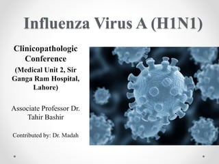
- 1. Influenza Virus A (H1N1) Clinicopathologic Conference (Medical Unit 2, Sir Ganga Ram Hospital, Lahore) Associate Professor Dr. Tahir Bashir Contributed by: Dr. Madah
- 2. Influenza• Seasonal influenza is an acute respiratory infection caused by influenza viruses that affect mainly the nose, throat, bronchi & occasionaly the lungs Classification: (on the basis of core Ribonucleoprotein & Matrix protein) Influenza virus A, B, C & D The subtypes of influenza A viruses are determined by envelope glycoproteins possessing either haemagglutinin (HA) or neuraminidase (NA) activity • There are 16 (HA) and 9 (NA) Antigens • Subtypes H1, H2 or H3, and N1 or N2 infect humans • Currently circulating in humans are subtype A(H1N1) and A(H3N2) influenza viruses. • The A(H1N1) caused the pandemic in 2009 and subsequently replaced the seasonal influenza A(H1N1) virus which had circulated prior to 2009.
- 4. Influenza viruses belong to the family Orthomyxoviridae (A family of RNA viruses) • Minor point mutations (“antigenic drift”) enables the virus to evade immune recognition, resulting in repeated influenza outbreaks during interpandemic years.
- 5. • Major changes in the HA antigen (“antigenic shift”) are caused by reassortment of genetic material from different A subtypes. • Result in new pandemic strains • Rare events • Occur through reassortment between animal and human subtypes.
- 7. Influenza A (H1N1) virus: emerged in 2009. It is a new reassortment that has never before circulated among humans, not closely related to previous or current human seasonal influenza viruses Influenza B viruses: sporadic cases Influenza C virus: detected less frequently and usually causes mild infections Influenza D viruses primarily affect cattle and are not known to infect or cause illness in people
- 9. Mode of transmission: • Droplet/ aerosol/ airborne infection: via coughing & sneezing • Contact: Direct (Hand to hand) Indirect (Hand to surface to hand) Incubation Period:1-7 days Communicability: • From 1 day before to 7 days after the onset of symptoms • Chances may persist till complete resolution of symptoms
- 10. Epidemiology/ Agent Factors Reservoir of infection: • Humans- Primary reservoir for human infections • Major reservoir: animals & birds (Swine, horses, dogs, cats, domestic poultry, water & wild birds) Source of infection: • A case or a subclinical case
- 11. Pathogenesis • Infects the respiratory tract • Even 3 or few viral particles can infect • Neuraminidase facilitates infection by reducing the viscosity of mucous • Ciliated cells are infected in the respiratory tract • When superficial layers are damaged, exposes the basal layers & causes bacterial infections
- 12. Case Definitions Suspected Case • A person with acute febrile illness(fever) with sorethroat, cough, headache & myalgias Probable Case • In addition to meeting the criteria of suspected case, one of the following: • H/o close contact with a confirmed case in last 7 days • H/O travel to a community having 1 or more confirmed cases in last 7 days or residence in it • Individual who died of an unexplained acute respiratory illness despite having a clinically compatible illness Confirmed Case •A person with an acute febrile respiratory illness with laboratory confirmed pandemic influenza A (H1N1) infection by 1 or more: •Real Time- PCR •Viral Culture •Four fold rise in Pandemc influenza A (H1N1) virus specific neutralizing antibodies
- 13. In 2009, cases of influenza like illness were first reported in Mexico on March 18; the outbreak was subsequently confirmed as H1N1 influenza A On June 11, 2009, WHO raised the pandemic alert level to phase 6 (indicating a global pandemic) because of widespread infection beyond North America to Australia, the United Kingdom, Argentina, Chile, Spain, and Japan. As of September 1, 2009, the WHO reported that H1N1 influenza had been confirmed in over 200,000 people in more than 100 countries and that they are aware of at least 2185 confirmed deaths.
- 14. Clinical Features • Asymptomatic infection • Uncomplicated upper respiratory tract disease Influenza‐like illness (ILI) symptoms include: Fever ≥ 38°C cough, sore throat, nasal congestion or rhinorrhea headache muscle pain and malaise NO DYSPNEA/ SOB Patients may present with some or all of these symptoms Gastrointestinal illness may also be present, such as diarrhoea and/or vomiting, especially in children, but WITHOUT EVIDENCE OF DEHYDRATION. Some patients with uncomplicated illness may experience atypical symptoms and may not have fever (e.g. elderly or immunosuppressed patients)
- 16. Complicated or severe influenza o Presenting features (dyspnoea, tachypnoea, hypoxia) and/or radiological signs of lower respiratory tract disease (e.g. pneumonia) o CNS involvement (e.g. encephalopathy, encephalitis) o Severe dehydration o Presenting secondary complications, such as renal failure, multi-organ failure and septic shock o Other complications can include rhabdomyolysis and myocarditis. o Exacerbation of underlying chronic disease, including asthma, chronic obstructive pulmonary disease (COPD), chronic hepatic or renal insufficiency, diabetes, or other cardiovascular conditions (e.g. congestive cardiac failure). Signs and symptoms of progressive disease Patients who present initially with uncomplicated influenza may progress to more severe disease: rapid (i.e. within 24 hours).
- 17. Indicators of progression Symptoms and signs suggesting oxygen impairment or cardiopulmonary insufficiency: Shortness of breath (with activity or at rest) Tachypnoea Cyanosis Bloody or coloured sputum, chest pain, and low blood pressure Hypoxia as indicated by pulse oximetry or arterial blood gases Symptoms and signs suggesting CNS complications: Altered mental status Recurring or persistent convulsions (seizures) Confusion, unconsciousness Weakness or paralysis Severe dehydration: decreased activity, dizziness, decreased urine output and lethargy Evidence of sustained virus replication or invasive secondary bacterial infection: based on laboratory testing or clinical signs persistent or recurrent high fever and other symptoms beyond 3 days
- 18. Workup/ Diagnostic Modalities • Hematological: CBC counts nonspecific but leukopenia & relative lymphopenia are nonspecific. Thrombocytopenia maybe present • Microbiological: Isolation of the viral culture (Confirmatory: may take more than 7 days) • Biochemical: Raised hepatic enzymes • Radiological: Chest Xrays • Confirmation of pandemic Influenza A H1N1 is through Nasal, Nasopharyngeal & throat swabs, wash/ aspirate Tracheal aspirate RT-PCR, RIDTs Paired serum for serological testing at an interval of 14 days: four fold rise in influenza specific antibodies
- 19. Rapid influenza diagnostic tests (RIDTs) are antigen detection tests that detect influenza viral nucleoprotein antigen Can provide results within 30 minutes or less Cannot distinguish between influenza A virus subtypes cannot provide any information about antiviral drug susceptibility low sensitivity than viral culture or RT-PCR Real-time reverse transcriptase-polymerase chain reaction (rRT-PCR): Detection of influenza-specific RNA Can provide results within 4 hours
- 21. Sample Collection • The sample should be collected by a trained physician/ microbiologist preferably before administration of the antiviral drug • Swab with synthetic tip made of calcium alginate is ideal within 5 days of the onset of symptoms • Keep at 4°C • in viral transport media upto 24 hours • If more than that, store at -70°C
- 22. Treatment guidelines • Infection control precautions: Early implementation • Prompt treatment • Early identification & follow up of persons at risk • Infrastructure/ Manpower/ Material Support: Isolation facilities: Wards with beds kept 1 metre apart Manpower: Dedicated doctors & paramedics Equipment: Portable Xray machines, ventilators & pulse oximeters Supplies: Adequate quantities of personal protection equipment, disinfectants, medication
- 23. • Standard Operating Procedures (SOPS): Reinforce standard infection control precautions: high efficacy gowns, masks, goggles, gloves, cap, shoe cover Restrict no. of visitors Antiviral prophylaxis to healthcare personnel managing the cases Proper waste disposal
- 24. Supportive Treatment IV fluids, plenty of oral fluids: Hydration & maintenance of electrolyte balance Supplemental O2 therapy: tachypnea, dyspnea, respiratory distress, O2 saturation <90% Mechanical ventilatory support: Severe pneumonia/ Acute respiratory failure (SpO2 < 90% & PaO2 <60% with O2 therapy Antibiotics for secondary infection, Vasopressors for shock Paracetamol/ ibuprofen for fever, myalgia & headache Avoidance of smoking For sorethroat: short course of topical decongestants, saline nasal drops, throat lozenges & steam inhalation Salicylate/ aspirin- contraindicated in children (potential to cause Reye’s syndrome) Constant monitoring of suspected cases for clinical/ radiologic evidence of LRTI & for hypoxia (Respiratory rate, O2 saturation, level of consciousness)
- 25. Antiviral Treatment Prompt initiation within 48 hours of the onset of symptoms Recommended duration of treatment: 5 days Effective against Influenza A virus: Amantadine & Rimantadine Effective against Influenza A & B viruses: Oseltamivir (Tamiflu) & Zenamivir (Relenza): Neuraminidase inhibitors
- 26. Pharmacological Management (WHO guideline August 20th, 2009) • Healthy people who catch mild to moderate cases of swine flu don’t need the drug at all • CDC has warned that the indiscriminate use of antiviral medications to prevent & treat influenza could ease the way for drug resistance to emerge
- 27. Antiviral susceptibilities of Circulating viruses:
- 28. Severe or Progressive symptoms • Confirmed or Strongly suspected • Severe or progressive • Antivirals are available • Treatment should be initiated as soon as possible • Treat with oseltamivir • Treat with Zanamivir if oseltamivir is not available
- 29. Uncomplicated • Confirmed or strongly suspected • Uncomplicated • Antivirals are available • Need not be treated with antivirals (patient not in “at risk” groups) • Treated with oseltamivir or zanamivir (patients in “at risk” groups)
- 30. Uncomplicated • At Risk means • Infants & children aged less than 5 • Elderly (more than 65) • Nursing home residents • Pregnant women • Patients with chronic comorbid conditions • Immunosuppression
- 31. Oseltamivir is the recommended drug of choice both for prophylaxis & treatment. However in view of increasing resistance Relenza (Zenamivir) in the dosage of 5mg BD may be preferred. Dosage of Oseltamivir: (By weight) For weight <15 kg : 30 mg BD for 5 days For weight 15-23 kg : 45 mg BD for 5 days For weight 24- <40 kg : 60 mg BD for 5 days For weight>40kg : 75 mg BD for 5 days
- 32. Inhaled Zanamivir (Relenza) Dosage Influenza A & B, treatment • Start within 2 days of symptom onset: administer 2 doses on day 1, at least 2 hours apart • 10 mg inhaled 12 hourly for 5 days. May consider longer treatment for patients severely ill after 5 days Influenza A & B (Prophylaxis) House hold settings: 10mg inhaled qDay for 10 days Initiate within 36 hours of exposure Community Outbreaks: Begin within 5 days of outbreak; May administer for upto 28 days
- 33. Oseltamivir/ Prophylaxis dosage • H1N1 influenza (swine flu) prophylaxis: 75mg PO qDay • Post exposure prophylaxis: Start within 7 days and continue for at least 10 days Incase of community outbreak: may be administered for upto 6 weeks
- 34. Chemoprophylaxis Indications • Risk of human to human transmission is high or low but the likelihood of complications is high • Oseltamivir or zanamivir might be used • For health care personnel at high risk only if not previously vaccinated • For contacts only if clinically indicated • Zanamivir can also be given for chemoprophylaxis (10mg, 2 inhalations OD)
- 35. Chemoprophylaxis Contraindications If the likelihood of complications of infection is low even if • “At risk” group • Healthcare workers • This recommendation applies independent of human to human transmission
- 36. Adverse Effects of Oseltamivir: Gastrointestinal Side Effects ( Transient Nausea, Vomiting) Occasionally, insomnia & vertigo Rare reports of anaphylaxis & rash Infrequently, abdominal pain, epistaxis, otitis media, dermatitis, conjunctivitis Should be given cautiously in patients with advanced renal disease Discharge Policy: Response to treatment after 2-3 days of initiation & patient becomes asymptomatic should be discharged after 5 days of treatment
- 40. Guidelines on Infection Control Measures During Pre-hospital care: • PATIENT SHOULD WEAR A SURGICAL MASK • Aerosol generating procedures should be avoided during transportation • The personnel in the patient’s cabin of ambulance should wear full complement of PPE. Driver should wear 3 layered surgical mask • The interior & exterior of the ambulance plus reusable patient care equipment needs to be sanitized using sodium hypochlorite / quaternary ammonium compounds During Hospital Care: • Admitted directly to isolation facility, MUST WEAR A MASK • Medic/ paramedic personnel attending the suspected, propbable or confirmed case should wear full complement of PPE
- 41. • Aerosol generating procedures such as Endotracheal intubation, suctioning, nebulization, chest physiotherapyshould be performed with the health personnel wearing the full complement of PPE with N95 mask on • Hand hygiene, Hand washing: Alcohol based sanitizers • Infection control precautions should continue for 7 days in adults and 14 days in children<12 years after the resolution of symptoms • Disinfection of contaminated surfaces and equipments should be done after discharge (Disinfectants used: 70% ethanol, 10% Sodium hypochlorite) • All waste generated from influenza patients in isolation wards/ rooms should be considered as infections waste & treated & disposed off.
- 42. Personal Protection Equipments (SOPS) • Gloves (Nonsterile) • Mask (High efficiency/surgical masks) • Long-sleeved, cuffed gown • Protective eyewear (goggles, face shields) • Cap (Maybe used in high-risk situations/ increased aerosols) • Plastic apron if splashing of blood, body fluids, excretions & secretions is anticipated • Shoe covers
- 43. Correct procedure for applying PPE 1. Hand wash 2. Wear coverall 3. Wear goggles 4. Shoe-cover 5. Head-cover 6. Face mask 7. Gloves Correct procedure for removing PPE 1. Remove gown 2. Gloves 3. Hand wash 4. Cap & face shield 5. Remove mask 6. Hand wash 7. Leave the room 8. Hand wash
- 44. • Guidelines for infection control practices: • 1. At individual level 1.1: Hand Hygiene: Frequent hand washing with soap & water/ alcohol based hand rubs/ antispetic hand wash & dry thoroughly
- 46. Or do you wanna end up like them?
- 47. 1.2 Respiratory Hygiene/ Cough etiquette: Cover the nose or mouth with a handkerchief/ tissue paper when coughing/ sneezing Use tissues to dispose respiratory secretions & dispose of them in the nearest waste receptacle after use Perform hand hygiene 1.3: Staying away: Stay arms length away from individuals showing influenza like illness 1.4. Use of mask: N95 mask for health personnel working in screening areas& isolation facilities
- 48. • 2. At Health Facility: • 2.1 Droplet precaution: • Wearing a surgical/ procedure mask • 2.2: Waste Disposal: • All waste from isolation wards, been treated as infectious waste, to be decontaminated. • Articles like swabs and gauzes discarded in yellow bag & gloves, face masks & disposable syringes discarded in blue/ white autoclavable biosafety bags.
- 50. Putting on PPE
- 51. Taking off PPE