Model Call Girl in Bikash Puri Delhi reach out to us at 🔝9953056974🔝
Inflammation of the lymph nodes, blood and lymph vessels. Inflammation of glandular organs. (lecture 11).pdf
1. Inflammation of the lymph nodes, blood and lymph vessels.
Inflammation of glandular organs.
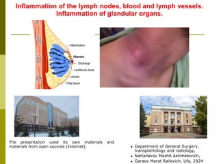
The presentation used its own materials and
materials from open sources (Internet). ◼ Department of General Surgery,
transplantology and radiology,
◼ Nartailakov Mazhit Akhmetovich,
◼ Garaev Marat Railevich, Ufa, 2024
2. - inflammation of the veins with their thrombosis.
Etiology: Factors leading to or contributing to venous thrombosis include
the following. Three main factors are considered classic (Virchow's triad).
1. The state of hypercoagulability (increased blood clotting). It can
occur as a result of injuries, operations, pregnancy and childbirth, against
the background of diabetes, excess fat intake, oral contraceptives,
dehydration; when an infection enters the walls of the veins during their
injuries, contact with inflammatory processes in soft tissues (for example,
with erysipelas, phlegmon), etc.
2. Injury to the vascular endothelium. Injury to adults during injections
into a vein, vein catheterization, trauma, radiation and chemotherapy, etc.
d.
3. Slow down blood flow. It can develop with varicose veins, pregnancy,
obesity, prolonged immobilization of a limb (immobilization in case of
injuries, for example), heart failure, forced position of the body (for
example, during long flights), compression of veins by tumors, etc.
Thrombosis can occur both under the action of one factor, and when they are
combined. Most often, thrombosis occurs in the veins of the lower
extremities.
There are thrombophlebitis of superficial and deep veins.`
Thrombophlebitis
Rudolf Ludwig Carl Virchow
(13/10/1821 - 5/09/1902)
3. Clinical picture: develops with pain in the projection of the veins (most often varicose veins - on the lower
leg), hyperemia of the skin, fever. Inflammation and thrombosis of the veins is accompanied by
inflammation of the surrounding tissues, hence the terms "thrombophlebitis" (phlebitis - inflammation of the
veins), "varicothrombophlebitis" (inflammation of a varicose vein) can often be heard. On palpation, a sharp
pain is determined along the thrombosed veins, which are palpated in the form of dense strands and nodes.
Hyperemia and hyperthermia of the skin is also observed along the course of the veins. Edema of the limb
is not expressed. The general condition of the patient, as a rule, does not particularly suffer. Sometimes
there is an increase in body temperature to subfebrile indicators.
Thrombophlebitis of superficial veins
4. With adequate treatment, inflammation subsides, the patency of the veins is partially or completely restored
over time. Cicatricle changes, changes in the valvular apparatus, and a decrease in the elasticity of the wall
may persist, which in the future increases the risk of recurrence of the inflammation process and chronicity.
Thrombophlebitis of superficial veins (continued)
5. Изменения вен в виде варикозного расширения являются благоприятствующим фоном для развития
тромбофлебита.
Thrombophlebitis of superficial veins (continued)
6. Complications:
- septicopyemia - the formation of abscesses in other organs and tissues as a result of the hematogenous
spread of fragments of an infected thrombus;
- ascending thrombophlebitis - thrombus growth in the proximal direction, the “head” of the thrombus can
grow to the proximal parts of the vessel and floats (“dangling”) with the threat of its detachment;
- pulmonary embolism - the most dangerous complication of thrombophlebitis: in case of inflammation of
the great saphenous vein on the thigh and the small saphenous vein in the popliteal region, complicated by
vein thrombosis, a thrombus fragment can also be torn off by the blood flow (the way the thrombus travels:
saphenous veins of the leg - great saphenous vein of the thigh - deep femoral vein - inferior vena cava -
right atrium - right ventricle - pulmonary artery) enter the pulmonary artery. There is an embolism
(blockage) of the branches or the main trunk (depending on the size of the emboli) of the pulmonary artery.
Massive pulmonary embolism in most cases ends in death.
Thrombophlebitis of superficial veins (continued)
7. A complication of thrombophlebitis is PE
PE on the right side in the X-ray, example No. 1
8. A complication of thrombophlebitis is PE (continued)
PE on the right side in the X-ray, example No. 2
10. A complication of thrombophlebitis is PE (continued)
PE on the angiopulmonogram (arrow indicates embolus)
11. Diagnostics:
1. Ultrasound of the veins - ultrasound of the vessels.
2. Ultrasound of the veins - unlike ultrasound, allows you to see and evaluate the condition of the vessel
wall.
3. Ultrasound with color mapping (triplex scanning) - simultaneously assesses the condition of the vessel
and blood flow through it in three modes (the most modern method).
Thrombophlebitis of superficial veins (continued)
12. Treatment: In acute thrombophlebitis, hospitalization of the patient to the hospital, bed rest, giving the
limb an elevated position is indicated. Drug therapy includes: - antibacterial drugs (for infectious
inflammation),
- direct anticoagulants - heparin (Fraksiparin, Enoxaparin, etc.), which change coagulants do not
require coagulogram control.
Local treatment: consists in the careful imposition of heparin-containing ointments on the area
of inflamed veins.
The process of putting on a
stocking with a butler
Thrombophlebitis of superficial veins (continued)
13. Surgical treatment:
With ascending thrombophlebitis, an urgent operation is performed under regional (spinal) anesthesia:
ligation of the great saphenous vein of the thigh at the place of its confluence with the deep femoral
vein (Troyanov operation) or ligation and removal of the altered vein (Troyanov-Trendlenburg
operation. This option is preferable). The purpose of the operation is to prevent the separation of a
blood clot and pulmonary embolism.
In the initial stages or limited forms of acute thrombophlebitis of the saphenous veins, sometimes an
operation is performed to excise the inflamed veins (venectomy).
Venectomy
Thrombophlebitis of superficial veins (continued)
14. - thrombosis and inflammation of deep veins (usually of the lower extremities) at different levels: deep
veins of the lower leg (thrombophlebitis of the sural veins), popliteal and deep femoral veins, iliac vein
(ileofemoral thrombosis), pelvic veins.
Clinical picture: the main clinical manifestation of deep vein thrombophlebitis is limb edema,
accompanied by arching pains, pastosity of the skin of the limb, tissue tension, and sometimes high
body temperature. Hyperemia of the skin is usually not observed. The main danger of deep vein
thrombophlebitis is pulmonary embolism.
Thrombophlebitis (thrombosis) of deep veins
Deep vein thrombosis on the left
15. With adequate treatment, the patency of the veins is partially or completely restored over time (the veins
are recanalized).
In connection with the redistribution of blood flow during the period of obstruction of deep veins, failure of
perforating veins develops (veins connecting the superficial and deep vein system), varicose
saphenous veins, which in turn lead to trophic changes in the skin and subcutaneous tissue in the
form of brown induration of the skin, alopecia and the appearance of ulcers. This process is called
post-thrombotic disease.
Thrombophlebitis (thrombosis) of deep veins (continuation)
Post-thrombotic disease of the right lower limb complicated by a trophic ulcer
16. Diagnostics: similar to measures for thrombophlebitis of superficial veins. Phlebography can sometimes be
used (the method is relatively complex, invasive and available only in large hospitals, which, combined
with the high capabilities of ultrasound methods, limits its use).
Treatment: With deep vein thrombosis, emergency hospitalization of the patient to the hospital, strict bed
rest, giving the limb an elevated position is indicated. Drug therapy includes: drugs to improve the
rheological properties of blood, anticoagulants of direct and indirect action, sometimes drugs for
thrombolysis. Anticoagulant therapy is carried out under the control of a coagulogram. Treatment
continues up to 2-3 weeks, until the organization of a thrombus and recanalization of the vein.
Surgical treatment: carried out with ascending thrombosis (thrombus grows rapidly in the proximal
direction), with the threat of separation of the thrombus with the risk of pulmonary embolism or
already developed thromboembolism. In such cases, under X-ray control, an endovascular installation
of a temporary or permanent cava filter is performed.
into the inferior vena cava. Temporary
the filter can be removed within
up to 3 months after surgery.
Stages of cava filter removal under
x-ray control
Сava filter
Thrombophlebitis (thrombosis) of deep veins
(continuation)
17. - inflammation of the lymphatic vessels, is of a secondary nature due to the penetration of infection from
the primary inflammatory focus.
Clinical picture: lymphangitis is manifested by a bright red path running from the primary inflammatory
focus to regional lymph nodes, fever, pain along the inflamed lymphatic vessels. On palpation, pain is
determined along the course of the inflamed vessel, which can be palpated in the form of a seal. As a
rule, inflammation of the regional lymph node (lymphadenitis) also occurs. Edema of the limb is not
expressed. Lymphangitis should be considered as a lymphogenous spread of infection and a threat to
the development of a generalization of the infectious process.
Diagnostics: similar to measures for thrombophlebitis of superficial veins.
Treatment: rest is necessary - the immobilization of the limb is performed. Antibacterial therapy is carried
out, sanitation of the primary inflammatory focus, sometimes physiotherapy.
Lymphangitis
18. An example of a subcutaneous panaritium of the 2nd finger of the right hand
complicated by lymphangaitis
19. - inflammation of the lymph nodes, often of a secondary nature, develops during the transition of
inflammation from the primary focus to the lymph nodes (often regional).
Classification (by stages):
1. Reactive (serous) lymphadenitis
2. Purulent lymphadenitis
3. Adenophlegmon.
Clinical picture: Lymphadenitis in the first days is manifested by an increase in lymph nodes, local pain,
swelling, general subfebrile temperature - this is the stage of reactive (serous) lymphadenitis,
with timely treatment it undergoes a reverse development.
With untimely treatment, lack of protective forces, serous lymphadenitis passes into a purulent stage. At
the same time, there is a rise in temperature to 38-39 degrees, hyperemia and hyperthermia of the
skin in the area of inflammation, and severe pain. Further development of the process leads to
purulent fusion of the lymph node and the transition of inflammation to the surrounding tissue -
adenophlegmon develops.
Lymphadenitis
Purulent lymphadenitis of
submandibular limf. nodes
Serous lymphadenitis of the
posterior cervical limf. node
Adenophlegmon
23. Diagnostics: Preliminary - based on the clinical picture. Additionally: Ultrasound examination, puncture,
sampling for histological, cytological, microbiological studies (depending on the form of the disease).
With an increase in lymph nodes, it is necessary to carry out differential diagnostics with such processes as:
- metastatic lesion of the lymph. nodes in malignant tumors;
- systemic diseases with defeat limf. tissues (lymphogranulomatosis, leukemia, etc.);
- strangulated femoral hernia (in the groin);
- chronic infectious diseases with defeat limf. nodes (tuberculosis, syphilis);
- HIV infection.
Лимфаденит (продолжение)
Arrows indicate
lymph nodes on
ultrasound.
25. Lymphadenitis (continued)
Adenophlegmon: before
and after surgery
Treatment:
With reactive lymphadenitis, anti-inflammatory therapy, sanitation of the primary focus, half-alcohol
compress, antibiotic therapy, and physiotherapy are carried out.
With purulent lymphadenitis, surgical treatment - opening, excision of lymph. knot and wound
drainage.
With adenophlegmon, after opening the abscess, it is necessary to excise the lymph node with adjacent
necrotic tissue. The wound is then kept open until the inflammation is relieved.
Drug therapy - as in other purulent processes.
27. - breast inflammation.
Classifications:
1. Depending on the functional activity of the mammary gland:
1. Lactation - against the background of lactation. Occurs mainly against the background of lactostasis in
nulliparous women. Also play a role: insufficient care for the mammary gland, microtrauma, improper
feeding;
2. Non-lactational (not associated with lactation).
2. By localization:
1. Subareolar - in the peripapillary zone;
2. Intramammary - in the thickness of the mammary gland;
3. Retromammary - behind the mammary gland;
(between the gland and the pectoralis major muscle);
4. Galacto-oophoritis - localization of inflammation
in the milk ducts;
5. Subcutaneous - in the subcutaneous tissue in
projections of the mammary gland.
Mastitis
28. Classifications (continued):
3. By stages:
1. infiltration;
2. Abscess formation.
4. By the nature of exudate and inflammation:
1. Serous;
2. Purulent;
3. Gangrenous.
5. According to the clinical course:
1. Acute;
2. Chronic (optional precancerous condition, often against the background of mastopathy).
Mastitis (continued)
Acute purulent lactational mastitis
with the formation of an
intramammary abscess in the outer
quadrants of the left breast.
Chronic purulent non-lactational
mastitis with the formation of a
fistula in the paraareolar zone
29. Clinical picture: characterized by edema, local arching pains, fever, hyperemia, general subfebrile
temperature - this is the stage of serous mastitis, with timely treatment it can reverse.
With untimely or insufficient treatment, weakness of the body's immune forces, serous mastitis passes into
a purulent stage. At the same time, there is a rise in temperature to 38-40 degrees and above,
hyperemia and hyperthermia of the skin in the area of inflammation, and sharp pain. During abscess
formation, fluctuation (softening in the center of the inflammation zone) can be detected.
Mastitis (continued)
Acute purulent lactational mastitis with the formation of an intramammary abscess in the outer quadrants of the left breast.
30. Diagnostics: Mammography (in chronic processes), ultrasound (in any form), fistulography (in the
presence of fistulas), thermography (mainly in research work), puncture, sampling for histological,
cytological, microbiological studies (depending on the form of the disease ). Rarely - MRI.
Mastitis (continued)
Purulent mastitis with ultrasound
Chronic mastitis on mammogram
32. Treatment:
In the 1st stage (infiltrative, serous) - conservative: immobilization of the gland (rest is created by
tight bandaging), milk evacuation (manually and with the help of an electric suction), penicillin-
novocaine blockade in the retromammary space (an outdated, rarely used method today),
antimicrobial drugs, drugs to suppress lactation (with lactational mastitis).
Mastitis (continued)
Milk evacuation
33. Treatment:
In the 2nd stage (abscess formation, purulent) - surgical treatment under general anesthesia:
1) the abscess is opened with one or more incisions, taking into account the location and size, the cavity is
treated with antiseptics and drained;
2) with retromammary mastitis, a semilunar incision is performed under the mammary gland in the
transitional fold, the further course of the operation is similar;
3) in the gangrenous form - multiple incisions with opening and drainage of all foci or (with total damage or
necrosis and the threat of generalization of the infectious process) amputation of the mammary gland;
4) in chronic forms - sectoral resection of the affected part of the gland;
5) at present, puncture and drainage of breast abscesses under ultrasound guidance are also practiced.
Mastitis (continued)
Typical incision sites for mastitis
34. Treatment: (with purulent chronic mastitis)
Mastitis (continued)
Scheme of sectoral resection of the
mammary gland
Sectoral resection process
35. An example of the treatment of chronic purulent mastitis
The condition before the operation
Condition after sectoral breast
resection
36. - inflammation of one or more salivary glands. Most often, large salivary glands become inflamed:
parotid and submandibular, less often - sublingual.
Since inflammation of the parotid salivary gland (parotitis) is the most common in general surgical practice,
we will consider it further.
Sialadenitis
Etiology: parotitis is a purulent
inflammation of the parotid
salivary gland that develops
when microorganisms penetrate
into it, most often from the oral
cavity along the excretory duct
of the gland. The penetration of
the infection is facilitated by a
decrease or cessation of
salivation. For example, in
dehydrated patients with
common infectious diseases or
after major operations. Also
(less often) microorganisms can
penetrate into the parotid gland
by the lymphogenous or
hematogenous route. Sometimes
inflammation occurs against the
background of cicatricial
strictures, the formation of
stones in the excretory ducts
(calculous sialadenitis).
Classifications (there are many): distinguish
Depending on the form:
edematous-infiltrative and
destructive (abscess, phlegmon, necrosis) forms of acute
parotitis.
According to the clinical course: acute and chronic
37. Examples of severe forms of mastitis and mastitis-like situations
Necrosis of the soft tissues of the breast with
deep phlegmon of the breast on the background
of immunodeficiency.
Malignant neoplasm of the breast with disintegration
and infection.
38. Parotitis (continuation)
Clinical picture: An increasing painful infiltrate appears in the region of the gland, with increased pain on
palpation. Body temperature rises to 39-40ºС. Pain limits chewing and swallowing. Tissue edema increases
every day, the skin over the gland becomes thinner, reddens, fluctuation is determined during abscess
formation. The general condition of the patient worsens as the disease develops, swelling can spread to the
neck, cheek, submandibular region, with purulent forms, swelling of the soft palate and the side wall of the
pharynx is sometimes also noted. Opening the mouth is sharply difficult due to the spread of inflammation
and edema to the masticatory muscles. On palpation of the gland area, saliva mixed with pus may be
released from the duct into the oral cavity. Some patients may develop facial paresis. In the absence of
surgical treatment for purulent mumps, pus can destroy the capsule of the gland with the spread of the
process to the subcutaneous tissue (development of phlegmon) and, sometimes, breaks out through the skin
forming a purulent fistula. Through the fistula, pus, sequesters (free-lying dead tissue fragments),
parenchyma and gland capsules come out.
Parotitis on external examination Parotitis when viewed from the
mucosal side
Abscessing mumps with outburst
41. Parotitis (continuation)
Diagnostics: In the blood test, there is an increase in leukocytes, a shift of the leukoformula to the
left. Depending on the form of the disease, the alleged causes, the patient's condition, the following
hardware methods can be used: contrast sialography (contrast injection into the gland duct followed by
radiography), CT, MRI, sialosonography (ultrasound of the salivary gland); thermosialography (graphical
representation of the temperature of the salivary gland). Additional modern methods include puncture
biopsy of the salivary gland under ultrasound guidance and sialoendoscopy (used mainly for suspected
cysts and neoplasms).
Contrast sialogram (the arrows on
the right indicate the area
of narrowing of the duct - the cause
of inflammation)
Inflammation of the salivary gland
on ultrasound
43. Parotitis (continuation)
Treatment: Depending on the form of parotitis, treatment can be conservative or surgical.
Operation for purulent parotitis
With the use of broad-spectrum antibiotics, most parotitis in the initial phases of development is
cured conservatively. When the initial symptoms of parotitis appear, antibiotics, thermal and
physiotherapeutic procedures (warming compresses, UHF therapy, etc.) are used. Showing increased
salivation, rinsing the mouth with antiseptics. If treatment is started in a timely manner, then in most cases
the inflammatory process in the parotid gland undergoes a reverse development, suppuration does not
develop.
With the ineffectiveness of conservative measures / the development of purulent parotitis, surgical
treatment is indicated - revision of the parotid gland, opening of purulent foci, creating conditions for the
outflow of exudate. Anesthesia is usually general. If there is a fluctuation, then the incision is made in the
place of the greatest softening of the tissues, the abscess cavity is inspected and drained. The incisions are
made taking into account the direction of the main branches of the facial nerve - parallel to them. When an
abscess breaks into the peripharyngeal space, it is drained through the bed of the submandibular salivary
gland. Radical surgery usually quickly leads to positive dynamics: inflammatory symptoms quickly stop, the
general condition of the patient improves. Exudate and sequesters flow through the main wound and
through the drains, the wound is cleaned and granulates.
44. Parotitis (continuation)
Complications:
Skin fistulas of the salivary glands
1. Bleeding from arrosted vessels located in the parenchyma of the parotid gland, carotid artery with their
purulent fusion.
2. Development of phlegmon of the peripharyngeal space.
3. With advanced purulent parotitis, the process may spread along the vascular bundle of the neck with
the formation of deep phlegmon of the neck and the development of mediastinitis.
4. With the breakthrough of the abscess, as well as after surgery for sialadenitis, formation of external
salivary fistulas is possible.
Prognosis (forecast): With early diagnosed parotitis and rational antibiotic therapy, the prognosis is
favorable: in most patients, it is possible to prevent the development of a purulent process. Purulent
parotitis with timely and radical surgical treatment is usually not accompanied by severe complications.
45. Thank you for your attention!
Ready to answer your questions.