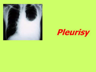
2. Pleurisy is an infectious or aseptic inflammation of
pleural leafs with formation of fibrin matters and/or
accumulation of fluid effusion (serous, purulent) in pleural
cavity.
Pleurisy should be distinguished from following
conditions:
•Pleural effusions – accumulation of pleural fluid of any cause in
general, including non-inflammatory cause.
•Adhesions and commissures in pleural cavity which are result of
pleurisy and identified as “adhesive pleurisy”.
3. Pleura:
- parietal
- visceral
Thickness of layer 6 - 15
micron
Volume of fluid 0.3 ml/kg –
on body mass = 18 - 22 ml;
protein - 1 g / dL
pressure = -10 cmH20;
4. Pleural effusions: pathogenesis
transudate – influence of systemic factors:
1. Change of systemic or pulmonary capillary pressure (increase of capillary
pressure in visceral pleura in left ventricle heart failure or increase of capillary
pressure in parietal pleura in right ventricle heart failure,
2. Decrease of oncotic plasma pressure : hypoproteinemia of various causes.
exudative – lesion of pleura itself:
1. Inflammatory pleura lesion increases its permeability, especially for protein.
2. Decrease of lymphatic outflow from pleural cavity (often observed in
tumorous lesion of pleura).
3. Increase of hydrostatic pressure gradient caused by decrease of intrapleural
pressure (in significant lesions of lung parenchyma).
5. Pathophysiology.
•Compression atelectasis
•Compressive mediastinum shift in intact side.
•Gas exchange impairment with hypoxemia.
•Hemodynamics impairment caused by heart shift and
compression of vena cava
Outcome of pleurisy
- complete resolution of exudation or
- formation of pleural adhesions.
- calcification (incrustation) of pleura.
Purulent exudation can not be resolved independently under any
circumstances.
10. DRY (FIBRINOUS) PLEURISY
(LOCAL, DISSEMINATED, BILATERAL)
Clinical manifestation
(are combined with manifestation of underlying condition)
- Chest pain associated with breathing motions;
- pleural friction rub, usually heard in both phases of
respiration, reminds snow crunch, paper rustle or skin scratch,
changes when pressed with phonendoscope, does not change
after cough.
Laboratory signs are not specific.
Radiological and ultrasound study may reveal pleura reaction
and also signs of underlying condition.
3 times sputum analysis.
11. Clinical manifestation:
- pain reduction and onset of chest congestion and dyspnea;
- forced semi-sitting position with bending on affected side,
-limitation of respiratory excursions,
-flatness (protrusion) of intercostal spaces on the affected side.
- percussion reveals dullness over zone of effusion
- Auscultation reveals diminished vesicular resonance, in large
effusions resonance can not heard.
SEROUS (SEROFIBRINOUS)
EXUDATIVE PLEURISY
12. INSTRUMENTAL DIAGNOSTICS
1. Radiological study (including side position) - fluid that excesses 100
ml causes opacity of posterior costodiaphragmatic recess or indistinct
contours of diaphragm.
SEROUS (SEROFIBRINOUS)
EXUDATIVE PLEURISY (continuation)
13. When volume of effusion increases shadow with oblique or
horizontal level appears.
When effusion is significant, symptoms of mediastinum shift
appear.
14.
15. 2. Ultrasound study of pleural cavities
- Is highly informative for encysted pleurisy, pericissuritis,
puncture control and dynamic follow-up.
- is painless, does not cause radiation exposure and can be
repeated several times.
3. Diagnostic thoracocentesis – procedure for verification of
effusion, determination of its type and finding etiologically
significant substances (microorganisms, tumor cells, etc.).
.
Complications of the procedure:
•Lung injury with development of pneumothorax, hemoptysis
and rarely, gas embolism.
•Injury of neurovascular bundle.
16. Sign Transudate Exudate
Relative density <1,015 > 1,018
Protein <20 g/l > 30g/l
Effusion protein/ Serum pro-
tein
<0,5 >0,5
Rivalt’s test negative positive
РН >7,3 <7,3
Cytology Mesenchyme cells, neutrophiles, lymphocytes,
erythrocytes, eosinophiles, tumor cells.
Laboratory criteria of types of pleural effusion
Detection of tumor cells, microorganisms
and parasites during microscopy of pleural
effusion sediment is an absolute diagnostic
sign which means final verification of
diagnosis.
Mesothelioma : tumor cells in
effusion sediment
21. SUPPURATIVE PLEURISY (PLEURAL EMPYEMA,
PYOTHORAX)
ACUTE pleural empyema:
- sudden onset, quickly progressing symptoms of purulent
intoxication (high grade fever up to 39-40, chills and excessive
sweating, significant shift of laboratory indices);
- physical and radiological symptoms are similar to those in
exudative (serofibrinous) pleurisy;
- Pus is found in thoracocentesis. Pus should be studied by means of
bacterioscopy, microbiology and cytology methods!
22. CHRONIC pleural empyema:
Purulent effusion can not be absorbed !
If purulent effusion breaks into bronchus then
pleuro-bronchial fistula is formed.
If purulent effusion breaks through chest wall
then pleuro-cutaneous fistula is formed.
If purulent effusion breaks both into bronchus
and through chest wall then bronchial-pleuro-
cutaneous fistula is formed.
23. TREATMENT
Treatment of pleurisy is treatment of underlying
condition.
Symptomatic treatment of dry pleurisy includes
analgesics and anti-inflammatory agents.
Symptomatic treatment of exudative pleurisy
includes anti-inflammatory and diuretic agents,
evacuation of effusion by means of puncture.
24. Acute pleural empyema:
- Daily punctures with evacuation of pus and
sanation of pleural cavity or
-Constant draining with active aspiration or
water seal drainage by Byulau
TREATMENT
-Antibacterial therapy
-Disintoxication and general restorative therapy.
Treatment of chronic empyema is operative
treatment.
Main goal — elimination of residual cavity,
fistula, lung expanding and restoration of lung
function.
25. Prophylaxis of pleural empyema
1. Rational and timely treatment of inflammation processes in
lung.
2. Timely evacuation of any effusions, accompanied by
intrapleural introduction of antibiotics if necessary.
3. If gas or blood are present in pleural cavity (chest trauma,
postoperational condition of lung, etc.) then contact of pleural
leafs and full expansion of lungs should be maintained.