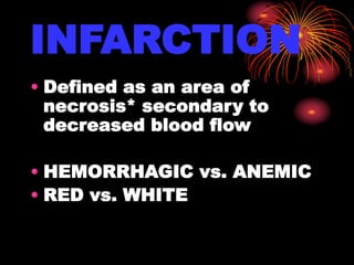
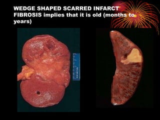
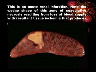
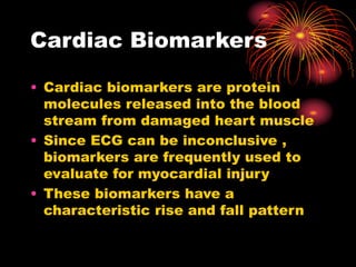
INFARCTION
BY
DR ABDUL AZIZ SHAIKH
INFARCTION
• Defined as an area of
necrosis* secondary to
decreased blood flow
• HEMORRHAGIC vs. ANEMIC
• RED vs. WHITE
WEDGE SHAPED SCARRED INFARCT
FIBROSIS implies that it is old (months to
years)
Microscopic features
This is an acute renal infarction. Note the
wedge shape of this zone of coagulative
necrosis resulting from loss of blood supply
with resultant tissue ischemia that produces
the pale infarct.
splenic infarcts
Hemorrhagic infarction
Cardiac Biomarkers
• Cardiac biomarkers are protein
molecules released into the blood
stream from damaged heart muscle
• Since ECG can be inconclusive ,
biomarkers are frequently used to
evaluate for myocardial injury
• These biomarkers have a
characteristic rise and fall pattern
Troponin T and I
• These isoforms are very
specific for cardiac injury
• Preferred markers for detecting
myocardial cell injury
• Rise 2-6 hours after injury
Peak in 12-16 hours
Stay elevated for 5-14 days
Creatinine Kinase ( CK-
MB)
• Creatinine Kinase is found in heart
muscle (MB), skeletal muscle (MM),
and brain (BB)
• Increased in over 90% of myocardial
infraction
• However, it can be increased in
muscle trauma, physical exertion,
post-op, convulsions, and other
conditions
Creatine Kinase (MB)
• Time sequence after myocardial
infarction
Begins to rise 4-6 hours
Peaks 24 hours
returns to normal in 2 days
• MB2 released from heart muscle and
converted to MB1.
• A level of MB2 > or = 1 and a ratio of
MB2/MB1 > 1.5 indicates myocardial
injury
Myoglobin
• Damage to skeletal or cardiac
muscle release myoglobin into
circulation
• Time sequence after infarction
Rises fast 2hours
Peaks at 6-8 hours
Returns to normal in 20-36 hours
• Have false positives with skeletal
muscle injury and renal failure
Renal Failure and Renal
Transplantation
• Diagnostic accuracy of serum
markers of cardiac injury are
altered in patients with renal
failure
• Cardiac troponins decreased
diagnostic sensitivity and
specificity in patients receiving
renal replacement therapy
CBC
• CBC is indicated if anemia is
suspected as precipitant
• Leukocytosis may be observed
within several hours after
myocardial injury and returns
returns to levels within the
reference range within one week
Chemistry Profile
• Potassium and magnesium levels
should be monitored and corrected
• Creatinine levels must be considered
before using contrast dye for
coronary angiography and
percutanous revascularization
C-reactive Protein (CRP)
• C- reactive protein is a marker
of acute inflammation
• Patients without evidence of
myocardial necrosis but with
elevated CRP are at increased
risk of an event
Chest X-Ray
• Chest radiography also reveals
complications of myocardial
infarction such as heart failure
• Echocardiography
• Echocardiography
• Therapy
•The goals of therapy in
AMI are the expedient
restoration of normal
coronary flow and the
maximum salvage of
functional myocardium
Infarction

Infarction

  • 2.
  • 3.
    INFARCTION • Defined asan area of necrosis* secondary to decreased blood flow • HEMORRHAGIC vs. ANEMIC • RED vs. WHITE
  • 4.
    WEDGE SHAPED SCARREDINFARCT FIBROSIS implies that it is old (months to years)
  • 5.
  • 6.
    This is anacute renal infarction. Note the wedge shape of this zone of coagulative necrosis resulting from loss of blood supply with resultant tissue ischemia that produces the pale infarct.
  • 7.
  • 8.
  • 9.
    Cardiac Biomarkers • Cardiacbiomarkers are protein molecules released into the blood stream from damaged heart muscle • Since ECG can be inconclusive , biomarkers are frequently used to evaluate for myocardial injury • These biomarkers have a characteristic rise and fall pattern
  • 10.
    Troponin T andI • These isoforms are very specific for cardiac injury • Preferred markers for detecting myocardial cell injury • Rise 2-6 hours after injury Peak in 12-16 hours Stay elevated for 5-14 days
  • 11.
    Creatinine Kinase (CK- MB) • Creatinine Kinase is found in heart muscle (MB), skeletal muscle (MM), and brain (BB) • Increased in over 90% of myocardial infraction • However, it can be increased in muscle trauma, physical exertion, post-op, convulsions, and other conditions
  • 12.
    Creatine Kinase (MB) •Time sequence after myocardial infarction Begins to rise 4-6 hours Peaks 24 hours returns to normal in 2 days • MB2 released from heart muscle and converted to MB1. • A level of MB2 > or = 1 and a ratio of MB2/MB1 > 1.5 indicates myocardial injury
  • 13.
    Myoglobin • Damage toskeletal or cardiac muscle release myoglobin into circulation • Time sequence after infarction Rises fast 2hours Peaks at 6-8 hours Returns to normal in 20-36 hours • Have false positives with skeletal muscle injury and renal failure
  • 14.
    Renal Failure andRenal Transplantation • Diagnostic accuracy of serum markers of cardiac injury are altered in patients with renal failure • Cardiac troponins decreased diagnostic sensitivity and specificity in patients receiving renal replacement therapy
  • 15.
    CBC • CBC isindicated if anemia is suspected as precipitant • Leukocytosis may be observed within several hours after myocardial injury and returns returns to levels within the reference range within one week
  • 16.
    Chemistry Profile • Potassiumand magnesium levels should be monitored and corrected • Creatinine levels must be considered before using contrast dye for coronary angiography and percutanous revascularization
  • 17.
    C-reactive Protein (CRP) •C- reactive protein is a marker of acute inflammation • Patients without evidence of myocardial necrosis but with elevated CRP are at increased risk of an event
  • 18.
    Chest X-Ray • Chestradiography also reveals complications of myocardial infarction such as heart failure • Echocardiography • Echocardiography
  • 19.
    • Therapy •The goalsof therapy in AMI are the expedient restoration of normal coronary flow and the maximum salvage of functional myocardium