2. 254
M. T. Hussan and others
MATERIALS AND METHODS
The experiment was carried out in the Department of Anatomy and Histology, Faculty of Veterinary Science,
Bangladesh Agricultural University, Mymensingh.
Chickens
A total 15 (fifteen) day-old “Cobb-500” broiler chickens of both sexes were purchased from “Kazi Farm Ltd.”
Mymensingh. The chickens had no developmental disorders and detectable diseases that may influence in the
distribution of lymphocytes and plasma cells containing different classes of immunoglobulins (IgA, IgG and IgM)
in lymphoid organs (bursa of Fabricius, cecal tonsils) and mucosal organ (ileum) of chickens in their postnatal
development.
Management
The chickens were reared in litter system in the poultry shed of the Department of Medicine, Faculty of
Veterinary Science, BAU, Mymensingh. Biosecurity of the poultry shed was maintained strictly. Optimum
temperature, lighting and ventilation were maintained in the brooder. To avoid the stress of transport, vitamin-C
with water was supplied on the first day. For the first day, the chickens were maintained on suji (coarse flour of
wheat) which was then replaced with a commercial ready feed (Paragon Poultry Feed, Mymensingh) and at day 4,
BCRDV vaccine was administered through intraocular route for the prevention of Ranikhat Disease.
Collection of sample
For immunohistochemical staining purpose, the bursa of Fabricius, cecal tonsils and ileum was collected after
sacrificing the chickens through cervical subluxation method. Sampling was done every 7 days interval starting
from day 3 up to 32 days of age. Samples were collected from three chickens on each occasion.
Preparation of samples for immunohistochemical studies
For immunohistochemical studies the samples were collected, cut into pieces and then fixed in the “Bouin’s
fluid” for 12 hours. The selected samples were dehydrated in a series of ascending grades of alcohol (70%, 80%,
95% and 100%), cleared in several changes of xylene, and infiltrated with different grades of melted paraffin in
the oven. The tissues were then embedded in paraffin and finally the sections were cut at 6 µm thickness using
sliding microtome (MIC 509, Euromex, Japan). After sectioning, the sections were floated on luke-warm water in
a floatation bath at 370
C for stretching then the sections were attached on cleaned glass slides using eggs albumin
and dried on a hot plate of slide warmer boxes. The sections were then deparaffinized first in several changes of
xylene followed by rehydration in a series of descending grades of alcohol (100%, 95%, 80% and 70%) and
finally. The histological sections were stained using indirect immunoperoxidase method as described earlier
(Khan et al., 1997) to understand the development and frequency of plasma cells containing different classes of
immunoglobulins (IgA, IgG and IgM) in the lymphoid organs (bursa of Fabricius and cecal tonsils) and mucosal
organ (ileum) of chickens in the different ages of postnatal development.
Antibodies
The antibodies for detecting Igs-positive cells used in this experiment were normal rabbit serum (Biosource,
Camarillo, California, USA), goat anti-chicken IgA (Bethyl Lab, USA), goat anti-chicken IgG (Bethyl Lab, USA),
goat anti-chicken IgM (Bethyl Lab, USA) and HRP-conjugated rabbit anti-goat IgG (Bethyl Lab, USA).
Immunohistochemistry
The tissues were fixed in Bouin’s fluid and were embedded in paraffin according to the conventional method.
Paraffin sections of 6 µm in thickness were immunostained by the indirect immunoperoxidase method. In brief,
after endogenous peroxidase was inhibited with methanol and hydrogen peroxide (H2O2), the sections were
overlayed with 2% normal rabbit serum (Biosource, Camarillo, California, USA) diluted with 0.01M phosphate
buffered saline (PBS) for 2 hours, followed by incubation with goat anti-chicken IgG (1:1000) (Bethyl Lab. Inc.
USA), goat anti-chicken IgA (1:1000) (Bethyl Lab. Inc. USA) or goat anti-chicken IgM (1:1000) (Bethyl Lab.
Inc. USA) for 18h at 40
C. After brief washing with phosphate buffered saline (PBS), sections were treated with
1% peroxidase-conjugated rabbit anti-goat IgG (1:1000) (Bethyl Lab. Inc. USA) for 1h at room temperature. The
positive reactions for different classes of Igs were revealed by treating the sections with 0.2 mg 3, 3′-diamino-
3. 255
Development of lymphoid tissues and mucosa of broilers
benzidine (DAB)-tetrahydrochloride dehydrate (AppliChem, Darmstadt) per ml of Tris-hydrochloride (0.05 M,
pH 7.6) containing 0.03% H2O2 and then counterstained few dips with hematoxylin.
Histoplanimetry
The immunopositive cells (Igs-positive cells) in the lymphoid tissues (bursa of Fabricius and cecal tonsils) and
mucosa (ileum) of different ages of chickens were counted in 20 fields at a magnification of 40 according to
Weibel (1969) and their relative frequency per 0.1 mm2
was calculated using ocular micrometer.
Counting of Igs-containing plasma cells
The Igs-positive cells in the cecal tonsils and ileum were counted in the lamina propria and in the core of the
villi, where the Igs-positive cells were uniformly distributed. Distributions of Igs-positive cells in the lymphoid
follicles were omitted for counting purpose. Photographs from the selected specimens were prepared and placed
in the paper for better illustration of the results (Fig. 1).
RESULTS AND DISCUSSION
The bursa of Fabricius, thymus and spleen are considered as major lymphoid organs of chickens, which were
very small at hatching. Their size increase gradually with advancement of age and at about 15 weeks of age the
size of lymphoid organs attained a peak (Khan et al., 1998). In the bursa of Fabricius, the Igs positive cells (IgA,
IgG and IgM) were found principally beneath the capsule, around the follicles and in the cortex and also medulla.
Table1. The frequency of Igs positive cells in the bursa of Fabricius of postnatal development of different
ages of broilers (mean±SD)
DaysIgs
Day 3 Day 11 Day 18 Day 25 Day 32
IgG
62±1.99 88.52±0.58 102.00±1.1
5
100.00±2.3
1
135.00±1.73
IgA
36±2.31 79.00±1.15
b
85.00±2.89 115.00±1.1
5
118.00±1.15
IgM
95±2.85 135.00±1.1
5
149.00±0.5
8
165.00±1.1
5
219.00±1.15
Table 2. The frequency of Igs positive cells in the cecal tonsils of postnatal development of different ages of
broilers (mean±SD)
DaysIgs
Day 3 Day 11 Day 18 Day 25 Day 32
IgG
15±2.15 62.00±0.58 95.00±1.15 118.00±1.15 122.00±0.58
IgA
0.00±0.00 80.00±1.15 112.00±1.15 115.00±1.73 140.00±0.58
IgM
65±1.95 90.00±0.58 101.00±1.15 112.00±1.15 135.00±0.58
4. 256
M. T. Hussan and others
In all stages of development (from day 3 to day 32) IgM positive cells were more followed by IgG and IgA (Table
1). These findings were similar with the findings of Honjo and Hirota (1993) stated that most of the bursal
lymphocytes were IgM positive, a small population of bursal cells were IgG positive and a few cells were IgA
positive. These findings were also embryologically supported by Rose (1979) observed that shortly after stem
cells first appear in the bursa (about the 12th
day of embryogenesis), they express surface IgM, and about the time
of hating IgG followed by IgA. Seeding out of the immunocompetent B-lymphocytes to the peripheral lymphoid
tissue occurs in the same sequence, i.e. IgM bearing cells, IgG bearing cells and IgA bearing cells.
Table 3. The frequency of Igs positive cells in the ileum of postnatal development of
different ages of broilers (mean±SD)
DaysIgs
Day 3 Day 11 Day 18 Day 25 Day 32
IgG
0.00±0.00 55.00±2.89 62.00±0.58 70.00±1.15 105.00±1.15
IgA
0.00±0.00 65.00±1.73 134.00±1.15 140.00±0.58 147.00±1.15
IgM
0.00±0.00 70.00±2.89 75.00±2.89 94.00±0.58 125.00±2.89
In the cecal tonsils, Igs positive cells (IgA, IgG and IgM) were distributed within the lymphatic nodules,
lamina propria and in the core of the villi, these findings were similar with the findings of Khan et al. (2008) and
Islam et al. (2008). Early in the postnatal development (at day 3), no IgA positive cells were found in the cecal
tonsils and IgM positive cells were more than IgG positive cells, these findings were with the findings of Jeurissen
et al. (1989) stated that in 5 days-old chickens, cecal tonsils were microscopically visible and consisted of large
HIS-C7 positive leukocyte infiltrates. The leukocytes included B-cells with membrane bound IgM, IgG or IgA
and some IgM and IgG plasma cells. At day 32, IgA positive cells were more than IgG and IgM positive cells
these findings were similar with the findings of Khan et al. (2008) and Islam et al. (2008). With the advancement
of age, the Igs positive cells (IgA, IgG and IgM) were increased (Table 2), these findings were similar with the
findings of Jeurissen et al. (1989) stated that in older chickens, the size of cecal tonsils and the number of IgM,
IgG and IgA plasma cells gradually increased.
In the early postnatal development (at day 3) no immunoglobulin positive cells were found in the ileum. From
The later stage, Igs positive cells (IgA, IgG and IgM )were found distributed in the lamina propria, around the
Intestinal glands and in the core of the villi of ileum, these findings were similar with the findings of Khan et al.
(2008). Immunoglobulin positive cells (IgA, IgG and IgM) were increased gradually with increasing age and IgA
positive cells were more than IgG and IgM positive cells at day 32 (Table 3), these findings were similar with the
findings of Islam et al. (2008).
5. 257
Development of lymphoid tissues and mucosa of broilers
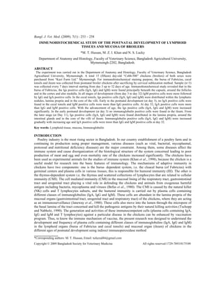
Fig. 1 (a-b). Immunostained sections from bursa of Fabricius in the chickens of 25 days old (a) and 32 days
old (b) showing IgG positive cells (arrow) x200.
Fig. 2 (a-b). Immunostained sections from cecal tonsils in the chickens of 3 days old (a) and 32 days old
(b) showing IgM positive cells (arrow) x200.
Fig. 3 (a-b). Immunostained sections from ileum in the chickens of 11 days old (a) and 32 days old (b)
showing IgA positive cells (arrow) x200.
a
a
a
b
b
b
6. 258
M. T. Hussan and others
REFERENCES
1. Befus AD, Johnston N, Leslie GA and Bienenstock J (1980). Gut-associated lymphoid tissue in the chicken. 1.
Morphology, ontogeny and some functional characteristics of Peyer’s patches. Journal of Immunology 125 (6): 2626-
2632.
2. Honjo K and Hirota Y (1993). Immunohistochemical investigations of lymphocytes in the lymphoid organs of
cyclophosphamide treated chickens. Journal of Veterinary Medical Science 55 (5): 895-897.
3. Islam MN, Khan MZI, Jahan MR, Karim MR and Yasuhiro Kon (2008). Comparative studies of mucosa and
immunoglobulin (Ig) containing plasma cells in the gastrointestinal tract of broiler and native chickens of Bangladesh.
Journal of Poultry Science 45: 125-131.
4. Janeway JCA, Jones B and Iayday A (1988). Specificity and function of T cell bearing δγ receptor. Immunology Today
9 : 73-76.
5. Jeurissen SHM, Janse EM, Koch G and Boer GF (1989). Postnatal development of mucosa-associated lymphoid
tissues in chickens. Cell and Tissue Research 258: 119-124.
6. Khan MZI, Hashimoto Y, Iwami Y and Iwanaga T (1997). Postnatal development of B-lymphocytes and
immunoglobulin containing plasma cells in the chicken oviduct: studies on the cellular distribution and influence of
sex hormones. Veterinary Immunology and Immunopathology 56:329-338.
7. Khan MZI, Hashimoto Y and Asaduzzaman M 1998. Development of T-cell sub-populations in postnatal chicken
lymphoid organs. Vetrinarski Arhiv 68(5): 183-189.
8. Khan MZI, Akter SH, Islam MN, Karim MR, Islam MR and Kon Y (2008). The effect of Selenium and Vitamin E on
the Lymphocytes and Immunoglobulin containing Plasma cells in the Lymphoid organ and Mucosa-Associated
Lymphatic Tissues of Broiler Chickens. Anatomia, Histologia, Embryologia 37: 52-59.
9. Rose ME (1979). Lymphatic System, Form and Function in Birds, Vol. 1., Ed. by King, K.S. and McLelland, I.,
Academic press, London, pp. 368: 324-372
10. Tschopp J and Nabholz M (1990). Perforin-mediated target cell lysis by cytolytic T lymphocyte. Annual Review of
Immunology 8: 279
11. Weibel ER (1969). Stereological principles for morphometry in electron microscopic cytology. International Review
for Cytology 26:235-302