2. 54 C. Fossum et al. / Veterinary Immunology and Immunopathology 158 (2014) 53–61
and phospholipid (Morein et al., 1984). The formulation
soon proved useful for a number of membrane-derived
antigens for induction of the protective immunity to
various microorganisms (Morein, 1987, 1988). Electron
microscopy demonstrated the formation of a spherical
cage-like structure, 40 nm in size, regardless of the source
of antigen. A standardized methodology and mixture of
Quil A, cholesterol and phophatidylcholine was established
for incorporation of amphipathic antigens (reviewed in
Lövgren Bengtsson and Morein, 2000). Later, it was realized
that incorporation of antigens into the ISCOM structure
was not necessary for potent immune stimulation (Lövgren
and Morein, 1988; Lövgren Bengtsson and Sjölander, 1996).
Pre-formed ISCOMs without incorporated antigen, called
ISCOM-Matrix and later referred to as Matrix, became used
as stand-alone adjuvant, simply mixed with antigens.
The glycosidic saponins, extracted from the bark of the
tree Quillaja saponaria Molina (Quil A), in free form, have
been used as adjuvant for almost a century (reviewed in
Dalsgaard et al., 1990). Quil A is a potent adjuvant; how-
ever, free saponins have haemolytic activity that may cause
side effects. By formulation of saponins with cholesterol
and phospholipids in ISCOMs or in Matrix particles, the
haemolytic activity is abolished and a more potent and less
reactogenic adjuvant is created. The Matrix made from Quil
A or similar preparations are currently called Matrix-Q.
Biochemical separation techniques revealed that the
lytic and structure forming capacities of Quil A were
mainly restricted to different fractions (Kensil et al., 1991;
Rönnberg et al., 1995) and that various combinations
of these components affected the adjuvant properties
considerably (Johansson and Lövgren-Bengtsson, 1999).
Detailed biochemical and functional characterisations of
the saponin fractions have resulted in a very well-
tolerated Matrix formulation called Matrix-M. Matrix-M
is a mixture of Matrix particles made from two dif-
ferent purified fractions of Quillaja derived saponins.
Matrix-MTM (Isconova AB, Uppsala, Sweden) is a potent
adjuvant that is licensed for several animal vaccines
and now also has entered human clinical trials (Lövgren
Bengtsson et al., 2011). Another similar, albeit differ-
ent product based on the ISCOM technology is the
ISCOMATRIX® adjuvant (CSL; Commonwealth Serum Lab-
oratories, Melbourne, Australia). MatrixTM formulations
are available in various forms and recommended for dif-
ferent species according to their saponin sensitivity and
there are two preparations available for research pur-
poses, AbISCO-100® (Matrix-M type) and AbISCO-300®
(Matrix-Q type).
Attempts to explore the power of Matrix formula-
tions on immune parameters have mainly been carried
out in mice. These studies have revealed a prominent
recruitment and activation of cells in the draining
lymph node/spleen, antigen delivery to dendritic cells
accompanied by cytokine and chemokine production.
This allows for cross-presentation with the subsequent
induction of cytotoxic T cells and a long-lasting antibody
response (Duewell et al., 2011; Morelli et al., 2012;
Reimer et al., 2012). However, which molecular path-
way(s) are activated by the Matrix of ISCOM is still not
clarified.
In the pig, early experimental ISCOM/Matrix-Q vac-
cines focused on Aujeszky’s Disease Virus (Tsuda et al.,
1991; Puentes et al., 1993; Tulman and Garmendia, 1994),
rotavirus (Iosef et al., 2002; Nguyen et al., 2003; González
et al., 2004; Nguyen et al., 2006a; Azevedo et al., 2010), and
the use of virus-like particles in combination with ISCOM-
Matrix (Tulman and Garmendia, 1994). The latter concept
has been used successfully in young pigs, inducing an active
immune response even in the presence of maternal immu-
nity (Nguyen et al., 2006b; McIntosh et al., manuscript) as
also shown for other ISCOM formulations applied in calves
(Hägglund et al., 2004) or mice (Morein et al., 2007).
Taken together, Matrix-M appears as a promising adju-
vant also in the pig where improved vaccines to several
diseases are desirable. The pig is also a valuable model for
studies of adjuvant effects in man because of many simi-
larities between the species in the organization of innate
immune cells and their cytokine production (Auray et al.,
2010; Bertho et al., 2011; Faibairn et al., 2011; Marquet
et al., 2011; Kapetanovic et al., 2012). To reflect the early
inflammatory response to Matrix-M in the pig, results
from in vitro studies using porcine blood mononuclear or
polymorphonuclear cells are reported here together with
results from an in vivo toxicity test carried out in piglets. The
findings are related to the global transcriptional response
to Matrix-M recorded in pigs at the site of injection and in
the draining lymph node (Ahlberg et al., 2012).
2. Materials and methods
2.1. Animals and experimental designs
Five experimental set ups were used to study the early
inflammatory response to the Matrix of ISCOMs in the pig.
An animal toxicity study was carried out in one-week old
PIC-crossbred piglets, housed in a 600 sow commercial
swine farrow-to-finish facility in Saskatchewan, Canada
(Set I). In vitro cellular toxicity studies were performed with
PBMC (Set II) and polymorphonuclear neutrophil leuko-
cytes (PMNL; Set III) purified from the blood of finishing
pigs housed in a specific pathogen free herd (Wallgren et al.,
1999; Swedish Livestock Research Centre, Lövsta-Uppsala,
Sweden). In vitro induced expression of cytokine mRNA (Set
IV) was determined using PBMC obtained from convention-
ally reared pigs at the University Research Station (Funbo
Lövsta, Uppsala, Sweden) whereas in vivo induced expres-
sion of mRNA for interferon-related genes (Set V) was
studied in lymph nodes obtained from 11 weeks old SPF-
pigs (Ahlberg et al., 2012). All procedures were conducted
in accordance with the University of Saskatchewan’s Com-
mittee for Animal Care and Supply (permit #20060004)
and with the Ethical Committee for Animal Experiments,
Uppsala, Sweden.
2.2. In vivo evaluation of Matrix-Q toxicity (Set I)
The potential for adverse reactions was tested by the
s. c. injection of variable doses of Matrix-Q (Isconova AB,
Uppsala, Sweden) into one-week-old piglets. Piglets were
injected with either 75 g (n = 3), 100 g (n = 3), or 150 g
(n = 3) of the Matrix-Q diluted in sterilized 0.01 M PBS to
3. C. Fossum et al. / Veterinary Immunology and Immunopathology 158 (2014) 53–61 55
a final volume of 200 L using a 1 mL-25 gauge needle. A
single injection was administered subcutaneously 1 cm off
the dorsal midline between the shoulders and piglets were
observed 7 times over a period of 30 h for rectal temper-
ature, injection site reactivity (swelling, redness, or pain),
and activity level (active or lethargic).
2.3. In vitro evaluation of Matrix-M toxicity (Set II)
The viability of PBMC was evaluated after 18 h incu-
bation in the presence of 0.3, 1 or 3 g Matrix-M
(AbISCO-100®; Isconova AB) per ml culture medium.
PBMCs were isolated from heparinized blood by density
gradient centrifugation on Ficoll-Paque (Pharmacia, Upp-
sala, Sweden). The cell viability was determined by flow
cytometry (FACSCanto flow cytometer; BD Biosciences,
San Jose, CA) using the Annexin V-FITC Apoptosis Detec-
tion Kit I with Propidium Iodide (PI) staining solution (BD
PharmingenTM, San Jose, CA) as recommended. Parallel cul-
tures with PBMC incubated in growth medium or treated
with UV-light (480 mJ for 90 s) before incubation were
included as negative and positive controls, respectively.
Ten thousand events per sample were collected and the
data were analyzed using the FACSDiva software (v. 5.0.2,
BD Biosciences).
2.4. In vitro effects of Matrix-M on PMNL activity (Set III)
Heparinized blood was diluted with an equal volume of
3% Dextran (T-2000, Pharmacia Biotech, Uppsala, Sweden)
and allowed to sediment for 30 min at RT. The leucocyte
rich fraction was washed once in PBS, resuspended and
layered on a discontinuous gradient of 70% and 80% Per-
coll (GE Healthcare, Uppsala, Sweden). After centrifugation
(300 × g) for 25 min, cells in the two bands generated were
collected, the cell numbers were counted and the purity
was determined on cytospin glasses stained with Diff Quick
(Vector Lab Inc., Burligame, CA). The cells recovered on the
80% Percoll cushion were enriched for PMNL (91.8 ± 6.1%,
n = 20) and used for studies of the formation of neutrophil
extracellular traps (NETs).
The PMNL were washed twice in PBS and resuspended in
RPMI 1640 supplemented with HEPES, l-glutamine antibi-
otics and 2% BSA (Sigma–Aldrich®). Two millilitre cell
suspension (107 neutrophils per ml) was seeded on cov-
erslips in six well plates (Costar®) and incubated for 1 h
at 37 ◦C. Thereafter, 400 l medium (negative control) or
phorbol 12-myristate 13-acetate (PMA; Sigma–Aldrich®)
at a final concentration of 100 nM (positive control) were
added to the wells. The effect of Matrix-M was tested at
the final concentrations of 0.3, 1 and 3 g/ml in parallel
cultures. After 4 h of incubation the medium was care-
fully removed from the wells and 1 ml of 50 nM Sytox
Green (Invitrogen) was added to each well and incu-
bated in the dark for 10 min. Thereafter formaldehyde
(ProlaboTM, Taipan, Malaysia) was added to a final con-
centration of 4% per well and incubated for 30 min before
the coverslip was carefully removed and allowed to dry
before mounted in Vectashield with DAPI (Vector Lab Inc.,
Burligame, CA). The morphology of cell nucleus and extra-
cellular DNA fragments were determined in a fluorescence
microscope (Nikon, Microphot-FX, Nikon Instruments Inc.,
Tokyo, Japan) equipped with a digital camera (Coolpix 990,
Nikon) for documentation.
2.5. In vitro Matrix-M induced expression of cytokine
genes (Set IV)
The effect of Matrix-M on the expression of cytokine
mRNA by porcine PBMC was tested in vitro and com-
pared to that induced by the CpG-ODN 2216 (Cybergene
AB, Huddinge, Sweden) or LPS (Sigma Aldrich, Stein-
heim, Germany), using methods detailed elsewhere (Bolind
Bågenholm, 2009; Wikström et al., 2011). All inducers were
diluted in RPMI 1640 medium (BioWhittaker, Cambrex
Bioscience, Verviers, Belgium) supplemented with 20 mM
HEPES buffer, 2 mM l-glutamine, 200 IU penicillin/ml,
100 g/ml streptomycin, 0.5 M 2-mercaptoethanol and
5% foetal calf serum (Invitrogen, Life Technologies, Carls-
bad, CA, USA). Three ml cell cultures (final concentration
5 × 10⁶ cells per ml) were established in 25 cm2 tissue cul-
ture flasks (Nunc, Roskilde, Denmark) containing Matrix-M
(1 g/ml), LPS (10 g/ml), ODN 2216 (5 g/ml) or only
growth medium. After 6 hours incubation at 37 ◦C, total
RNA was extracted, quality tested and DNA:se treated
before 2 g of RNA was used for cDNA synthesize as
described (Wikström et al., 2011).
Real-time TaqMan PCR was performed for IFN-␣, IFN-
␥, IL-6, IL-10, IL-12p40, IL-1, TNF-␣, TGF-, CXCR4,
RGS16 and two reference genes, Cyclophilin A (CyA)
and Hypoxanthine-guanine phosphoribosyl transferase
(HPRT), using previously published primers and protocols
(Wikström et al., 2011). The relative expression of target
genes induced by Matrix-M, LPS or ODN 2216 was com-
pared to their expression in PBMC cultured in plain growth
medium according to Livak and Schmittengen (2001), using
Cy A and HPRT as reference genes.
2.6. In vivo transcriptional response to Matrix-M (Set V)
Pigs were injected i.m. with either 150 g Matrix-M
suspended in 1 ml sterile endotoxin-free 0.9% NaCl solu-
tion (n = 6) or just saline (n = 6). Twenty four h after
injection, pigs were necropsied and tissues from the site
of injection and their draining lymph nodes were col-
lected and analyzed for early transcriptional response using
Affymetrix GeneChip® Porcine Genome Array as detailed
in Ahlberg et al. (2012). This was followed up by a more
comprehensive expression analysis focusing on IFN-␣, IFN-
, osteopontin (OPN) and stimulator of interferon genes
(STING) in the draining lymph node. Sample preparation,
RNA extraction and analysis, and cDNA construction are
detailed in Ahlberg et al. (2012).
Primer pairs for IFN-␣, IFN-, OPN, riboso-
mal protein L32 (RPL32), STING and tyrosine
3-monooxygenase/tryptophan 5-monooxygenase activa-
tion protein (YWHAZ) were chosen from published works
favouring those spanning an intron, and reoptimized in
house (Table 1). SYBR Green PCR was run as described
previously (Hjertner et al., 2013). Using PCR base line
normalized Cq values the expression of IFN-␣, IFN-,
OPN and STING was normalized to the geometric average
4. 56 C. Fossum et al. / Veterinary Immunology and Immunopathology 158 (2014) 53–61
Table 1
Primer details and gene specific optimized conditions.
Gene Primer sequence Primer location Target sequence Anneal
temp (◦
C)
Primer
conc (nM)
E (%)g
r2
Melt point
(◦
C)
IFN-␣a
F:AGCCTCCTGCACCAGTTCTG 346–365 NM 214393.1 60 500 100 0.997 84.5
R: TCACAGCCAGGATGGAGTCC 469–450
IFN-b F: TAGCACTGGCTGGAATGAAACC 288–309 NM 001003923.1 58 400 104 0.993 79.5
R: TCAGGTGAAGAATGGTCATGTCT 427–405
OPNc
F: TTGGACAGCCAAGAGAAGGACAGT 731–754 NM 214023.1 56 300 93 0.997 82.5
R: GCTCATTGCTCCCATCATAGGTCTTG 851–826
RPL32d F: CGGAAGTTTCTGGTACACAATGTAA 249–273 NM 001001636.1 55 300 97 0.997 77
R: TGGAAGAGACGTTGTGAGCAA 342–322
STINGe
F: TTACATCGGGTACCTGCGGC 489–508 NM 001142838.1 56 500 101 0.992 82
R: CCGAGTACGTTCTTGTGGCG 572–553
YWHAZf F: ATTGGGTCTGGCCCTTAACT 961–980 XM 001927228.4 58 400 101 0.997 78.5
R: GCGTGCTGTCTTTGTATGACTC 1106–1085
a
Wikström et al. (2011).
b
Lin et al. (2013).
c
White et al. (2005).
d
Dawson et al. (2004).
e
Xie et al. (2010).
f
Uddin et al. (2011).
g
Efficiency from serial dilutions of reference cDNA.
of RPL32 and YWHAZ (Vandesompele et al., 2002), and
calibrated to the average expression in all six control pigs.
2.7. Statistical analyses
Statistical differences in mRNA expression for IFN-␣,
IFN-, STING and OPN between pigs injected with Matrix-
M or saline were analyzed using the non-parametric
Mann–Whitney test whereas statistical differences for var-
ious in vitro treatments were determined using the paired
Student’s t-test (GraphPad Prism version 5.0 for Mac OS X,
GraphPad Software, San Diego, CA). p-Values ≤ 0.05 were
regarded as significant. For all statistical analyses detailed
in Ahlberg et al. (2012), q-values, i.e., p-values corrected for
multiple testing were used.
3. Results and discussion
3.1. In vivo evaluation of Matrix-Q toxicity (Set I)
The safety of saponin-based Matrix adjuvants in pigs
was studied with Matrix-Q in one-week-old piglets. No
adverse reactions were observed at the injection site
nor was the activity level of any piglet during the 30 h
period after injection with the Matrix-Q affected. The
expected normal rectal temperature of a suckling piglet
>24 h of age is 39.2 ± 0.3 ◦C (Straw et al., 1999). Taking
into consideration the baseline rectal temperature of each
piglet (temperature prior to injection), one piglet at 30 h
post-injection recorded a low grade fever of 39.6 ◦C (a tem-
perature greater than 0.3 ◦C above its baseline temperature
and greater than 39.5 ◦C). However, this piglet received the
lowest dose of Matrix-Q of 75 g.
3.2. In vitro evaluation of Matrix-M toxicity (Set II)
The effect of Matrix-M on cell survival was tested
in vitro using PBMC obtained from five pigs (Fig. 1). After
18 h of culture the proportion of Annexin labelled cells
M
edium
0.3
M
atrix
1
M
atrix
3
M
atrix
U
V
0
5
10
15
20
Percentage
a
M
edium
0.3
M
atrix
1
M
atrix
3
M
atrix
U
V
0
20
40
60
80
100
Percentage
b
Fig. 1. Percentage of apoptotic (a) or dead (b) porcine PBMC after 18 h of
incubation in the presence of 0.3, 1 or 3 g per ml of Matrix-M. The cor-
responding figures for PBMC incubated in medium or UV-treated before
incubation are included as negative and positive controls, respectively.
5. C. Fossum et al. / Veterinary Immunology and Immunopathology 158 (2014) 53–61 57
that were negative for PI was in mean 12.6 ± 4.6% for
cells incubated in the presence of Matrix-M, regardless
of concentration. This was similar to cells incubated in
growth medium (11.7 ± 2.7%) but higher than for those
that were UV-treated (6.7 ± 3.5%) prior to incubation. A
great proportion (74.5 ± 16.4%) of the UV-treated PBMC
was however stained with both PI and Annexin indicat-
ing a low cell survival. The corresponding figure for PBMC
grown in various concentrations of Matrix M (12.2 ± 3.7%)
was again very similar to that for PBMC grown in culture
medium (12.6 ± 2.3%). Thus, Matrix M did not induce apo-
ptosis or increase cell death during the present culture
conditions.
3.3. In vitro effects of Matrix-M on PMNL activity (Set III)
The ability of Matrix-M to induce formation of NETs was
tested in vitro using porcine PMNL obtained from pigs in a
SPF herd. The number of PMNL recovered per ml blood was
low due to the high health status, and prior activation of
them in vivo was less likely than for PMNL obtained from
conventionally reared pigs. In accordance, no signs of acti-
vation were evident after 4 h incubation in plain growth
medium (Fig. 3a) whereas a high proportion of neutrophils
with a condensed nucleus, showing signs of NETosis and
release of NETs was observed among the neutrophils incu-
bated with PMA (Fig. 3b and c).
The effect of Matrix-M on the neutrophils was less
evident. In the cultures supplemented with 0.3 or 1.0 g
Matrix-M per ml (Fig. 3d and e) the nucleus of the neu-
trophils showed the same shape as those incubated in plain
growth medium. Among neutrophils cultured in the pres-
ence of 3 g Matrix-M per ml (Fig. 3f) a slightly higher
proportion of the neutrophils had a condensed nuclei but
no NET formation was detected. Prolonged incubation, up
to 16 h, affected the survival of the neutrophils regardless
of culture set up. Still, no NET formation was observed in
cultures supplemented with Matrix-M or in the cultures
with plain growth medium. Instead, the nuclei of PMNL
grown over night in the presence of Matrix-M were dis-
integrated into small Sytox-stained fragments dispersed in
considerably swelled cells or around disrupted cells, giving
the impression of a pyroptotic cell death. The activation
of cathepsin genes (CTSB, CTSD, CTSH, CTSS and CTSZ) at
the injection site and the up regulation of the genes for
IL-1 and IL-18 (Table 5, Ahlberg et al., 2012) agree with
pyroptosis (Labbé and Saleh, 2011).
3.4. Early cytokine response of PBMC exposed to
Matrix-M in vitro (Set IV)
Analyses of the immediate cytokine response of PBMC
after in vitro exposure to Matrix-M revealed a low level
induction of TNF-␣ mRNA (p < 0.05) after 6 h incubation.
The mRNA expression for the other cytokines analyzed
did not differ from that of PBMC cultured in plain growth
medium. In comparison, mRNA specific for IL-1, IL-6 and
IL-10 (p < 0.001), and IL-12p40 and TNF-␣ (p < 0.01) was
induced after 6 h exposure of the PBMC to LPS, and IFN-
␣ mRNA (p < 0.01) was induced after 6 h in the presence
of the ODN 2216. Thus, Matrix-M did not evoke any strong
Table 2
Significant differences in relative mRNA expression determined by qPCR.
RNA was isolated after 6 h culture of porcine PBMC in the presence
of Matrix-M (1 g/ml), LPS (10 g/ml), ODN 2216 (5 g/ml) or in plain
growth medium.
Target gene Matrix-M vs.
medium (p
value)
LPS vs. medium
(p value)
ODN 2216 vs.
medium (p
value)
IFN-␣ ns * **
IFN-␥ ns ns *
IL-6 ns *** *
IL-10 ns *** *
IL-1 ns ***
ns
TNF-␣ * ** *
TGF- ns *
ns
IL-12p40 ns ** *
CXCR4 ns ns **
RGS16 ns ns ns
ns: not significant.
*
p ≤ 0.05.
**
p ≤ 0.01.
***
p ≤ 0.001.
immediate pro-inflammatory cytokine response in porcine
PBMC when tested in vitro. These results are in agreement
with the absence of fever and local reactions in the tox-
icity tests but caution should be taken regarding the fact
that these studies were performed in cultures of purified
lymphoid cells, devoid of PMNL and possibly also lacking
important soluble factors produced by other cells.
3.5. Global transcriptional response to Matrix-M at the
site of administration and in draining lymph node (Set V)
In order to better reflect the immune stimulatory prop-
erties of Matrix-M the global transcriptional response was
analyzed following i.m. administration of 150 g Matrix-M
in pigs aged eleven weeks (Ahlberg et al., 2012). Histo-
logical examination revealed an infiltration of leukocytes,
haemorrhage and necrosis in muscle after 24 h. At this
time, different grades of reactive lymphoid hyperplasia
were recorded in the draining lymph node that showed a
Fig. 2. Interferon-regulated genes (IRGs) upregulated at the injection site
(n = 36) and/or draining lymph node (n = 38) 24 h after Matrix-M admin-
istration. The ten most up-regulated IRGs in each group are listed. IRGs
were defined by the database Interferome (http://www.interferome.org).
6. 58 C. Fossum et al. / Veterinary Immunology and Immunopathology 158 (2014) 53–61
moderate enlargement in three of the six pigs injected with
Matrix-M. These observations (Table 2 and Fig. 1; Ahlberg
et al., 2012) were well in line with the absence of visi-
ble signs of inflammation when pigs were injected with
a similar dose of Matrix-Q and the marginal indication of
a pro-inflammatory cytokine response in porcine PBMC
exposed to Matrix-M in vitro.
As reported in Ahlberg et al. (2012), an inflammatory
response to Matrix-M at the injection site was evident
from the enrichment of genes in gene ontology (GO)
terms such as ‘immune response’ (q = 1.4E−10), ‘response
to wounding’ (q = 1.4E−9), ‘defence response’ (q = 5.1E−9),
‘inflammatory response’ (q = 7.4E−9), ‘positive regula-
tion of immune system process’ (q = 2.1E−4) and ‘innate
immune response’ (q = 7.2E−4). The corresponding analy-
sis of gene regulation in the draining lymph node (Ahlberg
et al., 2012) revealed an enrichment of up regulated genes
in the GO-terms ‘immune response’ (q = 9.9E−6), ‘response
to virus’ (q = 4.6E−4), ‘defence response’ (q = 2.3E−3), ‘reg-
ulation of cell proliferation’ (q = 1.1E−2) and ‘behaviour’
(q = 4.8E−2).
Cell migration patterns induced by Matrix-M were
characterized by enrichment for “myeloid cells” and in
particular by plasmacytoid dendritic cells (pDC). This
profile, together with the prominent up regulation of a
considerable number of interferon regulated genes (IRGs),
both at the site of injection and in the draining lymph node
(Fig. 2) initiated studies on possible mechanisms for induc-
tion of IFN production by Matrix-M. Because the genes
for two cytosolic RNA-sensing receptors (RIG-1 and MDA5)
both were upregulated as well as MyD88 that mediates
signalling via TLR7 and TLR9, it was tempting to specu-
late that nucleic acid played a part in the induction of
IRGs.
In patients with the autoimmune disorder systemic
lupus erythematosus (SLE), self-DNA released from PMNL
in the form of NETs can trigger pDC to produce IFN-␣
(Lande et al., 2011). In that case autoantibodies to DNA
are thought to mediate the uptake and delivery of DNA to
endosomal TLR9. One of the advantages with the ISCOMs
is their ability to deliver antigen to the cytosol allowing
presentation of antigen on MHC class I molecules and the
induction of a cytotoxic T cell response (Takahashi et al.,
1990). Therefore, Matrix-M could in theory mediate trans-
port of any associated nucleic acid over cell membrane(s)
and thereby initiate an IFN-response. The studies of PMNL
exposed to Matrix-M in vitro (Set III) did however not sup-
port the hypothesis that Matrix-M induced NET formation,
although a prominent influx of neutrophils were recorded
after in vivo administration.
Fig. 3. Porcine neutrophilic granulocytes incubated for 4 h in plain growth medium (a), 100 nM PMA, 20× magnification (b), 100 nM PMA, 40× magnification
(c) or 0.3 g (d), 1 g (e), 3 g (f) Matrix-M per ml.
7. C. Fossum et al. / Veterinary Immunology and Immunopathology 158 (2014) 53–61 59
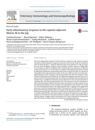
IFN-αα
1 2 3 4 5 6 7 8 9 10 11 12
0.0
0.5
1.0
1.5
2.0
Relativeexpression
IFN-ββ
1 2 3 4 5 6 7 8 9 10 11 12
0
10
20
30
40
OPN
1 2 3 4 5 6 7 8 9 10 11 12
0
5
10
15
Matrix Saline
Relativeexpression
STING
1 2 3 4 5 6 7 8 9 10 11 12
0
1
2
3
4
Matrix Saline
Fig. 4. Relative expression of IFN-␣, IFN-, OPN and STING in the iliac lymph node of pigs exposed to Matrix-M (nos. 1–6) or saline (nos. 7–12). The
expression was normalized to the geometric average of RPL32 and YWHAZ and calibrated to the average expression of all six control pigs. The difference
in expression between Matrix-M treated and saline treated groups was statistically significant (p < 0.05) for IFN- and OPN.
3.6. In vivo effects of Matrix-M on IFN-related responses
(Set V)
The indicated IFN response in pigs early after injection
with Matrix-M was followed up with an analysis of the
expression of IFN-␣ and -, as well as OPN (SPP1) and
STING in the draining lymph nodes of pigs that received
Matrix-M (n = 6) or just saline (n = 6). OPN and STING have
recently been identified as two modulators acting on two
different pathways that signal the expression of IFN-␣/
in response to viral infections as reviewed by Levy et al.
(2011). OPN, which had more than a hundred-fold increase
in expression at the site of injection (Ahlberg et al., 2012)
is an essential factor for endosomal TLR7/9 dependent
induction of IFN-␣/ expression via the adaptor molecule
MyD88 (Shinohara et al., 2006). STING is essential for
cytoplasmic foreign DNA sensor signalling, as well as RIG-
like receptor (RLR) signalling, acting through recruitment
of TBK1 (Ishikawa et al., 2009). Furthermore, recently a
new innate detection mechanism involving STING/TBK1
but independent of TLR and RLR pathways was described,
which was triggered by virus-cell membrane fusion only
(Holm et al., 2012).
Transcripts from IFN-␣, IFN-, OPN and STING could
be detected in the lymph node of all pigs except for two
control pigs, which had no detectable IFN- expression.
In this case a Cq value of 40 was assigned. The relative
expression of IFN-␣ was always less than two with no
difference in expression between Matrix M-treated and
control pigs (Fig. 4). However, four pigs (nos. 1, 2, 3 and 6)
out of the six Matrix-M-treated pigs showed elevated IFN-
 levels (p < 0.05) compared to the control group (Fig. 4).
In three of these pigs (nos. 1, 3 and 6) the relative expres-
sion of OPN was increased. The expression of OPN in the
Matrix-M treated group of pigs was significantly (p < 0.05)
higher than in the control group (Fig. 4). The fourth pig with
elevated IFN- expression (no. 2) had an OPN expression
level comparable to the control pigs but in this pig the rela-
tive expression of STING was increased threefold. However,
overall STING expression showed only a small increase in
two of the Matrix-M treated pigs and as a group, this was
not significantly different to the control group. Thus, four
pigs treated with Matrix-M showed an increase in IFN-
expression and these pigs also expressed an elevated level
of OPN and/or STING. For three of these pigs (nos. 1, 2, and 3)
the sampled draining lymph node was enlarged at autopsy,
further indicating a potent immune activation.
4. Conclusion
The adjuvant Matrix-M induces an inflammatory
response characterized by a rapid influx of neutrophilic
granulocytes and activation of genes regulating early
inflammation and other immune response genes in
8. 60 C. Fossum et al. / Veterinary Immunology and Immunopathology 158 (2014) 53–61
the pig. Functional annotation analysis and gene set
enrichment analysis specified a significant enrichment of
“myeloid cell” and pDC. The latter suggestion is well in
line with the distinct interferon-regulated gene profile
recorded both at the site of injection and in the draining
lymph node. In accordance the relative expression of
IFN- was increased in the draining lymph node 24 h after
injection with Matrix-M. As the IFN- expression was mir-
rored by an increased expression of OPN and STING both
the endosomal TLR7/TLR9 pathway including MyD88/OPN
and the cytosolic STING/TBK1 pathways could be involved
in Matrix-M mediated IFN activation, although the former
pathway seems like the preferred one. The Matrix com-
ponent inducing the interferon response still has to be
identified but the present Matrix-M formulation was well
tolerated in young piglets, signifying that the magnitude of
the inflammation was appropriate to initiate an immune
response without causing any adverse side reactions.
Acknowledgements
We are thankful for the excellent technical assistance of
the staff of the Prairie Diagnostic Services and the Prairie
Swine Centre of Saskatoon, Canada and by Ulla Schmidt
at the University Research Station, Funbo Lövsta, Upp-
sala, Sweden. Funding for this project was provided by
the EU 6th framework programme Food-CT-2004-513928,
Control of Porcine Circovirus Diseases (PCVDs): Towards
Improved Food Quality and Safety and by the Swedish
Research Council for Environment, Agricultural and Spatial
Planning (FORMAS).
References
Ahlberg, V., Lövgren Bengtsson, K., Wallgren, P., Fossum, C., 2012. Global
transcriptional response to ISCOM-Matrix adjuvant at the site of
administration and in the draining lymph node early after intramus-
cular injection in pigs. Dev. Comp. Immunol. 38, 17–26.
Auray, G., Facci, M.R., van Kessel, J., Buchanan, R., Babiuk, L.A., Gerdts,
V., 2010. Differential activation and maturation of two porcine DC
populations following TLR ligand stimulation. Mol. Immunol. 47,
2103–2111.
Azevedo, M.S., Gonzalez, A.M., Yuan, L., Jeong, K.I., Iosef, C., Van Nguyen,
T., Lövgren-Bengtsson, K., Morein, B., Saif, L.J., 2010. An oral versus
intranasal prime/boost regimen using attenuated human rotavirus
or VP2 and VP6 virus-like particles with immunostimulating com-
plexes influences protection and antibody-secreting cell responses to
rotavirus in a neonatal gnotobiotic pig model. Clin. Vaccine Immunol.
17, 420–428.
Bertho, N., Marquet, F., Pascale, F., Kang, C., Bonneau, M., Schwartz-
Cornil, I., 2011. Steady state pig dendritic cells migrating in skin
draining pseudo-afferent lymph are semi-mature. Vet. Immunol.
Immunopathol. 144, 430–436.
Bolind Bågenholm, A., 2009. Early immune response to and adjuvant
(AbISCO-100®
) tested in porcine peripheral blood mononuclear cells.
http://stud.epsilon.slu.se
Dalsgaard, K., Hilgers, L., Trouve, G., 1990. Classical and new approaches
to adjuvant use in domestic food animals. Adv. Vet. Sci. Comp. Med.
35, 121–159.
Dawson, H.D., Royaee, A.R., Nishi, S., Kuhar, D., Schnitzlein, W.M., Zucker-
mann Jr., F., Urban, J., Lunney, J.K., 2004. Identification of key immune
mediators regulating T helper 1 responses in swine. Vet. Immunol.
Immunopathol. 100, 105–111.
Duewell, P., Kisser, U., Heckelsmiller, K., Hoves, S., Stoitzner, P., Koernig, S.,
Morelli, A.B., Clausen, B.E., Dauer, M., Eigler, A., Anz, D., Bourquin, C.,
Maraskovsky, E., Endres, S., Schnurr, M., 2011. ISCOMATRIX adjuvant
combines immune activation with antigen delivery to dendritic cells
in vivo leading to effective cross-priming of CD8+ T cells. J. Immunol.
187, 55–63.
Fairbairn, L., Kapetanovic, R., Sester, D.P., Hume, D.A., 2011. The mononu-
clear phagocyte system of the pig as a model for understanding human
innate immunity and disease. J. Leukoc. Biol. 89, 855–871.
González, A.M., Nguyen, T.V., Azevedo, M.S., Jeong, K., Agarib, F., Iosef, C.,
Chang, K., Lövgren-Bengtsson, K., Morein, B., Saif, L.J., 2004. Antibody
responses to human rotavirus (HRV) in gnotobiotic pigs following a
new prime/boost vaccine strategy using oral attenuated HRV prim-
ing and intranasal VP2/6 rotavirus-like particle (VLP) boosting with
ISCOM. Clin. Exp. Immunol. 135, 361–372.
Hjertner, B., Olofsson, K.M., Lindberg, R., Fuxler, L., Fossum, C.,
2013. Expression of reference genes and T helper 17 asso-
ciated cytokine genes in the equine intestinal tract. Vet. J.,
http://dx.doi.org/10.1016/j.tvjl.2013.05.020 [Epub ahead of print].
Holm, C.K., Jensen, S.B., Jakobsen, M.R., Cheshenko, N., Horan, K.A., Moeller,
H.B., Gonzalez-Dosal, R., Rasmusse, S.B., Christensen, M.H., Yarovin-
sky, T.O., Rixon, F.J., Herold, B.C., Fitzgerald, K.A., Paludan, S.R., 2012.
Virus-cell fusion as a trigger of innate immunity dependent on the
adaptor STING. Nat. Immunol. 13, 343–737.
Hägglund, S., Hu, K.F., Larsen, L.E., Hakhverdyan, M., Valarcher, J.F., Taylor,
G., Morein, B., Belák, S., Alenius, S., 2004. Bovine respiratory syncy-
tial virus ISCOMs—protection in the presence of maternal antibodies.
Vaccine 23, 646–655.
Iosef, C., Van Nguyen, T., Jeong, K., Bengtsson, K., Morein, B., Kim, Y., Chang,
K.O., Azevedo, M.S., Yuan, L., Nielsen, P., Saif, L.J., 2002. Systemic and
intestinal antibody secreting cell responses and protection in gnotobi-
otic pigs immunized orally with attenuated Wa human rotavirus and
Wa 2/6-rotavirus-like-particles associated with immunostimulating
complexes. Vaccine 20, 1741–1753.
Ishikawa, H., Ma, Z., Barber, G.N., 2009. STING regulates intracellular DNA-
mediated, type I interferon-dependent innate immunity. Nature 461,
788–792.
Johansson, M., Lövgren-Bengtsson, K., 1999. Icoms with different quil-
laja saponin components differ in their immunomodulating activities.
Vaccine 17, 2894–2900.
Kapetanovic, R., Fairbairn, L., Beraldi, D., Sester, D.P., Archibald, A.L., Tug-
gle, C.K., Hume, D.A., 2012. Pig bone marrow-derived macrophages
resemble human macrophages in their response to bacterial
lipopolysaccharide. J. Immunol. 188, 3382–3394.
Kensil, C.R., Patel, U., Lennick, M., Marciani, D., 1991. Separation and char-
acterization of saponins with adjuvant activity from Quillaja saponaria
Molina cortex. J. Immunol. 146, 431–437.
Labbé, K., Saleh, M., 2011. Pyroptosis A Caspase-1-dependent pro-
grammed cells death and a barrier to infection. In: Couillin, I., Pétrilli,
V., Martinon, F. (Eds.), The Inflammasome, Progress in Inflammation
Research. Springer, Basel AG, pp. 17–36.
Lande, R., Ganguly, D., Facchinetti, V., Frasca, L., Conrad, C., Gregorio,
J., Meller, S., Chamilos, G., Sebasigari, R., Riccieri, V., Bassett, R.,
Amuro, H., Fukuhara, S., Ito, T., Liu, Y.J., Gilliet, M., 2011. Neutrophils
activate plasmacytoid dendritic cells by releasing self-DNA-peptide
complexes in systemic lupus erythematosus. Sci. Transl. Med. 3,
73ra19.
Levy, D.E., Marié, I.J., Durbin, J.E., 2011. Induction and function of type I
and III interferon in response to viral infection. Curr. Opin. Virol. 1,
476–486.
Lin, W., Qiu, Z., Liu, Q., Cui, S., 2013. Interferon induction and suppres-
sion in swine testicle cells by porcine parvovirus and its proteins. Vet.
Microbiol. 163, 157–161.
Livak, K.J., Schmittgen, T.D., 2001. Analysis of relative gene expression data
using real-time quantitative PCR and the 2(−Delta Delta C(T)) method.
Methods 25, 402–408.
Lövgren Bengtsson, K., Morein, B., 2000. The ISCOMTM
technology. In:
O’Hagan, D.T. (Ed.), Methods in Molecular Medicine, Vaccine Adju-
vant: Preparation Methods and Research Protocols. Humana Press,
Inc., Totowa, NJ, pp. 239–258.
Lövgren, K., Morein, B., 1988. The requirement of lipids for the formation of
immunostimulating complexes (iscoms). Biotechnol. Appl. Biochem.
10, 161–172.
Lövgren Bengtsson, K., Morein, B., Osterhaus, A.D., 2011. ISCOM
technology-based Matrix MTM
adjuvant: success in future vaccines
relies on formulation. Expert Rev. Vaccines 10, 401–403.
Lövgren Bengtsson, K., Sjölander, A., 1996. Adjuvant activity of iscoms;
effect of ratio and co-incorporation of antigen and adjuvant. Vaccine
14, 753–760.
Marquet, F., Bonneau, M., Pascale, F., Urien, C., Kang, C., Schwartz-Cornil,
I., Bertho, N., 2011. Characterization of dendritic cells subpopula-
tions in skin and afferent lymph in the swine model. PLoS ONE 6,
e16320.
Morein, B., Sundquist, B., Höglund, S., Dalsgaard, K., Osterhaus, A., 1984.
Iscom a novel structure for antigenic presentation of membrane pro-
teins from enveloped viruses. Nature 308, 457–460.
9. C. Fossum et al. / Veterinary Immunology and Immunopathology 158 (2014) 53–61 61
Morein, B., 1987. Potentiation of the immune response by immunization
with antigens in defined multimeric physical forms. Vet. Immunol.
Immunopathol. 17, 153–159.
Morein, B., 1988. The iscom antigen-presenting system. Nature 332,
287–288.
Morein, B., Blomqvist, G., Hu, K., 2007. Immune responsiveness in the
neonatal period. J. Comp. Pathol. 137, S27–S31.
Morelli, A.B., Becher, D., Koernig, S., Silva, A., Drane, D., Maraskovsky,
E., 2012. ISCOMATRIX: a novel adjuvant for use in prophylactic and
therapeutic vaccines against infectious diseases. J. Med. Microbiol. 61,
935–943.
Nguyen, T.V., Iosef, C., Jeong, K., Kim, Y., Chang, K.O., Lövgren-Bengtsson, K.,
Morein, B., Azevedo, M.S., Lewis, P., Nielsen, P., Yuan, L., 2003. Vaccine
21, 4059–4070.
Nguyen, T.V., Yuan, L., Azevedo, M.S., Jeong, K.I., Gonzalez, A.M., Iosef,
C., Lövgren-Bengtsson, K., Morein, B., Lewis, P., Saif, L.J., 2006a. High
titers of circulating maternal antibodies suppress effector and mem-
ory B-cell responses induced by an attenuated rotavirus priming and
rotavirus-like particle-immunostimulating complex boosting vaccine
regimen. Clin. Vaccine Immunol. 13, 475–485.
Nguyen, T.V., Yuan, L., Azevedo, M.S., Jeong, K.I., Gonzalez, A.M., Iosef, C.,
Lövgren-Bengtsson, K., Morein, B., Lewis, P., Saif, L.J., 2006b. Low titer
maternal antibodies can both enhance and suppress B cell responses to
a combined live attenuated human rotavirus and VLP-ISCOM vaccine.
Vaccine 24, 2302–2316.
Puentes, E., Cancio, E., Eiras, A., Nores, M.V., Aguilera, A., Regueiro, B.J.,
Seoane, R., 1993. Efficacy of various non-oily adjuvants in immuniza-
tion against the Aujeszky’s disease (pseudorabies) virus. Zentralbl.
Veterinarmed. B 40, 353–365.
Reimer, J.M., Karlsson, K.H., Lövgren-Bengtsson, K., Magnusson, S.E.,
Fuentes, A., Stertman, L., 2012. Matrix-MTM
adjuvant induces local
recruitment, activation and maturation of central immune cells in
absence of antigen. PLoS ONE 7, e41451.
Rönnberg, B., Fekadu, M., Morein, B., 1995. Adjuvant activity of non-toxic
Quillaja saponaria Molina components for use in ISCOM matrix. Vac-
cine 13, 1375–1382.
Straw, B.E., DAllaire, S., Mengeling, W.L., Taylor, D.J., 1999. Diseases of
Swine, 8th ed. Iowa State University Press, Ames, IA.
Shinohara, M.L., Lu, L., Bu, J., Werneck, M.B., Kobayashi, K.S., Glimcher, L.H.,
Cantor, H., 2006. Osteopontin expression is essential for interferon-
alpha production by plasmacytoid dendritic cells. Nat. Immunol. 7,
498–506.
Takahashi, H., Takeshita, T., Morein, B., Putney, S., Germain, R.N., Berzofsky,
J.A., 1990. Induction of CD8+ cytotoxic T cells by immunization with
purified HIV-1 envelope protein in ISCOMs. Nature 344, 873–875.
Tsuda, T., Sugimura, T., Murakami, Y., 1991. Evaluation of glycoprotein
gII ISCOMs subunit vaccine for pseudorabies in pig. Vaccine 9, 648–
652.
Tulman, E.R., Garmendia, A.E., 1994. Delivery of pseudorabies virus
envelope antigens enclosed in immunostimulating complexes
(ISCOMs): elicitation of neutralizing antibody and lymphoprolif-
erative responses in swine and protection in mice. Vaccine 12,
1349–1354.
Uddin, M.J., Cinar, M.U., Tesfaye, D., Looft, C., Tholen, E., Schellander, K.,
2011. Age-related changes in relative expression stability of com-
monly used housekeeping genes in selected porcine tissues. BMC Res.
Notes 4, 1–13.
Vandesompele, J., De Preter, K., Pattyn, F., Poppe, B., Van Roy, N., De Paepe,
A., Speleman, F., 2002. Accurate normalization of real-time quantita-
tive RT-PCR data by geometric averaging of multiple internal control
genes. Genome Biol., 3, research0034.1–0034.11.
Wallgren, P., Segall, T., Pedersen Mörner, A., Gunnarsson, A., 1999. Exper-
imental infections with Actinobacillus pleuropneumoniae in pigs—I.
Comparison of five different parenteral antibiotic treatments. Zen-
tralbl. Veterinarmed. B 46, 249–260.
White, F.J., Ross, J.W., Joyce, M.M., Geisert, R.D., Burghardt, R.C., Johnson,
G.A., 2005. Steroid regulation of cell specific secreted phosphopro-
tein 1 (Osteopontin) expression in the pregnant porcine uterus. Biol.
Reprod. 73, 1294–1301.
Wikström, F.H., Fossum, C., Fuxler, L., Kruse, R., Lövgren, T., 2011. Cytokine
induction by immunostimulatory DNA in porcine PBMC is impaired
by a hairpin forming sequence motif from the genome of Porcine Cir-
covirus type 2 (PCV2). Vet. Immunol. Immunopathol. 139, 156–166.
Xie, L., Liu, M., Fang, L., Su, X., Cai, K., Wang, D., Xiao, S., 2010. Molecu-
lar cloning and functional characterization of porcine stimulator of
interferon genes (STING). Dev. Comp. Immunol. 34, 847–854.