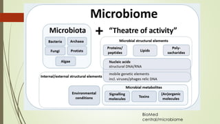
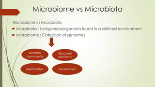
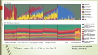
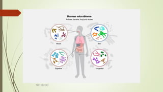
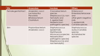
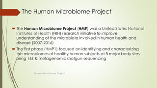
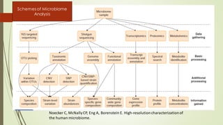
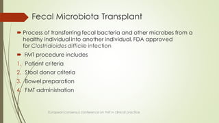
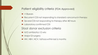
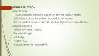
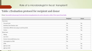
Human Microbiome
Dr Vivek Patil
PG JR
Dept. of Microbiology
AIIMS Raipur
Contents
 Introduction
 Microbiology of Normal Flora
 Role of Normal Flora
 The human microbiomeproject
 Faecal microbiota transplant
History of microbiome research
Biomedcentral.com
Defining ‘Microbiome’
 “The ecological community of commensal, symbiotic &
pathogenic microorganisms that literally share our body space &
have been all but ignored as determinants of health & disease.”
(Lederberg 2001)
 The microbiome comprises all of the genetic material within a
microbiota (HMP operational definition)
BioMed
central/microbiome
Microbiome vs Microbiota
 Microbiota : Living microorganisms found in a defined environment
 Microbiome : Collection of genomes
Microbe
community
Structural
elements
Metabolites Environment
Resident flora
 Constant populations which cannot be completely
removed permanently.
 Close association with particular area. (e.g. E.coli in
intestine)
 Harmless
 Re-establish themselves, when disturbed.
 Prevent permanent colonisation of the body by other
organisms
AnanthNarayan
Transient flora
 Temporary for short interval
 Produce disease when resident flora get disturbed
e.g. pneumococcus and meningococcus in
nasopharynx
 Patients may acquire MDR organisms from hospital
environment & HCW
 e.g. MRSA in nose and skin
MDR GNB (Klebsiella, E.coli, Pseudomonas,
Acinetobacter) in respiratory tract.
 Eliminated by proper hand hygiene
Ananthnarayan
Factors affecting the Microbiome
 Intrinsic factors:
1. Body environment : temperature, pH, oxygen concentration,
pressure, osmolarity, and nutrient source(sebum in skin; mucous in
gut: source of C)
2. Genetics, ethnicity, gender, age
 Extrinsic factors: Diet, Lifestyle, Medication, Geographic location,
Climate, Seasonality, Mode of delivery
 Mode of delivery: Vaginal vs Caesarean different similar
to adult around age 3 yrs.
 Bifidobacterium stimulates immunity decreased in old age
Naturereview article
Factors shaping neonatal microbiome
(From Tamburini S, Shen N, Wu HC, Clemente
JC. The microbiomein early life: implications for health outcomes. Nat Med.2016;22:713-722.)
 North-Central India, which was primarily consuming a plant-based
diet, was found to be associated with Prevotella and also showed
an enrichment of BCAA and lipopolysaccharide biosynthesis
pathways.
 In contrast, the gut microbiome of the cohort from Southern India,
which was consuming an omnivorous diet, showed associations
with Bacteroides, Ruminococcus, and Faecalibacterium and had
an enrichment of SCFA biosynthesis pathway & BCAA transporters.
The relative abundancesof the six
dominant bacterial phyla in different
body sites
Unravellingthe effects of the
environmentand host genotype on
the gut microbiome. Nat Rev
Microbiol. 2011;9:279-290.
Different composition but Same function!!!
(From Human Microbiome
Project Consortium)
NIH library
Oral cavity
 Mouth of an infant at birth is not sterile and it contains mother’s
vaginal flora i.e. micrococci, streptococci, coliforms & lactobacilli
 Diminish in 2-5 days after birth and are replaced
 GPC & GNC predominate; anaerobe: aerobe 100:1
 Anaerobes: Peptostreptococcus, Veillonella, Actinomyces,
Fusobacterium
 Aerobes: Streptococcus & Neisseria
Topley and ASM Manual of clinical microbiology
 Oral streptococci includes: S. salivarius (saliva & tongue)
Tooth surface: S. sanguis & S. mutans
Oral mucosa: S. vestibularis & S. sanguis
S. pneumoniae & B-haemolytic streptococci (grp A,C,F,G)
S. pyogenes can transiently colonise healthy individuals
Topley and ASM Manual of clinical microbiology
 20% of individuals are colonized with S. aureus.
 Other GPCs :Peptostreptococcus anaerobius, Micromonas,
Abiotrophia, Gemella spp.
 GNCs: Veillonella spp (15%), Neisseria, Moraxella, Kingella,
Cardiobacterium, Eikenella corrodens.
 GPBs : Actinomyces is predominant, Rothia
(A. israelli, A. naeslundii, A. odontolyticus, A. meyeri, A. georgiae)
They have fimbrie, adhere to mucosa, forms slime, entrap bacteria,
prevent their removal.
Topley and ASM Manual of clinical microbiology
 Other GPBs Bifidobacterium, Corynebacterium,
Eubacterium, Lactobacillus, Propionibacterium
B. denticum (dental plaque), Lactobacillus & streptococci
(tooth caries),
 GNB : Anaerobes like Fusobacterium, Bacteroides,
Porphyromonas, Prevotella, Selenomonas
Topley and ASM Manual of clinical microbiology
 Haemophilus spp (<5%) mc species is H. parainfluenza & non
capsulated strains of H. influenza
 Actinobacillus actinomycetomcomitans & Treponema spp present
in gingival crevices is associated with periodontal disease
 Enterobacterales, Psuedomonas & Acinetobacter in small numbers.
Increased transiently in hospitalised patients
 Parasites: Trichomonas tenax & Entamoeba gingivalis
Topley and ASM Manual of clinical microbiology
 Oral cavity is principle entry point of human body, spread to other
sites occurs
 Nitrates Nitrites NO (antimicrobial,
vasodilator) shown by Lactobacilli
 Streptococcus salivarius Bacteriocins; inhibits GNB & prevents
periodontitis
 Dental caries: Streptococcus mutans, Streptococcus sobrinus, and
Lactobacillus acidophilus
Carbohydrates Low pH tooth demineralisation
& Cavitation
Topley and ASM Manual of clinical microbiology
reduction
Fermentation
Nares & Nasopharynx
 Nares: S. aureus, CoNS, Corynebacterium,
Peptostreptococcus, Fusobacterium spp.
 Increased carriage of S. aureus in preadolescent
 Nasopharynx: streptococci & Neisseria spp.
 S. salivarius, S. parasanguis & S. pneumoniae
 Colonisation with N. meningitidis in young adults of
military (10-95)% mc Neisseria spp in them are N.
subflava, N. sicca, N. mucosa, N. lactamica
 GNCB: M. catarrhalis (sinusitis, bronchitis), H. influenza,
Cardiobacterium hominis (damaged heart valve)
Topley and ASM Manual of clinical microbiology
Trachea, larynx, bronchi & lungs
 Few organisms, transient
 Long term colonisation from upper airways occurs when
ciliated epithelial cells are damaged (influenza / COPD)
Topley and ASM Manual of clinical microbiology
Esophagus
 The esophagus is colonized by bacteria that are
introduced from the oropharynx by swallowing or from
the stomach by reflux.
 As in the oral cavity, Streptococcus is the dominant
genus in the healthy esophageal microbiome
 In Barrett’s esophagus and esophageal carcinoma,
gram negative anaerobes predominates
 In a diseased state, colonisation with Candida & viruses
(HSV, CMV)
Topley and ASM Manual of clinical microbiology
Stomach
 Inhospitable environment due to HCL, pepsinogen
 Discovery of H. pylori by Marshal & Warren (1882)
 Lactobacillus spp
 Streptococcus spp
 Helicobacter pylori
 Veillonella
 Clostridium
 H. pylori can modulate gastric environment & alter the habitat of
resident flora
 Increased risk for gastric cancer
Topley and ASM Manual of clinical microbiology
Intestine
 Bacteria to host cell ratio 1:1
 Majority in colon
 Main phyla are Firmicutes & Bacteroides (90%)
Others : Actinobacteria, Proteobacteria, Fusobacteria
 Fermicutes: 200 genera
Bacillus, Lactobacillus, Enterococcus, Clostridium, Ruminococcus
among them, S. aureus & C. perfringens are pathogenic
 Bacteroidota: Bacteroides, Prevotella
 Actinobacteria: Bifidobacterium
 Proteobacteria: Enterobacter, Helicobacter, Shigella, Salmonella,
Escherichia coli
Topley and ASM Manual of clinical microbiology
 Changes in 3 stages of life
1. Birth to weaning
2. Weaning to normal diet
3. Old age
 Birth : Vaginal delivery -- Vaginal & fecal
Caesarean -- Skin & environment
 Facultative anaerobes are first to colonise, they create anaerobic
conditions & promote growth of obligatory anaerobes Bifidobacterium,
Bacteroides spp. within 2 weeks
 At 3 days, naturally born infants have more Bifidobacteria than
Caesarean born babies
 Exclusively breastfed babies have more stable & less diverse microbiota
 After the start of solid food, Firmicutes are increased
Topley and ASM Manual of clinical microbiology
Normal colonisation in breast-fed term babies
Carlsson et al JCM, 1975
E. Coli,
streptococci
bifidobacteria
bacteroid
es
Lactobacilli
Enterobacteriaceae
0 5d 10 d 30 d 3-4 m 6m 1yr
Creates
anaerobic
environment
 At old age, decreased diversity, Bifidobacteria , Fermicutes
Increased Enterobacteriaceae, Bacteroidetes (>65years)
 Largest microbiota in large intestine: slow flow rate, mild acidic to
neutral pH
Large intestine contains several microenvironments like epithelial
surface & inner mucin layer mainly contain Obligate anaerobes
Mucin layer: Akkermansia muciniphila
Gut lumen: Ruminococcus spp. Depending upon dietary fiber intake
Topley and ASM Manual of clinical microbiology
 Small intestine: short transit time, high bile
Mainly facultative anaerobes Proteobacteria, Bacteroides,
Streptococci, Lactobacilli, Enterococci spp.
 Entamoeba spp: E. dispar, E. coli, E. hartmanii (large intestine)
 Intestinal flagellates: Pentatrichomonas hominis, Retortamonas
intestinalis, Chilomastix mesnili & Enteromonas hominis
Topley and ASM Manual of clinical microbiology
Role in health
 Regulate digestion- processing nutrients & metabolites; SCFA, bile
acids, amino acids
 Maintain intestinal epithelial integrity, immunity, prevent bacterial
invasion
 Supress pathogenic colonisation & growth
Topley and ASM Manual of clinical microbiology
Genitourinary tract
 Relatively sterile except in female urethra & vagina
 Microbicidal activity of bladder epithelium & flushing action of urine
 Ureters, kidneys, prostate & cervix are sterile
 Female urethra: lactobacilli, streptococci, CoNS
E. coli & Enterococcus spp are transient, can cause UTI
Topley and ASM Manual of clinical microbiology
 Anaerobes : Lactobacillus spp. (L.acidophilus,
L.fermentum, L.casei, L.cellobiosus)
Bifidobacterium spp., Porphyromonas, Prevotella,
Peptococcus spp., Propionibacterium, Mobiluncus(in small
numbers), Treponema spp.
 Facultative anaerobes : CoNS, Streptococci,
Corynebacterium spp, Viridans group of streptococci ,
Gardnerella vaginalis, Neisseria spp., Haemophilus
Topley and ASM Manual of clinical microbiology
 Mycoplasma (M. hominis, M. genitalium, M. fermentans, M.
primatum, M. spermatophilum, M. penetrans)
M. hominis & U. urealyticum have pathogenic potential
Topley and ASM Manual of clinical microbiology
Skin
 Anaerobes >100 folds>> aerobes ; gram positive > gram negatives
 Staphylococcus, Micrococcus, Corynebacterium, Peptococcus,
Finegoldia, Peptostreptococcus & Propionibacterium
 S. epidermidis is most common; other CoNS are S. hominis, S.
hemolyticus, S. warneri, S. capitis, S. saprophyticus, S. caprae, S.
saccharolyticus, S. pasteuri, S. lugdunensis, S. simulans & S. xylosus
Topley and ASM Manual of clinical microbiology
 Micrococcus spp (20%), most common is Micrococcus
luteus
Other GPCs are Dermacoccus, Kocuria kristinae, Kocuria
rosea, K. varians & kytococcus
 S. pyogenes are well suited for dry skin surfaces
 Anaerobic cocci survive in hair follicles & skin glands
Topley and ASM Manual of clinical microbiology
 Corynebacterium spp ; C. striatum, C. minutissimum, C.
psuedodiphtheriticum, C. xerosis, C.urealyticus (groin), C. jeikeium
(apocrine glands)
 Propionibacterium acnes (sebaceous gland), Propionibacterium
avidum (axilla & perineum)
 Other gram positive includes Dermabacter, Brevibacterium,
Turicella otitidis
 Transient members are Clostridium perfringens, Acinetobacter,
Burkholderia.
Topley and ASM Manual of clinical microbiology
Site Anaerobes Aerobes others
Mouth Anaerobic cocci
Actinomyces
Fusobacterium
Bifidobacterium
Prevotella
Spirochetes
Viridans streptococci Trichomonas tenax
Entamoeba gingivalis
Nasopharynx Prevotella species
Anaerobic cocci
Fusobacterium
Streptococci (α and
non-hemolytic)
Neisseria spp
Diphtheroids
Staphylococcus
epidermidis
Haemophilus
Meningococcus
Pneumococcus
Staphylococcus
aureus Gram-
negative bacilli
Yeasts
Gastro-intestinal tract Lactobacillus
Anaerobic cocci
Bacteroides fragilis
Fusobacterium
Bifidobacterium
Prevotella
Clostridium
Helicobacter pylori
Enterobacteriaceae
& other GNB
Enterococci,
Streptococci (α and
non hemolytic),
S. Agalactiae
Diphtheroids
Candida species
Staphylococcus
aureus
Entamoeba spp
intestinal
flagellates
Site Anaerobes Aerobes Others
Female genital tract Anaerobic cocci
Lactobacillus
Prevotella
Bifidobacterium
Clostridium
Corynebacterium
species
Streptococci (α, non-
hemolytic and
S. agalactiae)
Neisseria (non-
pathogenic species)
Enterococci
Enterobacteriaceae
and
other gram-negative
rods
S. epidermidis
Candida species
Skin Propionibacterium
Anaerobic cocci
Staphylococcus
epidermidis
Diphtheroids
Micrococcus species
Neisseria(Non-
pathogenic species)
Streptococci (α and
non hemolytic)
Staphylococcus
aureus Candida
species
Acinetobacter
species
The Human Microbiome Project
 The Human Microbiome Project (HMP) was a United States National
Institutes of Health (NIH) research initiative to improve
understanding of the microbiota involved in human health and
disease (2007-2016)
 The first phase (HMP1) focused on identifying and characterizing
the microbiomes of healthy human subjects at 5 major body sites
using 16S & metagenomic shotgun sequencing
Human Microbiome Project
 The second phase, known as the Integrative Human
Microbiome Project (iHMP) launched in 2014 with the
aim of characterization of microbiome & human host
from 3 cohorts of microbiome associated conditions
using multiple ‘omics’ strategies
 Study methods included 16S rRNA gene profiling,
whole metagenome shotgun sequencing,
metatranscriptomics, metabolomics
Culture independent molecular approaches to
study host-microbiome interaction
 DNA-based approaches- 16s rRNA, 18s rRNA
Who is there and what can they do?
 RNA-based approaches-
How do they respond?
What pathways are activated?
 Protein-based approaches-
How are they interacting with the host?
What proteins are being produced?
 Metabolite-basedapproaches-
What are the chemical outcomes of their activity?
Metagenomics
Metatranscriptomics
Metaproteomics
Metabolomics
the Human Microbiome, and Health Risk: A Research Strategy (2018)
Noecker C, McNallyCP, Eng A, Borenstein E. High-resolutioncharacterizationof
the human microbiome.
Schemes of Microbiome
Analysis
Fecal Microbiota Transplant
 Process of transferring fecal bacteria and other microbes from a
healthy individual into another individual. FDA approved
for Clostridioides difficile infection
 FMT procedure includes
1. Patient criteria
2. Stool donar criteria
3. Bowel preparation
4. FMT administration
European consensus conference on FMT in clinical practice
Patient eligibility criteria (FDA Approved)
 >18years
 Recurrent CDI not responding to standard vancomycin therapy
 SevereCDI not responding to therapy after 48 hours
 Laboratory confirmed CDI
Stool donor exclusion criteria
 H/O antibiotics 12 wks
 Major GI surgery
 HIV, HBV, HCV, tattooswithin last 6 months
DONAR SELECTION
Stool testing
(1) Clostridioides difficile (PCR or EIA test for Toxin A and B)
(2) Routine culture for enteric bacterial pathogens
(3) Complete Ova and Parasite studies, if pertinent travel history
Serologic testing
(4) anti-HIV type 1 and 2
(5) anti-HAV IgM
(6) HBsAg
(7) anti-HCV
(8) Rapid plasma reagin (RPR)
Specimen preparation
 Fresh stool (30-100 grams)
 Samples prepared within 6-8 hrs of defecation
 The sample is then diluted with 2.5–5 times the volume of the
sample with either normal saline, sterile water or 4% milk.
 Mix the solution with morter & pestle/ blender
 The suspension is then strained through a filter and transferred to an
administration container
 For later use it can be frozen after dilution using 10% glycerol
European consensus conference on FMT in clinical practice
Administration
European consensus conference on FMT in clinical practice
(A)Donor stool and normal
saline (1:3) ground in a
blender.
(B)Fecal suspension in 50-mL
syringes.
(C)Infusion using
colonoscopy.
*RECTAL
ENEMA
First oral pill for CDI, FDA
APPROVED on 16 APR 23
SER-109
Role of a microbiologist in fecal transplant!
Topley and ASM Manual of clinical microbiology
Thank You!

Human Microbiome Microbiota Faecal transplant.pdf

  • 1.
    Human Microbiome Dr VivekPatil PG JR Dept. of Microbiology AIIMS Raipur
  • 2.
    Contents  Introduction  Microbiologyof Normal Flora  Role of Normal Flora  The human microbiomeproject  Faecal microbiota transplant
  • 3.
    History of microbiomeresearch Biomedcentral.com
  • 4.
    Defining ‘Microbiome’  “Theecological community of commensal, symbiotic & pathogenic microorganisms that literally share our body space & have been all but ignored as determinants of health & disease.” (Lederberg 2001)  The microbiome comprises all of the genetic material within a microbiota (HMP operational definition)
  • 5.
  • 6.
    Microbiome vs Microbiota Microbiomevs Microbiota  Microbiota : Living microorganisms found in a defined environment  Microbiome : Collection of genomes Microbe community Structural elements Metabolites Environment
  • 7.
    Resident flora  Constantpopulations which cannot be completely removed permanently.  Close association with particular area. (e.g. E.coli in intestine)  Harmless  Re-establish themselves, when disturbed.  Prevent permanent colonisation of the body by other organisms AnanthNarayan
  • 8.
    Transient flora  Temporaryfor short interval  Produce disease when resident flora get disturbed e.g. pneumococcus and meningococcus in nasopharynx  Patients may acquire MDR organisms from hospital environment & HCW  e.g. MRSA in nose and skin MDR GNB (Klebsiella, E.coli, Pseudomonas, Acinetobacter) in respiratory tract.  Eliminated by proper hand hygiene Ananthnarayan
  • 9.
    Factors affecting theMicrobiome  Intrinsic factors: 1. Body environment : temperature, pH, oxygen concentration, pressure, osmolarity, and nutrient source(sebum in skin; mucous in gut: source of C) 2. Genetics, ethnicity, gender, age  Extrinsic factors: Diet, Lifestyle, Medication, Geographic location, Climate, Seasonality, Mode of delivery  Mode of delivery: Vaginal vs Caesarean different similar to adult around age 3 yrs.  Bifidobacterium stimulates immunity decreased in old age Naturereview article
  • 10.
    Factors shaping neonatalmicrobiome (From Tamburini S, Shen N, Wu HC, Clemente JC. The microbiomein early life: implications for health outcomes. Nat Med.2016;22:713-722.)
  • 12.
     North-Central India,which was primarily consuming a plant-based diet, was found to be associated with Prevotella and also showed an enrichment of BCAA and lipopolysaccharide biosynthesis pathways.  In contrast, the gut microbiome of the cohort from Southern India, which was consuming an omnivorous diet, showed associations with Bacteroides, Ruminococcus, and Faecalibacterium and had an enrichment of SCFA biosynthesis pathway & BCAA transporters.
  • 13.
    The relative abundancesofthe six dominant bacterial phyla in different body sites Unravellingthe effects of the environmentand host genotype on the gut microbiome. Nat Rev Microbiol. 2011;9:279-290.
  • 14.
    Different composition butSame function!!! (From Human Microbiome Project Consortium)
  • 15.
  • 16.
    Oral cavity  Mouthof an infant at birth is not sterile and it contains mother’s vaginal flora i.e. micrococci, streptococci, coliforms & lactobacilli  Diminish in 2-5 days after birth and are replaced  GPC & GNC predominate; anaerobe: aerobe 100:1  Anaerobes: Peptostreptococcus, Veillonella, Actinomyces, Fusobacterium  Aerobes: Streptococcus & Neisseria Topley and ASM Manual of clinical microbiology
  • 17.
     Oral streptococciincludes: S. salivarius (saliva & tongue) Tooth surface: S. sanguis & S. mutans Oral mucosa: S. vestibularis & S. sanguis S. pneumoniae & B-haemolytic streptococci (grp A,C,F,G) S. pyogenes can transiently colonise healthy individuals Topley and ASM Manual of clinical microbiology
  • 18.
     20% ofindividuals are colonized with S. aureus.  Other GPCs :Peptostreptococcus anaerobius, Micromonas, Abiotrophia, Gemella spp.  GNCs: Veillonella spp (15%), Neisseria, Moraxella, Kingella, Cardiobacterium, Eikenella corrodens.  GPBs : Actinomyces is predominant, Rothia (A. israelli, A. naeslundii, A. odontolyticus, A. meyeri, A. georgiae) They have fimbrie, adhere to mucosa, forms slime, entrap bacteria, prevent their removal. Topley and ASM Manual of clinical microbiology
  • 19.
     Other GPBsBifidobacterium, Corynebacterium, Eubacterium, Lactobacillus, Propionibacterium B. denticum (dental plaque), Lactobacillus & streptococci (tooth caries),  GNB : Anaerobes like Fusobacterium, Bacteroides, Porphyromonas, Prevotella, Selenomonas Topley and ASM Manual of clinical microbiology
  • 20.
     Haemophilus spp(<5%) mc species is H. parainfluenza & non capsulated strains of H. influenza  Actinobacillus actinomycetomcomitans & Treponema spp present in gingival crevices is associated with periodontal disease  Enterobacterales, Psuedomonas & Acinetobacter in small numbers. Increased transiently in hospitalised patients  Parasites: Trichomonas tenax & Entamoeba gingivalis Topley and ASM Manual of clinical microbiology
  • 21.
     Oral cavityis principle entry point of human body, spread to other sites occurs  Nitrates Nitrites NO (antimicrobial, vasodilator) shown by Lactobacilli  Streptococcus salivarius Bacteriocins; inhibits GNB & prevents periodontitis  Dental caries: Streptococcus mutans, Streptococcus sobrinus, and Lactobacillus acidophilus Carbohydrates Low pH tooth demineralisation & Cavitation Topley and ASM Manual of clinical microbiology reduction Fermentation
  • 22.
    Nares & Nasopharynx Nares: S. aureus, CoNS, Corynebacterium, Peptostreptococcus, Fusobacterium spp.  Increased carriage of S. aureus in preadolescent  Nasopharynx: streptococci & Neisseria spp.  S. salivarius, S. parasanguis & S. pneumoniae  Colonisation with N. meningitidis in young adults of military (10-95)% mc Neisseria spp in them are N. subflava, N. sicca, N. mucosa, N. lactamica  GNCB: M. catarrhalis (sinusitis, bronchitis), H. influenza, Cardiobacterium hominis (damaged heart valve) Topley and ASM Manual of clinical microbiology
  • 23.
    Trachea, larynx, bronchi& lungs  Few organisms, transient  Long term colonisation from upper airways occurs when ciliated epithelial cells are damaged (influenza / COPD) Topley and ASM Manual of clinical microbiology
  • 24.
    Esophagus  The esophagusis colonized by bacteria that are introduced from the oropharynx by swallowing or from the stomach by reflux.  As in the oral cavity, Streptococcus is the dominant genus in the healthy esophageal microbiome  In Barrett’s esophagus and esophageal carcinoma, gram negative anaerobes predominates  In a diseased state, colonisation with Candida & viruses (HSV, CMV) Topley and ASM Manual of clinical microbiology
  • 25.
    Stomach  Inhospitable environmentdue to HCL, pepsinogen  Discovery of H. pylori by Marshal & Warren (1882)  Lactobacillus spp  Streptococcus spp  Helicobacter pylori  Veillonella  Clostridium  H. pylori can modulate gastric environment & alter the habitat of resident flora  Increased risk for gastric cancer Topley and ASM Manual of clinical microbiology
  • 26.
    Intestine  Bacteria tohost cell ratio 1:1  Majority in colon  Main phyla are Firmicutes & Bacteroides (90%) Others : Actinobacteria, Proteobacteria, Fusobacteria  Fermicutes: 200 genera Bacillus, Lactobacillus, Enterococcus, Clostridium, Ruminococcus among them, S. aureus & C. perfringens are pathogenic  Bacteroidota: Bacteroides, Prevotella  Actinobacteria: Bifidobacterium  Proteobacteria: Enterobacter, Helicobacter, Shigella, Salmonella, Escherichia coli Topley and ASM Manual of clinical microbiology
  • 27.
     Changes in3 stages of life 1. Birth to weaning 2. Weaning to normal diet 3. Old age  Birth : Vaginal delivery -- Vaginal & fecal Caesarean -- Skin & environment  Facultative anaerobes are first to colonise, they create anaerobic conditions & promote growth of obligatory anaerobes Bifidobacterium, Bacteroides spp. within 2 weeks  At 3 days, naturally born infants have more Bifidobacteria than Caesarean born babies  Exclusively breastfed babies have more stable & less diverse microbiota  After the start of solid food, Firmicutes are increased Topley and ASM Manual of clinical microbiology
  • 28.
    Normal colonisation inbreast-fed term babies Carlsson et al JCM, 1975 E. Coli, streptococci bifidobacteria bacteroid es Lactobacilli Enterobacteriaceae 0 5d 10 d 30 d 3-4 m 6m 1yr Creates anaerobic environment
  • 29.
     At oldage, decreased diversity, Bifidobacteria , Fermicutes Increased Enterobacteriaceae, Bacteroidetes (>65years)  Largest microbiota in large intestine: slow flow rate, mild acidic to neutral pH Large intestine contains several microenvironments like epithelial surface & inner mucin layer mainly contain Obligate anaerobes Mucin layer: Akkermansia muciniphila Gut lumen: Ruminococcus spp. Depending upon dietary fiber intake Topley and ASM Manual of clinical microbiology
  • 30.
     Small intestine:short transit time, high bile Mainly facultative anaerobes Proteobacteria, Bacteroides, Streptococci, Lactobacilli, Enterococci spp.  Entamoeba spp: E. dispar, E. coli, E. hartmanii (large intestine)  Intestinal flagellates: Pentatrichomonas hominis, Retortamonas intestinalis, Chilomastix mesnili & Enteromonas hominis Topley and ASM Manual of clinical microbiology
  • 31.
    Role in health Regulate digestion- processing nutrients & metabolites; SCFA, bile acids, amino acids  Maintain intestinal epithelial integrity, immunity, prevent bacterial invasion  Supress pathogenic colonisation & growth Topley and ASM Manual of clinical microbiology
  • 32.
    Genitourinary tract  Relativelysterile except in female urethra & vagina  Microbicidal activity of bladder epithelium & flushing action of urine  Ureters, kidneys, prostate & cervix are sterile  Female urethra: lactobacilli, streptococci, CoNS E. coli & Enterococcus spp are transient, can cause UTI Topley and ASM Manual of clinical microbiology
  • 33.
     Anaerobes :Lactobacillus spp. (L.acidophilus, L.fermentum, L.casei, L.cellobiosus) Bifidobacterium spp., Porphyromonas, Prevotella, Peptococcus spp., Propionibacterium, Mobiluncus(in small numbers), Treponema spp.  Facultative anaerobes : CoNS, Streptococci, Corynebacterium spp, Viridans group of streptococci , Gardnerella vaginalis, Neisseria spp., Haemophilus Topley and ASM Manual of clinical microbiology
  • 34.
     Mycoplasma (M.hominis, M. genitalium, M. fermentans, M. primatum, M. spermatophilum, M. penetrans) M. hominis & U. urealyticum have pathogenic potential Topley and ASM Manual of clinical microbiology
  • 35.
    Skin  Anaerobes >100folds>> aerobes ; gram positive > gram negatives  Staphylococcus, Micrococcus, Corynebacterium, Peptococcus, Finegoldia, Peptostreptococcus & Propionibacterium  S. epidermidis is most common; other CoNS are S. hominis, S. hemolyticus, S. warneri, S. capitis, S. saprophyticus, S. caprae, S. saccharolyticus, S. pasteuri, S. lugdunensis, S. simulans & S. xylosus Topley and ASM Manual of clinical microbiology
  • 36.
     Micrococcus spp(20%), most common is Micrococcus luteus Other GPCs are Dermacoccus, Kocuria kristinae, Kocuria rosea, K. varians & kytococcus  S. pyogenes are well suited for dry skin surfaces  Anaerobic cocci survive in hair follicles & skin glands Topley and ASM Manual of clinical microbiology
  • 37.
     Corynebacterium spp; C. striatum, C. minutissimum, C. psuedodiphtheriticum, C. xerosis, C.urealyticus (groin), C. jeikeium (apocrine glands)  Propionibacterium acnes (sebaceous gland), Propionibacterium avidum (axilla & perineum)  Other gram positive includes Dermabacter, Brevibacterium, Turicella otitidis  Transient members are Clostridium perfringens, Acinetobacter, Burkholderia. Topley and ASM Manual of clinical microbiology
  • 38.
    Site Anaerobes Aerobesothers Mouth Anaerobic cocci Actinomyces Fusobacterium Bifidobacterium Prevotella Spirochetes Viridans streptococci Trichomonas tenax Entamoeba gingivalis Nasopharynx Prevotella species Anaerobic cocci Fusobacterium Streptococci (α and non-hemolytic) Neisseria spp Diphtheroids Staphylococcus epidermidis Haemophilus Meningococcus Pneumococcus Staphylococcus aureus Gram- negative bacilli Yeasts Gastro-intestinal tract Lactobacillus Anaerobic cocci Bacteroides fragilis Fusobacterium Bifidobacterium Prevotella Clostridium Helicobacter pylori Enterobacteriaceae & other GNB Enterococci, Streptococci (α and non hemolytic), S. Agalactiae Diphtheroids Candida species Staphylococcus aureus Entamoeba spp intestinal flagellates
  • 39.
    Site Anaerobes AerobesOthers Female genital tract Anaerobic cocci Lactobacillus Prevotella Bifidobacterium Clostridium Corynebacterium species Streptococci (α, non- hemolytic and S. agalactiae) Neisseria (non- pathogenic species) Enterococci Enterobacteriaceae and other gram-negative rods S. epidermidis Candida species Skin Propionibacterium Anaerobic cocci Staphylococcus epidermidis Diphtheroids Micrococcus species Neisseria(Non- pathogenic species) Streptococci (α and non hemolytic) Staphylococcus aureus Candida species Acinetobacter species
  • 40.
    The Human MicrobiomeProject  The Human Microbiome Project (HMP) was a United States National Institutes of Health (NIH) research initiative to improve understanding of the microbiota involved in human health and disease (2007-2016)  The first phase (HMP1) focused on identifying and characterizing the microbiomes of healthy human subjects at 5 major body sites using 16S & metagenomic shotgun sequencing Human Microbiome Project
  • 41.
     The secondphase, known as the Integrative Human Microbiome Project (iHMP) launched in 2014 with the aim of characterization of microbiome & human host from 3 cohorts of microbiome associated conditions using multiple ‘omics’ strategies  Study methods included 16S rRNA gene profiling, whole metagenome shotgun sequencing, metatranscriptomics, metabolomics
  • 42.
    Culture independent molecularapproaches to study host-microbiome interaction  DNA-based approaches- 16s rRNA, 18s rRNA Who is there and what can they do?  RNA-based approaches- How do they respond? What pathways are activated?  Protein-based approaches- How are they interacting with the host? What proteins are being produced?  Metabolite-basedapproaches- What are the chemical outcomes of their activity? Metagenomics Metatranscriptomics Metaproteomics Metabolomics the Human Microbiome, and Health Risk: A Research Strategy (2018)
  • 43.
    Noecker C, McNallyCP,Eng A, Borenstein E. High-resolutioncharacterizationof the human microbiome. Schemes of Microbiome Analysis
  • 44.
    Fecal Microbiota Transplant Process of transferring fecal bacteria and other microbes from a healthy individual into another individual. FDA approved for Clostridioides difficile infection  FMT procedure includes 1. Patient criteria 2. Stool donar criteria 3. Bowel preparation 4. FMT administration European consensus conference on FMT in clinical practice
  • 45.
    Patient eligibility criteria(FDA Approved)  >18years  Recurrent CDI not responding to standard vancomycin therapy  SevereCDI not responding to therapy after 48 hours  Laboratory confirmed CDI Stool donor exclusion criteria  H/O antibiotics 12 wks  Major GI surgery  HIV, HBV, HCV, tattooswithin last 6 months
  • 46.
    DONAR SELECTION Stool testing (1)Clostridioides difficile (PCR or EIA test for Toxin A and B) (2) Routine culture for enteric bacterial pathogens (3) Complete Ova and Parasite studies, if pertinent travel history Serologic testing (4) anti-HIV type 1 and 2 (5) anti-HAV IgM (6) HBsAg (7) anti-HCV (8) Rapid plasma reagin (RPR)
  • 47.
    Specimen preparation  Freshstool (30-100 grams)  Samples prepared within 6-8 hrs of defecation  The sample is then diluted with 2.5–5 times the volume of the sample with either normal saline, sterile water or 4% milk.  Mix the solution with morter & pestle/ blender  The suspension is then strained through a filter and transferred to an administration container  For later use it can be frozen after dilution using 10% glycerol European consensus conference on FMT in clinical practice
  • 49.
    Administration European consensus conferenceon FMT in clinical practice (A)Donor stool and normal saline (1:3) ground in a blender. (B)Fecal suspension in 50-mL syringes. (C)Infusion using colonoscopy.
  • 50.
  • 51.
    First oral pillfor CDI, FDA APPROVED on 16 APR 23 SER-109
  • 53.
    Role of amicrobiologist in fecal transplant! Topley and ASM Manual of clinical microbiology
  • 54.