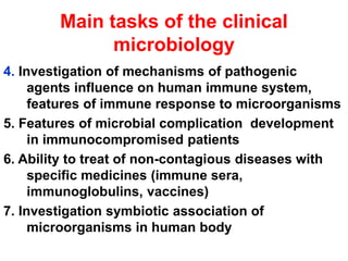
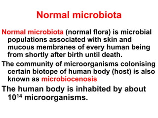
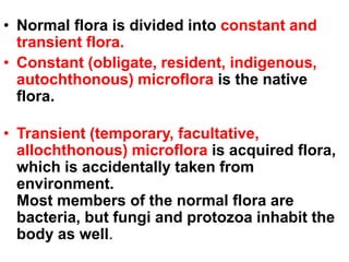
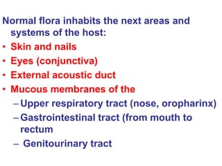
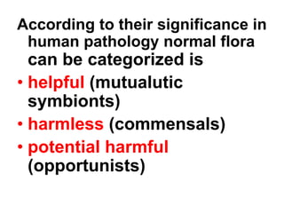
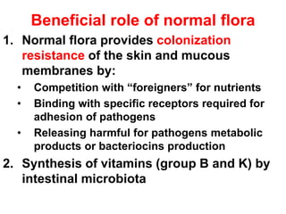
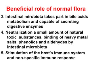
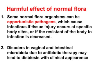
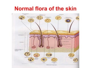
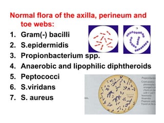
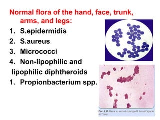
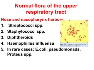
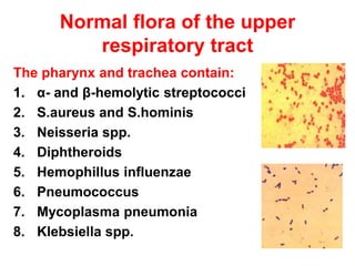
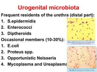
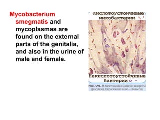
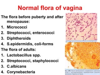
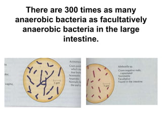
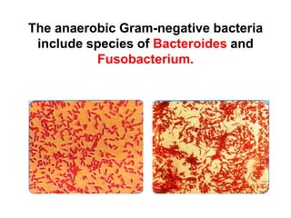
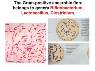
This document discusses clinical microbiology and normal human microbiota. It describes the major tasks of clinical microbiology as investigating opportunistic microorganisms that can cause hospital-acquired infections and developing rapid diagnostic tests. It then characterizes the normal microbiota found in different areas of the human body like the skin, respiratory tract, and gastrointestinal tract. Finally, it discusses hospital-acquired infections, identifying common pathogens and emphasizing prevention through proper sterilization, hand washing, and rational antibiotic use.
Premium Bangalore Call Girls Jigani Dail 6378878445 Escort Service For Hot Ma...
clinical microbiology
1. Clinical microbiology. Major
tasks and purposes.
Opportunistic microbiota as
major cause of hospital
infections. Short characteristics
of microbiota representatives
from different biotopes of human
body
Vinnitsa National Pirogov Memorial Medical
University/ Department of microbiology
2. Clinical microbiology
Clinical microbiology is the part of medical
microbiology
Clinical microbiology investigates
microbiological aspects non-epidemic
microbial complications (for example,
nosocomial infections) and elaborates
laboratory diagnostics of them
3. Main tasks of the clinical
microbiology
1. Investigation of morphology, physiology,
genetics and ecology of opportunistic germs and
their significance in human being
2. Elaboration rapid diagnostic tests for infectious
complications and improvement present
methods of laboratory diagnostics
3. Study the pathogenic properties of opportunistic
microorganisms and their role in pathogenesis
of microbial complications of somatic illness
4. Main tasks of the clinical
microbiology
4. Investigation of mechanisms of pathogenic
agents influence on human immune system,
features of immune response to microorganisms
5. Features of microbial complication development
in immunocompromised patients
6. Ability to treat of non-contagious diseases with
specific medicines (immune sera,
immunoglobulins, vaccines)
7. Investigation symbiotic association of
microorganisms in human body
5. Normal microbiota
Normal microbiota (normal flora) is microbial
populations associated with skin and
mucous membranes of every human being
from shortly after birth until death.
The community of microorganisms colonising
certain biotope of human body (host) is also
known as microbiocenosis
The human body is inhabited by about
1014 microorganisms.
6. • Normal flora is divided into constant and
transient flora.
• Constant (obligate, resident, indigenous,
autochthonous) microflora is the native
flora.
• Transient (temporary, facultative,
allochthonous) microflora is acquired flora,
which is accidentally taken from
environment.
Most members of the normal flora are
bacteria, but fungi and protozoa inhabit the
body as well.
7. Normal flora inhabits the next areas and
systems of the host:
• Skin and nails
• Eyes (conjunctiva)
• External acoustic duct
• Mucous membranes of the
–Upper respiratory tract (nose, oropharinx)
–Gastrointestinal tract (from mouth to
rectum
– Genitourinary tract
8. According to their significance in
human pathology normal flora
can be categorized is
• helpful (mutualutic
symbionts)
• harmless (commensals)
• potential harmful
(opportunists)
9. Beneficial role of normal flora
1. Normal flora provides colonization
resistance of the skin and mucous
membranes by:
• Competition with “foreigners” for nutrients
• Binding with specific receptors required for
adhesion of pathogens
• Releasing harmful for pathogens metabolic
products or bacteriocins production
2. Synthesis of vitamins (group B and K) by
intestinal microbiota
10. Beneficial role of normal flora
3. Intestinal mirobiota takes part in bile acids
metabolism and capable of secreting
digestive enzymes
4. Neutralization a small amount of natural
toxic substances, binding of heavy metal
salts, phenolics and aldehydes by
intestinal microbiota
5. Stimulation of the host's immune system
and non-specific immune response
11. Harmful effect of normal flora
1. Some normal flora organisms can be
opportunistic pathogens, which cause
infectious if tissue injury occurs at specific
body sites, or if the resistant of the body to
infection is decreased.
2. Disoders in vaginal and intestinal
microbiota due to antibiotic therapy may
lead to disbiosis with clinical appearence
12. Harmful effect of normal flora
• Usage of broad spectrum antibiotics may
inhibit sensitive members and thereby
select resistant bacteria
• Normal flora may produce enzymes splitting
or inactivating antibiotics
• Some metabolic products of normal flora act
as cocarcinogenes (protein putrifaction
prducts, nitrites, nitrates,etc.)
13. Harmful effect of normal flora
• Normal germs may cause confusion in
diagnosis due to their presence in
clinical samples and their resemblance
to pathogens
• Some pathogens may belong to normal
microbiota of carriers and be
transmitted to healthy persons
15. Normal flora of the axilla, perineum and
toe webs:
1. Gram(-) bacilli
2. S.epidermidis
3. Propionbacterium spp.
4. Anaerobic and lipophilic diphtheroids
5. Peptococci
6. S.viridans
7. S. aureus
16. Normal flora of the hand, face, trunk,
arms, and legs:
1. S.epidermidis
2. S.aureus
3. Micrococci
4. Non-lipophilic and
lipophilic diphtheroids
1. Propionbacterium spp.
17. Normal flora of the upper
respiratory tract
Nose and nasopharynx harbors:
1. Streptococci spp.
2. Staphylococci spp.
3. Diphtheroids
4. Haemophillus influensa
5. In rare cases: E.coli, pseudomonads,
Proteus spp.
18. Normal flora of the upper
respiratory tract
The pharynx and trachea contain:
1. α- and β-hemolytic streptococci
2. S.aureus and S.hominis
3. Neisseria spp.
4. Diphtheroids
5. Hemophillus influenzae
6. Pneumococcus
7. Mycoplasma pneumonia
8. Klebsiella spp.
19. Urogenital microbiota
Frequent residents of the urethra (distal part):
1. S.epidermidis
2. Enterococci
3. Diptheroids
Occasional members (10-30%):
1. E.coli
2. Proteus spp.
3. Opportunistic Neisseria
4. Mycoplasma and Ureaplasma
21. Normal flora of vagina
The flora before puberty and after
menopause:
1. Micrococci
2. Streptococci, enterococci
3. Diphtheroids
4. S.epidermidis, coli-forms
The flora of adults:
1. Lactobacillus spp.
2. Streptococci, staphylococci
3. C.albicans
4. Corynebacteria
22. Microbiota of mouth cavity
• The oral cavity is colonize by microorganisms
from the surrounding environment within hours
after a human is born.
• Initially the microbiota consists mostly of the
genera (aerobes and anaerobes)
• Streptococcus (α-hemolytic and viridans group)
• Neisseria
• Actinomyces
• Veillonella
• Lactobacillus
• Candida
23. Microbiota of mouth cavity
• As the first teeth erupt, the anaerobes
(Porphyromonas, Prevotella, Fusobacterium)
become dominant due to the anaerobic
nature of the gingival groove.
• As teeth grow, S.parasanguis and S.mutans
attach to their enamel surfaces.
• S.salivarius attaches to the buccal and
gingival epithelial surfaces and colonizes the
saliva.
24.
25. Esophagus and stomach
The total amount is about 102-103 cells per
ml in the stomach
Representatives are:
1. Lactobacilli
2. Micrococci
3. Candida
4. Sarcina
5. Bacilli
6. Helicobacter pylori (in 30% of healthy
individuals)
26. Duodenum and jejunum
• The bacterial count in duodenum is 103-
105 per gram, in the jejunum and
proximal ileum 105-107 per gram
• Enterococci, fungi, lactobacilli,
coliforms and various other microbes
are predominant in the duodenum and
small intestine.
27. In the large intestine there are large
amounts of microorganisms (about 107-
109 per gram in the caecum and 1010-
1012 in the rectum). Almost one-third of
the dry weight of the faeces is made up
of microbes.
The faecal flora consists generally from
anaerobic microorganisms (96-99%)
28. There are 300 times as many
anaerobic bacteria as facultatively
anaerobic bacteria in the large
intestine.
31. The intestinal facultative anaerobs belong to
genera Escherichia, Proteus, Klebsiella,
Candida (yaest-like fungi). Anaerobic
Streptococcus are also present.
32. Hospital-acquired infection
Hospital-acquired infection (hospital-
associated infection, nosocomial infection)
is a infection developing in patients after 4
daysof admission to the hospital
Source of infection may be both:
1. Exogenous (pathogen is transmitted from
surrounding environment)
2. Endogenous (pathogen belongs to normal
microbiota)
33. Modes of exogenous infection
transmission
1. Direct contact with contaminated
equipment or hands of medical staff
carrying hospital pathogens
2. Inhalation of droplets, dust or aerosols
containing hospital strains
3. Food-borne route (hospital food may be
contaminated with antibiotic-resistant
enteral pathogens
4. Parenteral route
34. Hospital pathogens
The most often germs isolated from clinical
samples at HAI:
1. Meticillin-resistant S.aureus (MRSA) and
S.epidermidis
2. Streptococci group D (enterococci)
3. Enterobacteria (coliforms and others)
4. Non-fermentive Gram-negative bacteria
(Pseudomonads, Stenotrophomonas
maltophilia, Acinetobacter, Burkholderia
and others)
5. Hepatitis B and C viruses
35. Common causative agents of the HAI
1. Urinary tract infection are the most often
caused by E.coli, Proteus, Klebsiella,
Serratia, pseudomonas, Providencia,
coagulase(-) staphylococci, enterococci
and C.albicans
2. Respiratory infection.
Usual pathogens are S.aureus, Klebsiella,
Enterobacter, Serratia, Proteus, E.coli,
P.aeruginosa, Acinetobacter, Legionella
pneumophila and respiratory viruses
36. Common causative agents of the HAI
3. Wound and skin infections, sepsis
Predominant pathogens: S.aureus,
P.aeruginosa, Proteus, E.coli, coagulase(-)
staphylococci, enterococci
4. Bacteraemia and septicaemia
Cathter-associated contaminants – Gram-
negative bacteria
Associated with artificial heart valves
pathogens – coagulase (-) staphylococci
37. Common causative agents of the HAI
5. Burns:
S.aureus, P.aeruginosa, Acinetobacter,
S.pyogenes
6. Gastrointestinal infection includes food-
poisoning infection and neonatal
septicaemia associated more often with
Salmonella and Shigella
38. Prevention of the HAI
It is based on:
1. Finding and destroying hospital pathogens
2. Proper sterilisation and disinfection of
possible inanimate vechles
3. Disinfection of contaminated materials and
biological samples from the patients
4. Frequent washing of hands and changing
working clothes
39. Prevention of the HAI
5.Usage of disposable syringes, needles,
sterile gloves, dressing, face-masks and
proper control of fluids for infusion
6. Reliable antiseptic measures before
operation and “cutting-down” of the skin
7. Rational antibiotic therapy and prophilaxis
8. Investigation of nosocomial infection in
details and proper treatment of every
cases