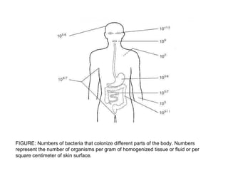
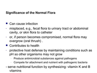
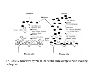
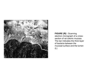
CHAPTER 6
NORMAL FLORA
 The term "normal microbial flora" denotes the population
of microorganisms that inhabit the skin and mucous
membranes of healthy normal persons
 The skin and mucous membranes always harbor a
variety of microorganisms that can be arranged into two
groups:
(1) The resident flora consists of relatively fixed types of
microorganisms regularly found in a given area at a
given age; if disturbed, it promptly reestablishes itself.
(2) The transient flora consists of nonpathogenic or
potentially pathogenic microorganisms that inhabit the
skin or mucous membranes for hours, days, or weeks; it
is derived from the environment, does not produce
disease, and does not establish itself permanently on
the surface.
 Members of the transient flora are generally of little
significance so long as the normal resident flora
remains intact. However, if the resident flora is
disturbed, transient microorganisms may colonize,
proliferate, and produce disease.
FIGURE: Numbers of bacteria that colonize different parts of the body. Numbers
represent the number of organisms per gram of homogenized tissue or fluid or per
square centimeter of skin surface.
Significance of the Normal Flora
 Can cause infection
- misplaced, e.g., fecal flora to urinary tract or abdominal
cavity, or skin flora to catheter
- or, if person becomes compromised, normal flora may
overgrow (oral thrush) .
 Contributes to health
- protective host defense by maintaining conditions such as
pH so other organisms may not grow
- Produce antimicrobial substances against pathogens
- Compete for attachment and nutrient with pathogenic bacteria
- serve nutritional function by synthesizing: vitamin K and B
vitamins
FIGURE: Mechanisms by which the normal flora competes with invading
pathogens..
FIGURE (A) : Scanning
electron micrograph of a cross-
section of rat colonic mucosa.
The bar indicates the thick layer
of bacteria between the
mucosal surface and the lumen
(L)
FIGURE: (B) Higher magnification of the
area indicated by the arrow in Fig. A,
showing a mass of bacteria (B)
immediately adjacent to colonized
intestinal tissue (T), (X2,624.)
Normal Flora of the Skin
 Because of its constant exposure to and contact with the
environment, the skin is particularly apt to contain
transient microorganisms. Nevertheless, there is a
constant and well-defined resident flora, modified in
different anatomic areas by secretions, habitual wearing
of clothing, or proximity to mucous membranes (mouth,
nose, and perineal areas).
 The predominant resident microorganisms of the skin
are:
- aerobic and anaerobic diphtheroid bacilli (eg,
corynebacterium,propionibacterium);
- Nonhemolytic aerobic and anaerobic staphylococci
(Staphylococcus epidermidis and other coagulase-
negative staphylococci, occasionally S aureus, and
Peptostreptococcus species);
- gram-positive, aerobic, spore-forming bacilli that are
ubiquitous in air, water, and soil; alpha-hemolytic
streptococci (viridans streptococci) and enterococci
(Enterococcus species); and gram-negative coliform
bacilli and acinetobacter.
- Fungi and yeasts are often present in skin folds; acid-
fast, nonpathogenic mycobacteria occur in areas rich in
sebaceous secretions (genitalia, external ear).
 Among the factors that may be important in eliminating
nonresident microorganisms from the skin are the low
pH, the fatty acids in sebaceous secretions, and the
presence of lysozyme.
 Neither profuse sweating nor washing and bathing can
eliminate or significantly modify the normal resident flora.
 Placement of an occlusive dressing on skin tends to
result in a large increase in the total microbial population
and may also produce qualitative alterations in the flora.
 Anaerobes and aerobic bacteria often join to form
synergistic infections (gangrene, necrotizing fasciitis,
cellulitis) of skin and soft tissues. The bacteria are
frequently part of the normal microbial flora. It is usually
difficult to pinpoint one specific organism as being
responsible for the progressive lesion, since mixtures of
organisms are usually involved.
Normal Flora of the Mouth & Upper
Respiratory Tract
 The flora of the nose consists of prominent
corynebacteria, staphylococci (S epidermidis, S aureus),
and streptococci.
 The mucous membranes of the mouth and pharynx are
often sterile at birth but may be contaminated by
passage through the birth canal. Within 4–12 hours after
birth, viridans streptococci become established as the
most prominent members of the resident flora and
remain so for life. They probably originate in the
respiratory tracts of the mother and attendants.
 Early in life, aerobic and anaerobic staphylococci, gram-
negative diplococci (neisseriae, Moraxella catarrhalis),
diphtheroids, and occasional lactobacilli are added.
 When teeth begin to erupt, the anaerobic spirochetes,
Prevotella species (especially P melaninogenica),
Fusobacterium species, Rothia species, and
Capnocytophaga species establish themselves, along
with some anaerobic vibrios and lactobacilli.
 Actinomyces species are normally present in tonsillar
tissue and on the gingivae in adults, and various
protozoa may also be present. Yeasts (Candida species)
occur in the mouth.
 In the pharynx and trachea, a similar flora establishes
itself, whereas few bacteria are found in normal bronchi.
Small bronchi and alveoli are normally sterile. The
predominant organisms in the upper respiratory tract,
particularly the pharynx, are nonhemolytic and alpha-
hemolytic streptococci and neisseriae. Staphylococci,
diphtheroids, haemophili, pneumococci, mycoplasmas,
and prevotellae are also encountered.
 Infections of the mouth and respiratory tract are usually
caused by mixed oronasal flora, including anaerobes.
Periodontal infections, perioral abscesses, sinusitis, and
mastoiditis may involve predominantly Prevotella
melaninogenica, fusobacteria, and peptostreptococci.
Aspiration of saliva (containing up to 102
of these
organisms and aerobes) may result in necrotizing
pneumonia, lung abscess, and empyema.
Normal Flora of the Intestinal Tract
 At birth the intestine is sterile, but organisms are soon
introduced with food.
 In breast-fed children, the intestine contains large
numbers of lactic acid streptococci and lactobacilli.
These aerobic and anaerobic, gram-positive, nonmotile
organisms (eg, Bifidobacterium species) produce acid
from carbohydrates and tolerate pH 5.0.
 In bottle-fed children, a more mixed flora exists in the
bowel, and lactobacilli are less prominent. As food habits
develop toward the adult pattern, the bowel flora
changes.
 Diet has a marked influence on the relative composition
of the intestinal and fecal flora. Bowels of newborns in
intensive care nurseries tend to be colonized by
Enterobacteriaceae, eg, klebsiella, citrobacter, and
enterobacter.
 In the normal adult, the esophagus contains
microorganisms arriving with saliva and food.
 The stomach's acidity keeps the number of
microorganisms at a minimum (103
–105
/g of contents)
unless obstruction at the pylorus favors the proliferation
of gram-positive cocci and bacilli.
 The normal acid pH of the stomach markedly protects
against infection with some enteric pathogens, eg,
cholera.
 As the pH of intestinal contents becomes alkaline, the
resident flora gradually increases.
 In the adult duodenum, there are 103
–106
bacteria per
gram of contents;
 In the jejunum and ileum, 105
–108
bacteria per gram;
 In the cecum and transverse colon, 108
–1010
bacteria per
gram.
 In the upper intestine, lactobacilli and enterococci
predominate, but in the lower ileum and cecum, the flora
is fecal.
 In the sigmoid colon and rectum, there are about 1011
bacteria per gram of contents, constituting 10–30% of
the fecal mass.
 Anaerobes outnumber facultative organisms by 1000-
fold.
 In diarrhea, the bacterial content may diminish greatly,
whereas in intestinal stasis the count rises.
 In the normal adult colon, 96–99% of the resident
bacterial flora consists of anaerobes: Bacteroides
species, especially B fragilis; Fusobacterium species;
anaerobic lactobacilli, eg, bifidobacteria; clostridia (C
perfringens, 103
–105
/g); and anaerobic gram-positive
cocci (Peptostreptococcus species).
 Only 1–4% are facultative aerobes (gram-negative
coliform bacteria, enterococci, and small numbers of
protei, pseudomonads, lactobacilli, candidae, and other
organisms).
 Intestinal bacteria are important in synthesis of vitamin
K, conversion of bile pigments and bile acids, absorption
of nutrients and breakdown products, and antagonism to
microbial pathogens.
 The intestinal flora produces ammonia and other
breakdown products that are absorbed and can
contribute to hepatic coma.
 Among aerobic coliform bacteria, only a few serotypes
persist in the colon for prolonged periods, and most
serotypes of Escherichia coli are present only over a
period of a few days.
 Antimicrobial drugs taken orally can, in humans,
temporarily suppress the drug-susceptible components
of the fecal flora. This is commonly done by the
preoperative oral administration of insoluble drugs.
- For example, neomycin plus erythromycin can in 1–2
days suppress part of the bowel flora, especially
aerobes. Metronidazole accomplishes that for
anaerobes.
 The feeding of large quantities of Lactobacillus
acidophilus may result in the temporary establishment of
this organism in the gut and the concomitant partial
suppression of other gut microflora.
 The anaerobic flora of the colon, including B fragilis,
clostridia, and peptostreptococci, plays a main role in
abscess formation originating in perforation of the bowel.
Prevotella bivia and P disiens are important in
abscesses of the pelvis originating in the female genital
organs.
Normal Flora of the Urethra
 The anterior urethra of both sexes contains small
numbers of the same types of organisms found on the
skin and perineum. These organisms regularly appear in
normal voided urine in numbers of 102
–104
/mL.
Normal Flora of the Vagina
 Soon after birth, aerobic lactobacilli appear in the vagina and
persist as long as the pH remains acid (several weeks).
 When the pH becomes neutral (remaining so until puberty), a
mixed flora of cocci and bacilli is present.
 At puberty, aerobic and anaerobic lactobacilli reappear in large
numbers and contribute to the maintenance of acid pH through
the production of acid from carbohydrates, particularly
glycogen.
 This appears to be an important mechanism in preventing the
establishment of other, possibly harmful microorganisms in the vagina.
- If lactobacilli are suppressed by the administration of
antimicrobial drugs, yeasts or various bacteria increase
in numbers and cause irritation and inflammation.
 After menopause, lactobacilli again diminish in number
and a mixed flora returns.
 The normal vaginal flora includes group B streptococci in
as many as 25% of women of childbearing age.
 During the birth process, a baby can acquire group B
streptococci, which subsequently may cause neonatal
sepsis and meningitis.
 The normal vaginal flora often includes also alpha
hemolytic streptococci, anaerobic streptococci
(peptostreptococci), Prevotella species, clostridia,
Gardnerella vaginalis, Ureaplasma urealyticum, and
sometimes listeria or Mobiluncus species.
 The cervical mucus has antibacterial activity and
contains lysozyme.
Normal Flora of the Conjunctiva
 The predominant organisms of the conjunctiva are
diphtheroids, S epidermidis, and nonhemolytic
streptococci.
 Neisseriae and gram-negative bacilli resembling
haemophili (Moraxella species) are also frequently
present.
 The conjunctival flora is normally held in check by the
flow of tears, which contain antibacterial lysozyme.

Chapter vii normal flora

  • 1.
  • 2.
     The term"normal microbial flora" denotes the population of microorganisms that inhabit the skin and mucous membranes of healthy normal persons
  • 3.
     The skinand mucous membranes always harbor a variety of microorganisms that can be arranged into two groups: (1) The resident flora consists of relatively fixed types of microorganisms regularly found in a given area at a given age; if disturbed, it promptly reestablishes itself. (2) The transient flora consists of nonpathogenic or potentially pathogenic microorganisms that inhabit the skin or mucous membranes for hours, days, or weeks; it is derived from the environment, does not produce disease, and does not establish itself permanently on the surface.
  • 4.
     Members ofthe transient flora are generally of little significance so long as the normal resident flora remains intact. However, if the resident flora is disturbed, transient microorganisms may colonize, proliferate, and produce disease.
  • 5.
    FIGURE: Numbers ofbacteria that colonize different parts of the body. Numbers represent the number of organisms per gram of homogenized tissue or fluid or per square centimeter of skin surface.
  • 6.
    Significance of theNormal Flora  Can cause infection - misplaced, e.g., fecal flora to urinary tract or abdominal cavity, or skin flora to catheter - or, if person becomes compromised, normal flora may overgrow (oral thrush) .  Contributes to health - protective host defense by maintaining conditions such as pH so other organisms may not grow - Produce antimicrobial substances against pathogens - Compete for attachment and nutrient with pathogenic bacteria - serve nutritional function by synthesizing: vitamin K and B vitamins
  • 7.
    FIGURE: Mechanisms bywhich the normal flora competes with invading pathogens..
  • 8.
    FIGURE (A) :Scanning electron micrograph of a cross- section of rat colonic mucosa. The bar indicates the thick layer of bacteria between the mucosal surface and the lumen (L)
  • 9.
    FIGURE: (B) Highermagnification of the area indicated by the arrow in Fig. A, showing a mass of bacteria (B) immediately adjacent to colonized intestinal tissue (T), (X2,624.)
  • 10.
    Normal Flora ofthe Skin  Because of its constant exposure to and contact with the environment, the skin is particularly apt to contain transient microorganisms. Nevertheless, there is a constant and well-defined resident flora, modified in different anatomic areas by secretions, habitual wearing of clothing, or proximity to mucous membranes (mouth, nose, and perineal areas).
  • 11.
     The predominantresident microorganisms of the skin are: - aerobic and anaerobic diphtheroid bacilli (eg, corynebacterium,propionibacterium); - Nonhemolytic aerobic and anaerobic staphylococci (Staphylococcus epidermidis and other coagulase- negative staphylococci, occasionally S aureus, and Peptostreptococcus species);
  • 12.
    - gram-positive, aerobic,spore-forming bacilli that are ubiquitous in air, water, and soil; alpha-hemolytic streptococci (viridans streptococci) and enterococci (Enterococcus species); and gram-negative coliform bacilli and acinetobacter. - Fungi and yeasts are often present in skin folds; acid- fast, nonpathogenic mycobacteria occur in areas rich in sebaceous secretions (genitalia, external ear).
  • 13.
     Among thefactors that may be important in eliminating nonresident microorganisms from the skin are the low pH, the fatty acids in sebaceous secretions, and the presence of lysozyme.  Neither profuse sweating nor washing and bathing can eliminate or significantly modify the normal resident flora.  Placement of an occlusive dressing on skin tends to result in a large increase in the total microbial population and may also produce qualitative alterations in the flora.
  • 14.
     Anaerobes andaerobic bacteria often join to form synergistic infections (gangrene, necrotizing fasciitis, cellulitis) of skin and soft tissues. The bacteria are frequently part of the normal microbial flora. It is usually difficult to pinpoint one specific organism as being responsible for the progressive lesion, since mixtures of organisms are usually involved.
  • 15.
    Normal Flora ofthe Mouth & Upper Respiratory Tract  The flora of the nose consists of prominent corynebacteria, staphylococci (S epidermidis, S aureus), and streptococci.  The mucous membranes of the mouth and pharynx are often sterile at birth but may be contaminated by passage through the birth canal. Within 4–12 hours after birth, viridans streptococci become established as the most prominent members of the resident flora and remain so for life. They probably originate in the respiratory tracts of the mother and attendants.
  • 16.
     Early inlife, aerobic and anaerobic staphylococci, gram- negative diplococci (neisseriae, Moraxella catarrhalis), diphtheroids, and occasional lactobacilli are added.  When teeth begin to erupt, the anaerobic spirochetes, Prevotella species (especially P melaninogenica), Fusobacterium species, Rothia species, and Capnocytophaga species establish themselves, along with some anaerobic vibrios and lactobacilli.  Actinomyces species are normally present in tonsillar tissue and on the gingivae in adults, and various protozoa may also be present. Yeasts (Candida species) occur in the mouth.
  • 17.
     In thepharynx and trachea, a similar flora establishes itself, whereas few bacteria are found in normal bronchi. Small bronchi and alveoli are normally sterile. The predominant organisms in the upper respiratory tract, particularly the pharynx, are nonhemolytic and alpha- hemolytic streptococci and neisseriae. Staphylococci, diphtheroids, haemophili, pneumococci, mycoplasmas, and prevotellae are also encountered.
  • 18.
     Infections ofthe mouth and respiratory tract are usually caused by mixed oronasal flora, including anaerobes. Periodontal infections, perioral abscesses, sinusitis, and mastoiditis may involve predominantly Prevotella melaninogenica, fusobacteria, and peptostreptococci. Aspiration of saliva (containing up to 102 of these organisms and aerobes) may result in necrotizing pneumonia, lung abscess, and empyema.
  • 19.
    Normal Flora ofthe Intestinal Tract  At birth the intestine is sterile, but organisms are soon introduced with food.  In breast-fed children, the intestine contains large numbers of lactic acid streptococci and lactobacilli. These aerobic and anaerobic, gram-positive, nonmotile organisms (eg, Bifidobacterium species) produce acid from carbohydrates and tolerate pH 5.0.
  • 20.
     In bottle-fedchildren, a more mixed flora exists in the bowel, and lactobacilli are less prominent. As food habits develop toward the adult pattern, the bowel flora changes.  Diet has a marked influence on the relative composition of the intestinal and fecal flora. Bowels of newborns in intensive care nurseries tend to be colonized by Enterobacteriaceae, eg, klebsiella, citrobacter, and enterobacter.
  • 21.
     In thenormal adult, the esophagus contains microorganisms arriving with saliva and food.  The stomach's acidity keeps the number of microorganisms at a minimum (103 –105 /g of contents) unless obstruction at the pylorus favors the proliferation of gram-positive cocci and bacilli.  The normal acid pH of the stomach markedly protects against infection with some enteric pathogens, eg, cholera.  As the pH of intestinal contents becomes alkaline, the resident flora gradually increases.
  • 22.
     In theadult duodenum, there are 103 –106 bacteria per gram of contents;  In the jejunum and ileum, 105 –108 bacteria per gram;  In the cecum and transverse colon, 108 –1010 bacteria per gram.  In the upper intestine, lactobacilli and enterococci predominate, but in the lower ileum and cecum, the flora is fecal.
  • 23.
     In thesigmoid colon and rectum, there are about 1011 bacteria per gram of contents, constituting 10–30% of the fecal mass.  Anaerobes outnumber facultative organisms by 1000- fold.  In diarrhea, the bacterial content may diminish greatly, whereas in intestinal stasis the count rises.
  • 24.
     In thenormal adult colon, 96–99% of the resident bacterial flora consists of anaerobes: Bacteroides species, especially B fragilis; Fusobacterium species; anaerobic lactobacilli, eg, bifidobacteria; clostridia (C perfringens, 103 –105 /g); and anaerobic gram-positive cocci (Peptostreptococcus species).  Only 1–4% are facultative aerobes (gram-negative coliform bacteria, enterococci, and small numbers of protei, pseudomonads, lactobacilli, candidae, and other organisms).
  • 25.
     Intestinal bacteriaare important in synthesis of vitamin K, conversion of bile pigments and bile acids, absorption of nutrients and breakdown products, and antagonism to microbial pathogens.  The intestinal flora produces ammonia and other breakdown products that are absorbed and can contribute to hepatic coma.  Among aerobic coliform bacteria, only a few serotypes persist in the colon for prolonged periods, and most serotypes of Escherichia coli are present only over a period of a few days.
  • 26.
     Antimicrobial drugstaken orally can, in humans, temporarily suppress the drug-susceptible components of the fecal flora. This is commonly done by the preoperative oral administration of insoluble drugs. - For example, neomycin plus erythromycin can in 1–2 days suppress part of the bowel flora, especially aerobes. Metronidazole accomplishes that for anaerobes.
  • 27.
     The feedingof large quantities of Lactobacillus acidophilus may result in the temporary establishment of this organism in the gut and the concomitant partial suppression of other gut microflora.  The anaerobic flora of the colon, including B fragilis, clostridia, and peptostreptococci, plays a main role in abscess formation originating in perforation of the bowel. Prevotella bivia and P disiens are important in abscesses of the pelvis originating in the female genital organs.
  • 28.
    Normal Flora ofthe Urethra  The anterior urethra of both sexes contains small numbers of the same types of organisms found on the skin and perineum. These organisms regularly appear in normal voided urine in numbers of 102 –104 /mL.
  • 29.
    Normal Flora ofthe Vagina  Soon after birth, aerobic lactobacilli appear in the vagina and persist as long as the pH remains acid (several weeks).  When the pH becomes neutral (remaining so until puberty), a mixed flora of cocci and bacilli is present.  At puberty, aerobic and anaerobic lactobacilli reappear in large numbers and contribute to the maintenance of acid pH through the production of acid from carbohydrates, particularly glycogen.  This appears to be an important mechanism in preventing the establishment of other, possibly harmful microorganisms in the vagina.
  • 30.
    - If lactobacilliare suppressed by the administration of antimicrobial drugs, yeasts or various bacteria increase in numbers and cause irritation and inflammation.
  • 31.
     After menopause,lactobacilli again diminish in number and a mixed flora returns.  The normal vaginal flora includes group B streptococci in as many as 25% of women of childbearing age.  During the birth process, a baby can acquire group B streptococci, which subsequently may cause neonatal sepsis and meningitis.
  • 32.
     The normalvaginal flora often includes also alpha hemolytic streptococci, anaerobic streptococci (peptostreptococci), Prevotella species, clostridia, Gardnerella vaginalis, Ureaplasma urealyticum, and sometimes listeria or Mobiluncus species.  The cervical mucus has antibacterial activity and contains lysozyme.
  • 33.
    Normal Flora ofthe Conjunctiva  The predominant organisms of the conjunctiva are diphtheroids, S epidermidis, and nonhemolytic streptococci.  Neisseriae and gram-negative bacilli resembling haemophili (Moraxella species) are also frequently present.  The conjunctival flora is normally held in check by the flow of tears, which contain antibacterial lysozyme.

Editor's Notes

  • #6 A diverse microbial flora is associated with the skin and mucous membranes of every human being from shortly after birth until death. The human body, which contains about 1013 cells, routinely harbors about 1014 bacteria (Fig.). This bacterial population constitutes the normal microbial flora. The normal microbial flora is relatively stable, with specific genera populating various body regions during particular periods in an individual's life. Microorganisms of the normal flora may aid the host (by competing for microenvironments more effectively than such pathogens as Salmonella spp or by producing nutrients the host can use), may harm the host (by causing dental caries, abscesses, or other infectious diseases), or may exist as commensals (inhabiting the host for long periods without causing detectable harm or benefit). Even though most elements of the normal microbial flora inhabiting the human skin, nails, eyes, oropharynx, genitalia, and gastrointestinal tract are harmless in healthy individuals, these organisms frequently cause disease in compromised hosts. Viruses and parasites are not considered members of the normal microbial flora by most investigators because they are not commensals and do not aid the host.
  • #7 Significance of the Normal Flora The fact that the normal flora substantially influences the well-being of the host was not well understood until germ-free animals became available. Germ-free animals were obtained by cesarean section and maintained in special isolators; this allowed the investigator to raise them in an environment free from detectable viruses, bacteria, and other organisms. Two interesting observations were made about animals raised under germ-free conditions. First, the germ-free animals lived almost twice as long as their conventionally maintained counterparts, and second, the major causes of death were different in the two groups. Infection often caused death in conventional animals, but intestinal atonia frequently killed germ-free animals. Other investigations showed that germ-free animals have anatomic, physiologic, and immunologic features not shared with conventional animals. For example, in germ-free animals, the alimentary lamina propria is underdeveloped, little or no immunoglobulin is present in sera or secretions, intestinal motility is reduced, and the intestinal epithelial cell renewal rate is approximately onehalf that of normal animals (4 rather than 2 days). The microorganisms that are constantly present on body surfaces are commensals. Their flourishing in a given area depends upon physiologic factors of temperature, moisture, and the presence of certain nutrients and inhibitory substances. Their presence is not essential to life, because "germ-free" animals can be reared in the complete absence of a normal microbial flora. Yet the resident flora of certain areas plays a definite role in maintaining health and normal function. Members of the resident flora in the intestinal tract synthesize vitamin K and aid in the absorption of nutrients. On mucous membranes and skin, the resident flora may prevent colonization by pathogens and possible disease through "bacterial interference." The mechanism of bacterial interference may involve competition for receptors or binding sites on host cells, competition for nutrients, mutual inhibition by metabolic or toxic products, mutual inhibition by antibiotic materials or bacteriocins, or other mechanisms. Suppression of the normal flora clearly creates a partial local void that tends to be filled by organisms from the environment or from other parts of the body. Such organisms behave as opportunists and may become pathogens. On the other hand, members of the normal flora may themselves produce disease under certain circumstances. These organisms are adapted to the noninvasive mode of life defined by the limitations of the environment. If forcefully removed from the restrictions of that environment and introduced into the bloodstream or tissues, these organisms may become pathogenic. For example, streptococci of the viridans group are the most common resident organisms of the upper respiratory tract. If large numbers of them are introduced into the bloodstream (eg, following tooth extraction or tonsillectomy), they may settle on deformed or prosthetic heart valves and produce infective endocarditis. Small numbers occur transiently in the bloodstream with minor trauma (eg, dental scaling or vigorous brushing). Bacteroides species are the most common resident bacteria of the large intestine and are quite harmless in that location. If introduced into the free peritoneal cavity or into pelvic tissues along with other bacteria as a result of trauma, they cause suppuration and bacteremia. There are many other examples, but the important point is that microbes of the normal resident flora are harmless and may be beneficial in their normal location in the host and in the absence of coincident abnormalities. They may produce disease if introduced into foreign locations in large numbers and if predisposing factors are present.
  • #11 Normal Flora of Skin Skin provides good examples of various microenvironments. Skin regions have been compared to geographic regions of Earth: the desert of the forearm, the cool woods of the scalp, and the tropical forest of the armpit. The composition of the dermal microflora varies from site to site according to the character of the microenvironment. A different bacterial flora characterizes each of three regions of skin: (1) axilla, perineum, and toe webs; (2) hand, face and trunk; and (3) upper arms and legs. Skin sites with partial occlusion (axilla, perineum, and toe webs) harbor more microorganisms than do less occluded areas (legs, arms, and trunk). These quantitative differences may relate to increased amount of moisture, higher body temperature, and greater concentrations of skin surface lipids. The axilla, perineum, and toe webs are more frequently colonized by Gram-negative bacilli than are drier areas of the skin. The number of bacteria on an individual's skin remains relatively constant; bacterial survival and the extent of colonization probably depend partly on the exposure of skin to a particular environment and partly on the innate and species-specific bactericidal activity in skin. Also, a high degree of specificity is involved in the adherence of bacteria to epithelial surfaces. Not all bacteria attach to skin; staphylococci, which are the major element of the nasal flora, possess a distinct advantage over viridans streptococci in colonizing the nasal mucosa. Conversely, viridans streptococci are not seen in large numbers on the skin or in the nose but dominate the oral flora. The microbiology literature is inconsistent about the density of bacteria on the skin; one reason for this is the variety of methods used to collect skin bacteria. The scrub method yields the highest and most accurate counts for a given skin area. Most microorganisms live in the superficial layers of the stratum corneum and in the upper parts of the hair follicles. Some bacteria, however, reside in the deeper areas of the hair follicles and are beyond the reach of ordinary disinfection procedures. These bacteria are a reservoir for recolonization after the surface bacteria are removed. Staphylococcus epidermidis S epidermidis is a major inhabitant of the skin, and in some areas it makes up more than 90 percent of the resident aerobic flora. Staphylococcus aureus The nose and perineum are the most common sites for S aureus colonization, which is present in 10 percent to more than 40 percent of normal adults. S aureus is prevalent (67 percent) on vulvar skin. Its occurrence in the nasal passages varies with age, being greater 130 in the newborn, less in adults. S aureus is extremely common (80 to 100 percent) on the skin of patients with certain dermatologic diseases such as atopic dermatitis, but the reason for this finding is unclear. Micrococci Micrococci are not as common as staphylococci and diphtheroids; however, they are frequently present on normal skin. Micrococcus luteus, the predominant species, usually accounts for 20 to 80 percent of the micrococci isolated from the skin. Diphtheroids (Coryneforms) The term diphtheroid denotes a wide range of bacteria belonging to the genus Corynebacterium. Classification of diphtheroids remains unsatisfactory; for convenience, cutaneous diphtheroids have been categorized into the following four groups: lipophilic or nonlipophilic diphtheroids; anaerobic diphtheroids; diphtheroids producing porphyrins (coral red fluorescence when viewed under ultraviolet light); and those that possess some keratinolytic enzymes and are associated with trichomycosis axillaris (infection of axillary hair). Lipophilic diphtheroids are extremely common in the axilla, whereas nonlipophilic strains are found more commonly on glabrous skin. Anaerobic diphtheroids are most common in areas rich in sebaceous glands. Although the name Corynebacterium acnes was originally used to describe skin anaerobic diphtheroids, these are now classified as Propionibacterium acnes and as P granulosum. P acnes is seen eight times more frequently than P granulosum in acne lesions and is probably involved in acne pathogenesis. Children younger than 10 years are rarely colonized with P acnes. The appearance of this organism on the skin is probably related to the onset of secretion of sebum (a semi-fluid substance composed of fatty acids and epithelial debris secreted from sebaceous glands) at puberty. P avidum, the third species of cutaneous anaerobic diphtheroids, is rare in acne lesions and is more often isolated from the axilla. Streptococci Streptococci, especially ß-hemolytic streptococci, are rarely seen on normal skin. The paucity of ß-hemolytic streptococci on the skin is attributed at least in part to the presence of lipids on the skin, as these lipids are lethal to streptococci. Other groups of streptococci, such as a-hemolytic streptococci, exist primarily in the mouth, from where they may, in rare instances, spread to the skin. Gram-Negative Bacilli Gram-negative bacteria make up a small proportion of the skin flora. In view of their extraordinary numbers in the gut and in the natural environment, their scarcity on skin is striking. They are seen in moist intertriginous areas, such as the toe webs and axilla, and not on dry skin. Desiccation is the major factor preventing the multiplication of Gram131 negative bacteria on intact skin. Enterobacter, Klebsiella, Escherichia coli, and Proteus spp are the predominant Gram-negative organisms found on the skin. Acinetobacter spp also occurs on the skin of normal individuals and, like other Gram-negative bacteria, is more common in the moist intertriginous areas. Nail Flora The microbiology of a normal nail is generally similar to that of the skin. Dust particles and other extraneous materials may get trapped under the nail, depending on what the nail contacts. In addition to resident skin flora, these dust particles may carry fungi and bacilli. Aspergillus, Penicillium, Cladosporium, and Mucor are the major types of fungi found under the nails.
  • #14 The number of superficial microorganisms may be diminished by vigorous daily scrubbing with soap containing hexachlorophene or other disinfectants, but the flora is rapidly replenished from sebaceous and sweat glands even when contact with other skin areas or with the environment is completely excluded.
  • #19 The Role of the Normal Mouth Flora in Dental Caries Caries is a disintegration of the teeth beginning at the surface and progressing inward. First the surface enamel, which is entirely noncellular, is demineralized. This has been attributed to the effect of acid products of bacterial fermentation. Subsequent decomposition of the dentin and cement involves bacterial digestion of the protein matrix. An essential first step in caries production appears to be the formation of plaque on the hard, smooth enamel surface. The plaque consists mainly of gelatinous deposits of high-molecular-weight glucans in which acid-producing bacteria adhere to the enamel. The carbohydrate polymers (glucans) are produced mainly by streptococci (Streptococcus mutans, peptostreptococci), perhaps in association with actinomycetes. There appears to be a strong correlation between the presence of S mutans and caries on specific enamel areas. The essential second step in caries production appears to be the formation of large amounts of acid (pH < 5.0) from carbohydrates by streptococci and lactobacilli in the plaque. High concentrations of acid demineralize the adjoining enamel and initiate caries. In experimental "germ-free" animals, cariogenic streptococci can induce the formation of plaque and caries. Adherence to smooth surfaces requires both the synthesis of water-insoluble glucan polymers by glucosyltransferases and the participation of binding sites on the surface of microbial cells. (Perhaps carbohydrate polymers also aid the attachment of some streptococci to endocardial surfaces.) Other members of the oral microflora, eg, veillonellae, may complex with glucosyltransferase of Streptococcus salivarius in saliva and then synthesize water-insoluble carbohydrate polymers to adhere to tooth surfaces. Adherence may be initiated by salivary IgA antibody to S mutans. Certain diphtheroids and streptococci that produce levans can induce specific soft tissue damage and bone resorption typical of periodontal disease. Proteolytic organisms, including actinomycetes and bacilli, play a role in the microbial action on dentin that follows damage to the enamel. The development of caries also depends on genetic, hormonal, nutritional, and many other factors. Control of caries involves physical removal of plaque, limitation of sucrose intake, good nutrition with adequate protein intake, and reduction of acid production in the mouth by limitation of available carbohydrates and frequent cleansing. The application of fluoride to teeth or its ingestion in water results in enhancement of acid resistance of the enamel. Control of periodontal disease requires removal of calculus (calcified deposit) and good mouth hygiene. Periodontal pockets in the gingiva are particularly rich sources of organisms, including anaerobes, that are rarely encountered elsewhere. While they may participate in periodontal disease and tissue destruction, attention is drawn to them when they are implanted elsewhere, eg, producing infective endocarditis or bacteremia in a granulopenic host. Examples are Capnocytophaga species and Rothia dentocariosa. Capnocytophaga species are fusiform, gram-negative, gliding anaerobes; Rothia species are pleomorphic, aerobic, gram-positive rods. Both probably participate in the complex microbial flora of periodontal disease with prominent bone destruction. In granulopenic immunodeficient patients, they can lead to serious opportunistic lesions in other organs.
  • #20 Gastrointestinal Tract Flora The stomach is a relatively hostile environment for bacteria. It contains bacteria swallowed with the food and those dislodged from the mouth. Acidity lowers the bacterial count, which is highest (approximately 103 to 106 organisms/g of contents) after meals and lowest (frequently undetectable) after digestion. Some Helicobacter species can colonize the stomach and are associated with type B gastritis and peptic ulcer disease. Aspirates of duodenal or jejunal fluid contain approximately 103 organisms/ml in most individuals. Most of the bacteria cultured (streptococci, lactobacilli, Bacteroides) are thought to be transients. Levels of 105 to about 107 bacteria/ml in such aspirates usually indicate an abnormality in the digestive system (for example, achlorhydria or malabsorption syndrome). Rapid peristalsis and the presence of bile may explain in part the paucity of organisms in the upper gastrointestinal tract. Further along the jejunum and into the ileum, bacterial populations begin to increase, and at the ileocecal junction they reach levels of 106 to 108 organisms/ml, with streptococci, lactobacilli, Bacteroides, and bifidobacteria predominating. Concentrations of 109 to 1011 bacteria/g of contents are frequently found in human colon and feces. This flora includes a bewildering array of bacteria (more than 400 species have been identified); nonetheless, 95 to 99 percent belong to anaerobic genera such as Bacteroides, Bifidobacterium, Eubacterium, Peptostreptococcus, and Clostridium. In this highly anaerobic region of the intestine, these genera proliferate, occupy most available niches, and produce metabolic waste products such as acetic, butyric, and lactic acids. The strict anaerobic conditions, physical exclusion (as is shown in many animal studies), and bacterial waste products are factors that inhibit the growth of other bacteria in the large bowel. Although the normal flora can inhibit pathogens, many of its members can produce disease in humans. Anaerobes in the intestinal tract are the primary agents of intraabdominal abscesses and peritonitis. Bowel perforations produced by appendicitis, cancer, infarction, surgery, or gunshot wounds almost always seed the peritoneal cavity and adjacent organs with the normal flora. Anaerobes can also cause problems within the gastrointestinal lumen. Treatment with antibiotics may allow certain anaerobic species to become predominant and cause disease. For example, Clostridium difficile, which can remain viable in a patient undergoing antimicrobial therapy, may produce pseudomembranous colitis. Other intestinal pathologic conditions or surgery can cause bacterial overgrowth in the upper small intestine. Anaerobic bacteria can then deconjugate bile acids in this region and bind available vitamin B12 so that the vitamin and fats are malabsorbed. In these situations, the patient usually has been compromised in some way; therefore, the infection caused by the normal intestinal flora is secondary to another problem. More information is available on the animal than the human microflora. Research on animals has revealed that unusual filamentous microorganisms attach to ileal epithelial cells and modify host membranes with few or no harmful effects. Microorganisms have been observed in thick layers on gastrointestinal surfaces (Fig. 6-3) and in the crypts of Lieberkuhn. Other studies indicate that the immune response can be modulated by the intestinal flora. Studies of the role of the intestinal flora in biosynthesis of vitamin K and other host-utilizable products, conversion of bile acids (perhaps to cocarcinogens), and ammonia production (which can play a role in hepatic coma) show the dual role of the microbial flora in influencing the health of the host. More basic studies of the human bowel flora are necessary to define their effect on humans.
  • #22 Administration of cimetidine for peptic ulcer leads to a great increase in microbial flora of the stomach, including many organisms usually prevalent in feces.
  • #25 More than 100 distinct types of organisms, which can be cultured routinely in the laboratory, occur regularly in normal fecal flora. There probably are more than 500 species of bacteria in the colon including many that are likely unidentified. Minor trauma (eg, sigmoidoscopy, barium enema) may induce transient bacteremia in about 10% of procedures.
  • #27 If lower bowel surgery is performed when the counts are at their lowest, some protection against infection by accidental spill can be achieved. However, soon thereafter the counts of fecal flora rise again to normal or higher than normal levels, principally of organisms selected out because of relative resistance to the drugs employed. The drug-susceptible microorganisms are replaced by drug-resistant ones, particularly staphylococci, enterobacters, enterococci, protei, pseudomonads, Clostridium difficile, and yeasts.
  • #33 In some women, the vaginal introitus contains a heavy flora resembling that of the perineum and perianal area. This may be a predisposing factor in recurrent urinary tract infections. Vaginal organisms present at time of delivery may infect the newborn (eg, group B streptococci).
  • #34 Conjunctival Flora The conjunctival flora is sparse. Approximately 17 to 49 percent of culture samples are negative. Lysozyme, secreted in tears, may play a role in controlling the bacteria by interfering with their cell wall formation. When positive samples show bacteria, corynebacteria, neisseriae, and moraxellae are cultured. Staphylococci and streptococci are also present, and recent reports indicate that Haemophilus parainfluenzae is present in 25 percent of conjunctival samples.