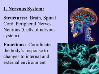
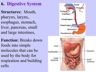
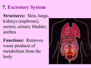
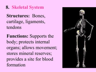
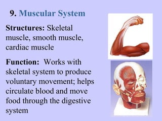
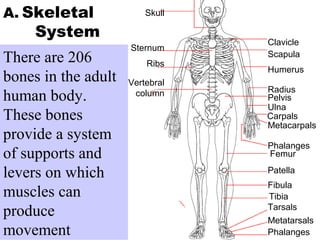
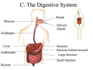
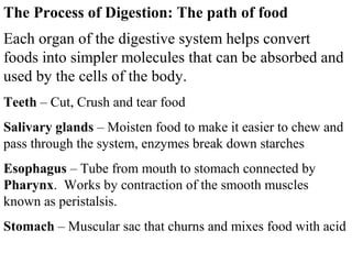
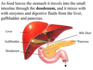
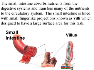
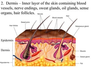
2. How do Humans and other complex mammals
maintain homeostasis? They must carry out all
needed life functions in a coordinated way.
What does our species need to accomplish?
Growth Repair of injuries
Get energy Get building materials
Get rid of waste
Keep away disease
Respond to changing environment
Reproduce
3. Eleven Body Systems work together to
maintain homeostasis and carry out these
tasks:
1. Nervous System 2. Endocrine System
3. Lymphatic System 4. Circulatory System
5. Respiratory System 6. Digestive System
7. Excretory System 8. Skeletal System
9. Muscular System 10. Integumentary System
11. Reproductive System
4. 1. Nervous System:
Structures: Brain, Spinal
Cord, Peripheral Nerves,
Neurons (Cells of nervous
system)
Functions: Coordinates
the body’s response to
changes to internal and
external environment
5. 2. Endocrine System – Hormone System
Structures: Glands -
Hypothalmus, pituitary, thyroid,
parathyroids, adrenals,
pancreas, ovaries (in females),
testes (in males)
Functions: Produce Hormones.
Controls growth, development,
metabolism, and reproduction
6. 3. Lymphatic System – Immune System
Structures: White blood
cells, thymus, spleen,
lymph nodes, lymph
vessels
Function: Helps protect
the body from disease;
collects fluid lost from
blood vessels; returns the
fluid to the circulatory
system
7. 4. Circulatory System
Structures: Heart,
Blood vessels, blood
Functions: Brings
oxygen, nutrients, and
hormones to cells;
fights infection;
regulates body
temperature
8. 5. Respiratory System
Structures: Nose,
pharynx, larynx, trachea,
bronchi, bronchioles, lungs
Functions: Provides
oxygen needed for cellular
respiration and removes
carbon dioxide from body
9. 6. Digestive System
Structures: Mouth,
pharynx, larynx,
esophagus, stomach,
liver, pancreas, small
and large intestines,
Function: Breaks down
foods into simple
molecules that can be
used by the body for
respiration and building
cells
10. 7. Excretory System
Structures: Skin, lungs,
kidneys (nephrons),
ureters, urinary bladder,
urethra
Functions: Removes
waste products of
metabolism from the
body
11. 8. Skeletal System
Structures: Bones,
cartilage, ligaments,
tendons
Functions: Supports the
body; protects internal
organs; allows movement;
stores mineral reserves;
provides a site for blood
formation
12. 9. Muscular System
Structures: Skeletal
muscle, smooth muscle,
cardiac muscle
Function: Works with
skeletal system to produce
voluntary movement; helps
circulate blood and move
food through the digestive
system
13. 10. Integumentary System (Skin)
Structures: Skin, Hair, Nails, Sweat
and Oil Glands
Functions: Serves as a barrier against
infection and injury; Helps to regulate
temperature; Protects against UV light
14. 11. Reproductive System
Structures:
Female- Uterus, Fallopian
tubes, ovary, cervical canal,
cervix, vagina
Male – Penis, Vas Deferens,
Prostate, Epididymis, Testes,
Scrotal Sac, Seminal Vesicle
Function: Produces
reproductive cells; in females
nurtures and protects
developing embryo
16. How does the human body
move from place to place and
have the ability to run, blink
or build things?
These things are all made
possible by the skeletal and
muscular systems.
17. Skull
Sternum
Ribs
Vertebral
column
Clavicle
Scapula
Humerus
Radius
Pelvis
Ulna
Carpals
Metacarpals
Phalanges
Femur
Patella
Fibula
Tibia
Tarsals
Metatarsals
Phalanges
A. Skeletal
System
There are 206
bones in the adult
human body.
These bones
provide a system
of supports and
levers on which
muscles can
produce
movement
18. Figure 36-3 The Bones are a solid network o fS tlriuvcintugr ec oefl lBso anned
protein fibers that are surrounded by calcium
deposits. Bones contain blood vessels and cavities
containing marrow. Red marrow produces red
blood cells, white blood cells and platelets
Bone
Marrow
19. Joints are places where one bone attaches to another.
Each type of joint is designed to allow movement
without damaging the other bones. Joints in the skull
allow no movement, joints in the spine allow a small
amount of movement and those shown below allow
movement in one or more directions.
Ball-and-Socket Joint
Hinge
Joint
Pivot Joint
Saddle Joint
Clavicle
Figure 36-4 Freely Movable Joints and Their
Movements
Section 36-1
Knee
Elbow
20. Muscle
Tendon
Femur
Patella
Bursa
Ligament
Cartilage
Fat
Fibula
Tibia
Knee
Section 36-1 Joint
Ligaments are a
tough connective
tissue that attach
bones to bones
Tendons are
connective tissue
which attach bones
and muscles to
allow bones to move
21. B. The Muscular System
More than 40% of the average human is muscle.
Muscles are involved in both voluntary actions and
involuntary actions. Three types of muscles cells are:
•Skeletal – Attached to bones for voluntary actions
and controlled by the central nervous system
•Smooth – Found in the digestive tract and the blood
vessels to move food and blood. Control involuntary
actions (you do not decide for them to work)
•Cardiac – Heart muscle cells are involuntary.
22. Figure 36-7 Skeletal Muscle Structure
Section 36-2 Skeletal muscles are made up of clusters of
filaments of proteins known as actin and myosin
which control muscle contraction and relaxation
23. Figure 36-8 Muscle Contraction
During Muscle
contraction Actin
filaments slide over
myosin filaments
shrinking the
muscle
25. Nervous system cells known as motor neurons
are attached to skeletal muscle cells to control
the voluntary movement.
Threadlike Nerve
or neuron
Axon or junction
26. Figure 36-11 Opposing Muscle Pairs
SkeSelcetiotna 3l6 -2muscles work in opposing pairs.
When one muscle contracts, the other relaxes.
Movement
Movement
Biceps
(relaxed)
Triceps
(contracted)
Biceps
(contracted)
Triceps
(relaxed)
27. Nutrition and Waste Removal
How does the human body take in required
nutrients and get rid of wastes?
The Digestive and Excretory Systems are
responsible for bringing in food and getting rid of
the leftovers.
28. Overview of Nutrients
Food supplies the raw materials for
building molecules your body needs, such
as:
• enzymes
• Lipids in cell membranes
• DNA
Food contains 45 substances your
body needs but cannot manufacture.
29. The nutrients your body needs are water,
carbohydrates, fats, proteins, vitamins and
minerals.
WATER – Most of the bodies reactions take place in
water. Humans need an average of 1 liter of water a
day. Dehydration causes many problems throughout
the body.
CARBOHYDRATES – Sugars and starches are used by the
body to provide the simple sugars needed for Respiration
(energy)
30. FATS- Deposits of fats protect body organs, insulate
the body and store energy, make up cell membranes and
coat the nervous system cells
PROTEINS – Supply the raw materials for
growth and repair in the form of enzymes
VITAMINS – Organic molecules that work with
enzymes to control body processes.
MINERALS – Inorganic nutrients that are
needed in small amounts. Calcium is needed to
build bones, Iron is needed for red blood cells.
32. C. The Digestive System
Pharynx
Esophagus
Liver
Gallbladder
Rectum
Mouth
Salivary
Glands
Stomach
Pancreas behind stomach
Large Intestine
Small Intestine
33. The Process of Digestion: The path of food
Each organ of the digestive system helps convert
foods into simpler molecules that can be absorbed and
used by the cells of the body.
Teeth – Cut, Crush and tear food
Salivary glands – Moisten food to make it easier to chew and
pass through the system, enzymes break down starches
Esophagus – Tube from mouth to stomach connected by
Pharynx. Works by contraction of the smooth muscles
known as peristalsis.
Stomach – Muscular sac that churns and mixes food with acid
34. As food leaves the stomach it travels into the small
intestine through the doudenum, and it mixes with
Section 38-2
with enzymes and digestive fluids from the liver,
gallbladder and pancreas.
Liver
Gallbladder
Doudenum
Bile Duct
Pancreas
35. Chemicals from the pancreas breakdown
carbohydrates, .
proteins, lipids and nucleic
acids. In addition chemicals from the pancreas
produce substances which neutralize stomach
acid.
If the acid travels too far without being
neutralized it will change the shape of
enzymes and cause problems in the
body.
The Liver makes bile which acts like a
detergent to break down fat. The gall
bladder is a storage area for the bile
36. The small intestine absorbs nutrients from the
digestive systems and transfers many of the nutrients
Section 38-2
to the circulatory system. The small intestine is lined
with small fingerlike projections known as villi which
designed to have a large surface area for this task.
Small
Intestine Villus
37. Large intestine (Colon)
Food that enters the large intestine is nutrient-free.
Usable nutrients have been absorbed leaving water
and undigestible substances. The large intestine
removes water from the waste. Once water is
eliminated the solid waste passes out of the body
through the rectum.
The appendix is located just below the entry
to the large intestine. In many animals it
helps digest difficult materials such as cellulose.
In humans the appendix is not used for any purpose
in digestion. When it gets infected it is
removed.
38. D. Excretory System:
In the process of obtaining nutrients and
carrying out chemical reactions the human body
produces wastes (CO2, Urea, Salts). If some of
these wastes are not removed they could
threaten homeostasis. Excess chemicals that
are not toxic also need to be removed.
The skin, lungs and kidneys all function to
get rid of excess or harmful products
produced in the body.
39. Kidneys contain millions of small filters called
nephrons, Section 38-3
which filter your entire blood supply
every 45 minutes. Filtration takes place because
blood pressure forces water, salt, glucose, amino acids
and urea into structures known as Bowman’s capsules.
(Protein and Blood are too large to enter)
Kidney Nephron
Bowman’s
Capsule
40. Processes of osmosis and active transport filter the
useful materials from the waste (urine). Substances
your body needs to keep are returned to the blood
stream. Substances your body needs to get rid of are
passed from the renal tubes to the urinary bladder.
Kidney
Vein Artery
Ureter
Urinary Bladder
Urethra
41. Other important parts of the bodies
excretory system
•The skin excretes excess water and
salts
•The lungs excrete carbon dioxide
produced from respiration
Both of these body organs serves multiple functions
in the human body.
42. The Respiratory System links to the
Circulatory System to provide cells with
Section 37-3
oxygen and remove carbon dioxide
Figure 37-13 The Respiratory System
Bronchi branch to air sacs
known as aveoli where
gas exchange occurs
43. Alveoli
Bronchiole
Capillary
Gas Exchange in
the lungs Section occurs
37-3
through the
process of
DIFFUSION
High concentration of
oxygen (O2) moves out
of lungs into blood to
balance concentration.
CO2 does the opposite
(moves from blood to
lungs)
44. Figure 37-15 The Mechanics The Lungs are only air sacs. In order foofr Bthreeamth itnog
move they must work together with a muscle
known as the Diaphragm
Air
inhaled
Rib cage
rises
Diaphragm
Air
exhaled
Rib
cage
lowers
Diaphragm
Inhalation
Exhalation
Section 37-3
45. Breathing is not an entirely
voluntary process. While
you have control of
breathing so that you can
briefly hold your breath or
blow up a balloon, your
body will not allow you not
to breathe. The brain has
control over the diaphragm
if the level of carbon dioxide
in the blood rises to high.
46. The circulatory system works together
with several other body systems to bring
needed supplies to cells which cannot move.
The circulatory system is a delivery system
that consists of the following:
• The Heart
• Blood Vessels
• Blood
47. The Heart:
The heart is about the size of a clenched fist. On
average the heart contracts about 72 times a
minute, pumping about 70 milliliters of blood with
each contraction.
The entire circulatory system
including Heart has structures
which prevent oxygen rich blood
and oxygen poor blood from
mixing. All blood is really red
but oxygen poor blood is shown
as blue in pictures for ease.
48. The heart has four main chambers with valves
that separate each to prevent backflow
Pulmonary Artery
Oxygen poor
blood to lungs
Left Atrium
Left Ventricle
49. The heart acts as two pumps. One for sending
oxygen poor blood to the lungs and the other
for sending oxygen rich blood to the body.
50. The heart’s contractions are controlled by a small
group of cardiac muscle cells known as the
sinoatrial node. These are the pacemakers of the
heart. These cells send an electrical message from
the atrium to the ventricles to contract
Sinoatrial
Node
Atrioventricular
node
Conducting
Fibers
51. Blood moves through 3 different types of vessels:
Arteries – Carry oxygen rich blood away from the heart
Capillaries – Tiny vessels only one cell thick. Bring oxygen
and nutrients to the cells. Remove waste products.
Veins – Carry blood back to the heart with wastes
ARTERY
VEIN
CAPILLARIES
52. Blood Pressure:
When the heart contracts it produces
pressure in the arteries. The force of
the blood on the arteries is known as
blood pressure. Blood pressure
decreases when the heart relaxes but it
does not disappear. The difference
between the two pressures is what is
measured when you go to the doctor
and have your blood pressure checked.
The average adult’s blood pressure is
120/80.
53. Blood
The Human body contains 4 to 6 liters of blood, which
is about 8% of the total body mass. Blood is made up
of a number of types of cells and substances:
55% Plasma – mostly water with dissolved
gases, salts, nutrients, enzymes, hormones, waste
products and plasma proteins.
45% Cells – Red Blood cells, White Blood
Cells, and platelets.
54. Whole
Blood
Sample
Plasma
Sample Placed in
Centrifuge
Cells
Blood Sample
That Has Been
Centrifuged
55. Red blood cells transport oxygen on an iron
containing protein called hemoglobin. They are
shaped like flat disks so that they have maximum
surface area and don’t get caught in the blood vessels.
Mature red
blood cells do
not have a
nucleus.
Your body produces red blood cells in bone marrow
and each one circulates for about 120 days. RBC’s
are destroyed by the liver and the spleen.
56. Platelets and plasma proteins work together to
make sure that too much blood is not lost.
1. Injured blood
vessel
2. Platelets
clump at site and
release Thrombin
3. Fibrin forms
from
Thrombin and
clot stops
blood loss
57. White Blood cells (also known as Leukocytes) are
also produced in the bone marrow but do not have
hemoglobin for carrying oxygen. They contain
nuclei and live anywhere from a few days to a few
months. They carry out an entirely different function
than the Red Blood Cells.
White blood cells are the army of the
circulatory system. They attack
foreign substances or organisms.
58. Normally the body contains 700 times more red
blood cells than white blood cells.
The body can increase the number of white blood
cells on demand if the body is threatened by a
foreign invader. Doctors often test for increased
White Blood Cell levels to check your health
59. Thymus
Heart
Thoracic
Duct
Spleen
Superior Vena
Cava
Lymph
Nodes
Lymph
Vessels
F. The Lymphatic
system collects
fluid that leaks
into body tissues
and returns it to
the circulatory
system.
Lymph nodes are
also filters that
collect invaders that
cause disease.
60. G. The Integumentary system or Skin is the bodies
largest organ. It serves many purposes and overlaps
with many of the body systems.
• Serves as a barrier against infection and injury
• Helps regulate body temperature
• removes excess salts and water
• Protects internal cells from UV radiation
• Serves as one link between the nervous system and
the environment (receives information on pressure,
temperature, pain)
61. The skin is made of 2 main layers
1. Epidermis – Outer layer of skin which comes in
contact with environment.
•The outermost layer is coated in dead cells.
•The inner epidermis rapidly divides to produce a
constant supply of new cells, constantly pushing
old cells to the top.
• As skin cells are forced upward they flatten and
organelles disappear and they form a layer of
waterproof covering
62. 2. Dermis – Inner layer of the skin containing blood
vessels, nerve endings, sweat glands, oil glands, sense
organs, hair follicles.
Epidermis
Dermis
63. Hair and Nails are made up of a
substance known as keratin. They are
both used to protect the skin from
damage:
• Fingernails and toe nails protect the
tips of your toes and fingers
•Hair on your head protects from UV
rays
•Eyelashes, Nose Hair and Ear Hair
prevent dirt and other particles from
entering the body.
65. How does the Human Body Control (Regulate)
all these body systems and make them work
together?
Electrical Impulses from the
Nervous System
Chemical Hormones from the
Endocrine System
66. The Nervous System:
The nervous system is the number one
communication center of the body. The basic cell
type that carries the communications is a network
of neurons that transmit electrical impulses.
Axon terminals
Myelin Sheath
Nodes
Cell Body
Nucleus
Dendrites
NEURON CELL
67. Electrical Impulses depend on the movement of
negatively charged electrons compared with the
positively charged ions across a cell membrane
68. Once and impulse begins it
moves along the axon in the
direction of the impulse.
To pass between neurons
the impulse must be sent
across a gap known as a
synapse which sends the
message from one neuron to
Synapse
the other.
When the impulse reaches the end of the axon it
transfers its impulse to another cell by releasing
chemicals known as neurotransmitters which pass the
message across the synapse.
69. The nervous system is divided into two
divisions:
• The Central Nervous System (CNS)–
Responsible for relaying messages, processing
and analyzing information.
• The Peripheral Nervous System – Receives
information from the environment and relays
commands from the CNS to the organs and glands
70. The brain is the main switching area of the central
nervous system.
Cerebrum – Responsible for voluntary activities of the
body (Intelligence, learning and judgement)
Cerebellum – Coordinates muscle movement
Brain Stem – Consists of the pons and the medulla
oblongata. Pass message between brain and body
Thalamus – Connects messages from the sense organs
to the Cerebrum
Hypothalamus - Controll center for hunger, thirst,
anger and body temperature.
72. The Endocrine System
Sends messages throughout the body by way of
chemicals known as hormones. Hormones travel
throughout the bloodstream to target cells which
contain matching receptors.
Hormone
Receptor
73. If a cell does not have a specific receptor the
hormone will not affect the cell.
Responses to hormones take longer and last
longer than nervous system messages.
Hormones can take minutes, hours or days to
influence cells.
Examples of functions controlled
by hormones:
Growth, Metabolism, Sleep,
Reaction to stress, Reproduction.
74. The Endocrine System works through a FEEDBACK
system. Glands of the Endocrine system determine
the level of a hormone in the blood and then changes
the rate of hormone production or sends out the
opposite hormone to counteract excess amounts of
hormone.
Examples of Feedback mechanisms:
Control of insulin/sugar levels in blood
Hypothalamus can measure water level in blood
and sends out hormones that tell the kidneys to
conserve water.
75. Example of
Feedback
Loop
Room temperature
increases
Thermostat senses
temperature change
and switches off
heating system
Thermostat senses
temperature change
and switches on
heating system
Room temperature
decreases
Section 35-1
76. Feedback Actions of Insulin and
Glucagon
Beta cells release
insulin into the blood
Body cells
absorb glucose
Blood glucose
level
decreases
Blood glucose
level increases
Liver converts
glycogen to glucose
Homeostasis: Normal
blood glucose level
Blood glucose
level
decreases
Alpha cells release
glucagon into blood
Blood glucose
level
increases
Liver converts
glycogen to
glucose
77. Important Glands and Hormones of the Human
Body
Gland Hormone Function
Pineal Melatonin Controls sleep and wake
cycle
Thyroid Thyroxine Controls appetite and
metabolism
Adrenal Adrenaline Deals with stressful
situations
Thymus Thymosin T-cell development (fight
diseases)
Ovary Estrogen Female reproduction
Testis Testosterone Male reproduction
79. K. The Reproductive System
Functions to make new individuals by
producing, storing and releasing specialized
sex cells known as gametes.
Cells from the male reproductive system,
known as sperm, must fuse with cells of the
female reproductive system, known as eggs.
80. Reproduction in both males and females is
regulated by hormones.
•In males - Testosterone is produced by the testes.
It is required for sperm production and
development of male physical characteristics.
•In females - Estrogen and progesterone are
female hormones produced by the ovaries.
Estrogen is required for the development of eggs
and female physical characteristics.
Progesterone prepare the uterus for the arrival of
a developing embryo.
81. Neither males or females are capable of
producing active reproductive cells until
puberty, which is a period of sexual
maturation.
Puberty begins when the hypothalmus
signals the pituitary to produce increased
levels of hormones that affect the sex
organs (gonads). The hormones are follicle
stimulating hormone (FSH) and luteinizing
hormone (LH).
82. In both the male and female reproductive
system sex cells are produced by the process
of Meiosis.
Meiosis involves producing a cell with
only 1 copy of each chromosome (haploid
nuclei). When the egg and sperm fuse, a
cell with two copies of each chromosome is
created. Every cell in the new individual
has two copies of each chromosome
(diploid nuclei).
83. The Male Reproductive System
Urinary
Bladder
Vas deferens
Pubic Bone
Urethra
Penis
Epididymis
Testis
Scrotum
Seminal
Vesicle
Rectum
Prostate
Bulbourethral
gland
84. Sperm development:
1. Sperm are made from special cells in testes that
undergo the process of meiosis.
2. Once sperm mature they move through a tube
known as the vas deferens upward from the
scrotal sacs into the abdomen into the seminal
vesicle. The sperm mixes with seminal fluid to
form semen. Between 50 and 130 million
sperm are present in one milliliter of semen.
The seminal vesicle merges with the urethra
(also connected to the urinary bladder)
85. 3. During sexual excitement the nervous system
of the male contracts the glands of the
reproductive tract. The release of semen is
controlled by the autonomic nervous system
so it is not entirely voluntary. The male
reproductive system is designed to deliver
sperm into the female reproductive system.
86. The Female Reproductive System Section 39-3
Fallopian Tube
Ovary
Uterus
Urinary
Bladder
Pubic Bone
Urethra
Cervix
Rectum
Vagina
87. 1. The ovaries produce one mature ova or egg per
month. Females are born with 400,000 immature
eggs which are stored in follicles. The follicle
assist in the maturing process of an egg from each
ovary once a month based on a signal from the FSH
hormone.
2. When the egg matures the follicle breaks open
releasing the egg into the fallopian tube. A process
known as ovulation. Fertilization occurs in the
fallopian tube if sperm is present.
3. The fallopian tube connects to the uterus which is a
cavity designed to protect and nourish a developing
embryo.
88. 4. If fertilized egg enters the uterus it is implanted into
the lining of the uterus and the embryo develops. If
fertilization does not occur the egg is discharged out
of the body along with the uterus lining through a
canal known as the vagina. Known as
MENSTRUATION
5. The endocrine system controls the cycle of events
surrounding ovulation through a feedback mechanism
that can signal the presence of a fertilized egg. While
the egg is maturing and preparing for release the
uterus is preparing for a fertilized egg by building up
the lining of the uterus. A new lining must be created
each month to prepare for nourishing the embryo.
95. Meiosis II
Prophase II Metaphase II Anaphase II Telophase II
Meiosis I results in two
haploid (N) daughter cells,
each with half the number of
chromosomes as the original.
The chromosomes line up in
a similar way to the
metaphase stage of mitosis.
The sister chromatids
separate and move toward
opposite ends of the cell.
Meiosis II results in four
haploid (N) daughter cells.
Section 11-4
Figure 11-17 Meiosis II
96. Meiosis II
Prophase II Metaphase II Anaphase II Telophase II
Meiosis I results in two
haploid (N) daughter cells,
each with half the number of
chromosomes as the original.
The chromosomes line up in
a similar way to the
metaphase stage of mitosis.
The sister chromatids
separate and move toward
opposite ends of the cell.
Meiosis II results in four
haploid (N) daughter cells.
Section 11-4
Figure 11-17 Meiosis II
97. Meiosis II
Prophase II Metaphase II Anaphase II Telophase II
Meiosis I results in two
haploid (N) daughter cells,
each with half the number of
chromosomes as the original.
The chromosomes line up in
a similar way to the
metaphase stage of mitosis.
The sister chromatids
separate and move toward
opposite ends of the cell.
Meiosis II results in four
haploid (N) daughter cells.
Section 11-4
Figure 11-17 Meiosis II
98. Meiosis II
Prophase II Metaphase II Anaphase II Telophase II
Meiosis I results in two
haploid (N) daughter cells,
each with half the number of
chromosomes as the original.
The chromosomes line up in
a similar way to the
metaphase stage of mitosis.
The sister chromatids
separate and move toward
opposite ends of the cell.
Meiosis II results in four
haploid (N) daughter cells.
Section 11-4
Figure 11-17 Meiosis II
99. Meiosis II
Prophase II Metaphase II Anaphase II Telophase II
Meiosis I results in two
haploid (N) daughter cells,
each with half the number of
chromosomes as the original.
The chromosomes line up in
a similar way to the
metaphase stage of mitosis.
The sister chromatids
separate and move toward
opposite ends of the cell.
Meiosis II results in four
haploid (N) daughter cells.
Section 11-4
Figure 11-17 Meiosis II
100. Figure 11-17 Meiosis II
Meiosis II
Section 11-4
Prophase II Metaphase II Anaphase II Telophase II
101.
102. Meiosis produces specialized cells
known as gametes
Each gamete must contain only half
the number of chromosomes that the
parent has. The gamete gets a mixture
of chromosomes from the parent cells.