Ophthalmic ultrasound report
Bscan Ultrasound
How to write a ultrasound report
Prof Dr Nagla Hassan Aly
Professor of ophthalmology
Memorial institute for ophthalmic research
Giza- Egypt
With understanding ofthe indications and proper examination
technique, one can gain a great amount of information which is not
possible with clinical examination alone
.
B-scan ultrasound is most useful when direct visualization of intraocular structures
is difficult
.
Situations that prevent normal examination includes
:
**
Lid problems (severe edema)
.
**
Corneal opacities ( scars, edema
** Lens inplace with well delineated posterior capsule
**Vitreous shows Partial PVD with low amplitude amorphous
echoes of vitreous floaters.
**Retina is acoustically in place.
**Normal thickness of choroid
**Normal acoustic appearance of optic nerve shadow.
Thank you
11.
what's your Expectationsif the patient is not normal
size of the globe
Normal -large -small
Contour of the globe
Normal or disturbed (rupture globe!)
Lens
-phakic ,pesudophakic
-lens in place with well delineated posterior capsule
- dropped lens -rupture capsule
-if pesudophakia, droped IOL!
Vitreous
-Clear
-opacities (Hgs or exudates)
12.
-PVD
-Sub hyaloid Hgs
-Endophthalmitis
-Uveitits
-Organizedvitreous Hgs
-vitreal membranes -traction bands on the surface of the retina!
Retina
-in place
-localized -total -subtotal RD
-good mobility on kinetic scanning -poor mobility with thickened
retina leaves
Choroid
-Normal thickness
- thickness
-choroidal mass! size-transverse and longitudinal diameters
ONH
-normal optic nerve head shadow
-coloboma-swelling-drusen-cupping (advanced)
; ID; Date; Time:
------------------------------------------------------------------------
A and B scans of the left eye showed ultrasonography.
* Normal ocular size and contour
* Lens could not be detected.
* Vitreous shows moderate to high echoes of amorphous vitreous opacities
denoting vitreous Hemorrhages or exudates; however, there is evidence of Loculi
* Retina is in place
*Acoustic evidence of thickened choroid
13.
* Normal acousticappearance of optic nerve shadow.
* NB clinical picture in endophthalmitis should be correlated
------------------------------------------------------------------------
A and B scan Ultrasonography of the right eye showed:
* Normal ocular size and contour.
* Lens acoustically in place.
* Vitreous shows mild amplitude amorphous echoes of vitreous floaters.
* Retina acoustically showed total retinal detachment in an open funnel with
thickened retinal leaves with good mobility on the kinetic scan.
* Normal thickness of the choroid
* Normal acoustic appearance of optic nerve shadow.
14.
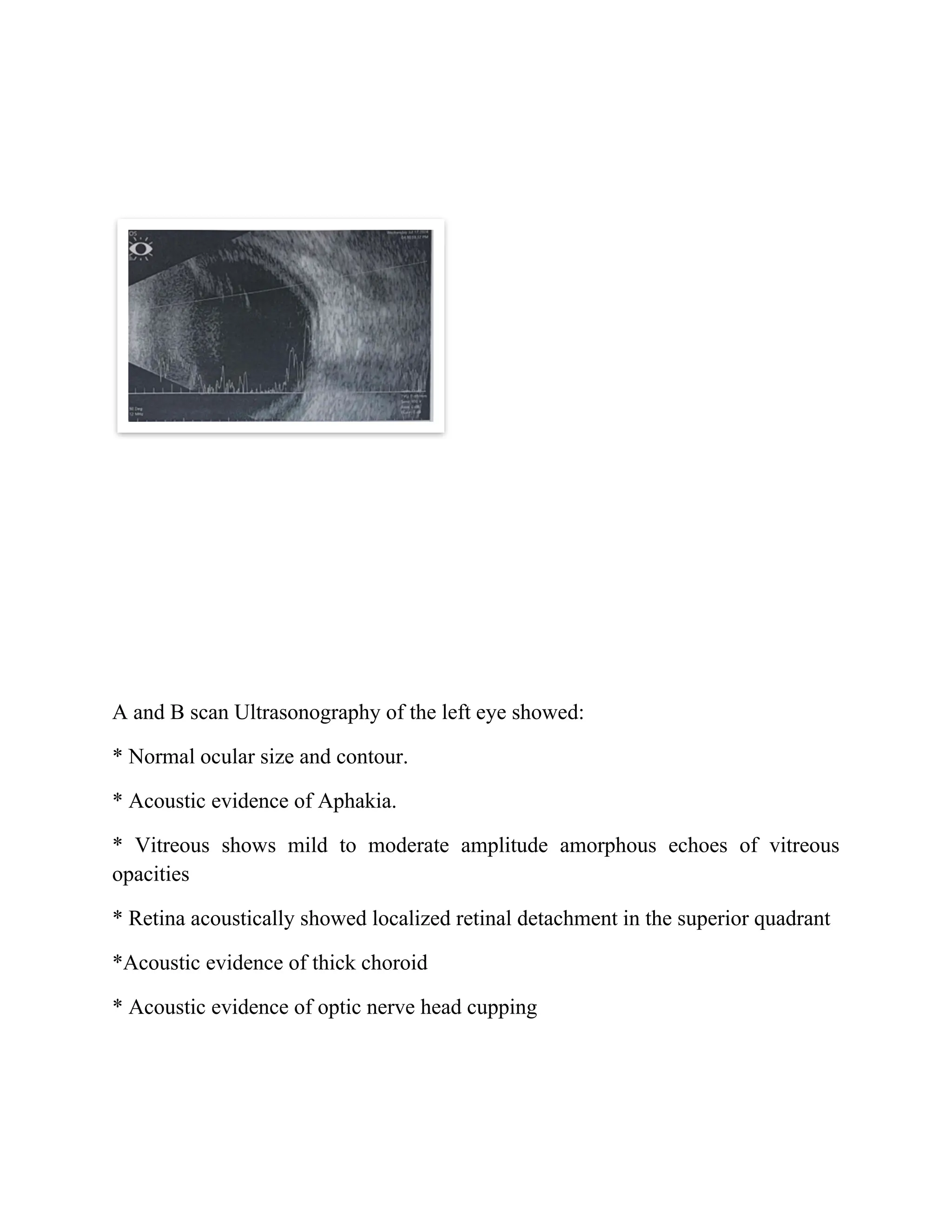
A and Bscan Ultrasonography of the left eye showed:
* Normal ocular size and contour.
* Acoustic evidence of Aphakia.
* Vitreous shows mild to moderate amplitude amorphous echoes of vitreous
opacities
* Retina acoustically showed localized retinal detachment in the superior quadrant
*Acoustic evidence of thick choroid
* Acoustic evidence of optic nerve head cupping
15.
A and Bscan Ultrasonography of the left eye showed:
* Normal ocular size and contour.
* Lens acoustically is in place.
*Vitreous shows moderate amplitude amorphous echoes of vitreous opacities.
* Retina acoustically showed total closed funnel retinal detachment with thickened
retinal leaves, which showed low mobility on the kinetic scan.
* Acoustic evidence of thick choroid