SlideShare a Scribd company logo
1 of 169
SONOGRAPHY OF LIVER
Dr.Abhilasha singh
DNB Resident
Radiodiagnosis
Liver-Introduction
• Also called ‘hepar’.
• Largest/heaviest solid organ in the
body.
• Weighs about 1600 gm in
males,1300 gm in females.
• Occupies the right hypochondrium,
epigastrium & left hypochondrium.
• Most part of the liver is covered by
ribs & costal cartilages.
• It is covered by network of
connective tissue (Glisson’s
Capsule)
Peritoneal ligaments
These ligaments connect the liver to the undersurface of the
diaphragm
Falciform ligament = It is a double fold of peritoneum from
umblicus to liver.Contains ligamentum teres,the remnant of
umblical vein,which attaches to the left portal vein.
Falciform ligaments split into coronary ligament ( which
becomes the right triangular ligament) and left triangular
ligament, between which lies the bare area of the liver.
Lobes of Liver
• Liver is divided into right
• & left lobes by falciform ligament .
• The right lobe , left lobe and caudate lobe.
Liver surfaces
• Divided into 2 anatomicalregions:
1.Diaphragmatic surface:
 Smooth and dome-shaped surface
 Inferior to diaphragm
 Separated from diaphragm by subphrenic recessandfrom
posterior organs {kidney and suprarenal glands} by
hepatorenal recess
 Covered by peritoneum except on the posterior surfaceof
liver which is not invested in peritoneum and is known as
the bare area of liver.
Diaphragmatic
surface
2. Visceralsurface
 Covered by visceral peritoneum except porta hepatis andgall
bladder bed.
• Thevisceral surface is related to:
 Right side of the stomach i.e. gastric and pyloric areas
 Superior part of the duodenum i.e. duodenal area
 Lesser omentum
 Gall bladder
 Right colic flexure and right transverse colon ;colic area
 Right kidney and suprarenal gland; Renalarea
Visceral relations :impression of neighbouring
viscera
Peritoneal relations of theLiver
The Lesser omentum
• Encloses the portal triad (bile duct, hepatic artery and portal vein )
• Passes from the liver to lesser curvature of the stomach + 2 cm of
duodenum.
• Thick free edge -- hepatoduodenal ligament
• Sheet like remainder – hepatogastric ligament
Development ofLiver
Liver development requires two linked
processes:
1. Differentiation of the various hepatic cell
types from their embryonic progenitors.
2. The arrangement of those cells into
structures that permit the distinctive
and excretorycirculatory, metabolic
functions of theliver.
Mediated by many essential regulators
which include signaling molecules, and
transcription factors.
Development of Liver:
Stages
• Specification
• Liver bud formation andexpansion
• Epithelial differentiation stage
Endoderm cells adjacent to the cardiogenic mesoderm begin
to differentiate into hepatoblasts.
Hepatoblasts proliferate and penetrate the endoderm basement
membrane to form the liver bud (25d). The liver bud then
expands in size, intercalating into the adjacent septum
transversummesenchyme.
During the epithelialdifferentiation stage,hepatoblasts mature
into hepatocytes or differentiate into cholangiocytes.
13/26
Development of liver and biliary
passages
• The hepatic diverticulum enlarges rapidly and divides into two parts
ie pars hepatica(cranial bud) and pars cystica(caudal bud) as it grows
between the layers of the ventralmesentery.
Superior
Caudalbud
Inferior
Gallbladder, cysticduct
Ventral pancreas
Pars hepatica
• It is thelarger cranial part of the hepatic diverticulum.
• Givesrise to:
1. Hepatocytes
2. Hepatic sinusoids
3. Kupffer cells and hematopoietic tissue
4. Intrahepatic bile ducts
• The liver grows rapidly to fill a large part of the
abdominal cavity.
• At first, the 2 lobes are of the same size but soon the
right become larger.
Pars cystica
• Becomes the gall bladder and the stem of the diverticulum
forms the cystic duct.
• The stalk connecting the hepatic andthe cystic ducts to the
duodenum becomes the common bileduct.
• The right and the left branches of the pars hepatica
canalized to form the right and the left hepaticducts.
• Bile begins to flow at about the 12th week.
16/26
Development of Liver
Parscystica
Endoderm
Parshepatica
Hepatocytes
Hepatic sinusoids
Kupffer cellsand
hematopoietic tissue
Intrahepatic biliarytree
Gallbladder
Extrahepatic bile ducts
(cystic duct ,CBD)
Ventralmesentery
Mesoderm
Visceralperitoneum of liver
Falciform ligament
SeptumTransversum
Cardiogenicmesoderm
Hepatoblast
Hepatic diverticulum
Kupffer cells derive from circulating monocytes and possibly yolk sacmacrophages.
Formation of the capsule &
ligaments of the liver
As the septum transversum is
penetrated by the growing pars
hepatica:
• The mesoderm of the septum
transversum between the liver
and the anterior abdominal
wall becomes the FALCIFORM
LIGAMENT.
• The mesoderm of the septum
transversum between the liver
and the foregut (stomach and
duodenum) forms the LESSER
OMENTUM.
• The mesoderm on the surface
of the liver differentiates into
CAPSULE AND PERITONEAL
COVERING.
Vascular Development
related to Liver
Extraembryonic
Major Venoussystem
Intraembryonic
Vitelline veins CardinalVeinsUmbilical Veins
Sinusvenosus
• During early development, there are 3 major venous systems in the
embryo—2 extraembryonic and 1 intraembryonic.
• The extraembryonic venous systems are the omphalomesenteric
(vitelline) and umbilical (placental) veins, and the intraembryonic
system includes the cardinal veins that drain the venous blood of the
embryo to theheart.
• All of these systems converge into the sinus venosus, a cavity that is
incorporated into the heart.
Development of Vitelline & umbilicalveins
A: 4th week B. 5th week. Note the plexus around the duodenum, formation of the
hepatic sinusoids, and initiation of left to right shunt between the vitelline veins.
Before entering the sinus
venosus, the vitelline veins form
a plexus around the duodenum
and pass through the septum
transversum.
The liver cords growing into the
septuminterrupt the course of the
veins and an extensive vascular
network, the hepatic
sinusoids,forms. There is also
initiation of the left to right
shunting between the vitelline
veins
Development of Vitelline & umbilicalveins
• Blood from the left side of the liver is
rechanneled towards the right, resulting in
an enlargement of the right vitelline vein
(right hepatocardiac channel).
• The right hepatocardiac channel forms the
hepatocardiac portion of the inferior vena
cava.
• The proximal part of the left vitelline vein
disappears. The anastomotic network
around the duodenum develops into a
single vessel, the portal vein.
• The superior mesenteric vein derives from
the right vitelline vein.
• The distal portion of the left vitelline
vein also disappears.
• The superior segment of vitelline veins
becomes the hepatic veins
A. 2nd mnth B.3rd mnth. Note formation of the ductus
venosus, portal vein , hepatic portion of IVC.The
splenic & SMVenter thePV.
Development of Vitelline & umbilical veins
A. 2nd mnth B.3rd mnth. Note formation of the
ductus venosus, portal vein , hepatic portion of IVC.
The splenic & SMVenterthe
PV.
22/26
• Theumbilical veins run from the placenta to the
heart and during fetal life are the predominant
afferentvessels that supply theliver.
• Initially the umbilical veins passon eachside of the
liver but someconnect to the hepaticsinusoids.
• Theproximal part of both umbilical veins and
the remainder of the right umbilical vein then
disappear. Theleft vein is the only one to carry
blood from the placenta to theliver.
• With the increaseof the placental circulation,a
direct communication forms between the left
umbilicalvein and the right hepatocardiac
channel, the ductus venosus.
• Theductus venosusbypassesthe sinusoidal
plexusof the liver.
After birth the left umbilical vein and the ductus venosus
are obliterated and form the ligamentum teres hepatic
or the round ligament and ligamentum venosum
respectively.
Arterial supply ofliver
The arterial supply of the liver begins as an off shoot from
the celiac trunk at around the eighth week ofgestation.
By the 10th week, the first arterial radicles are visible in the
central portion of the liver, and by the fifteenth week, they
reach the periphery of theliver.
Congenital anomalies ofliver
Riedel lobe is a tongue-like, inferior
projection of the right lobe of the liver
beyond the level of the most inferior
costal cartilage . It is notconsidered a
true accessory lobe of the liver but an
anatomical variant of the right lobe of the
liver.
• Congenital solitary nonparasitic
cysts of the liver
• Congenital Hepatic Fibrosis
• Congenital vascular malformation of the
liver
• Intrahepatic BiliaryAtresia
• Mesenchymal Hamartoma
• Accessoryand Ectopic Lobesof the Liver
Congenital anomalies ofliver
Anomalous supradiaphragmatic lobe
Ductul platemalformations
• Intrahepatic bile ducts (IHBDs) develop from bi-potential liver
progenitor cells(hepatoblasts) in contact with the mesenchyme of the
portal vein and thus form the “ductalplates.”
• The ductal plates are remodeled into mature tubular ducts. Lack of
remodeling results in “ductal platemalformation”.
• Aproposal is that virtually all congenital diseases of IHBDs represent
examples of DPM.
• DPM are developmental anomalies considered to result from lack of
ductal plate remodeling during bile ductmorphogenesis.
Classification of DPM
• Autosomal recessive polycystic kidney disease
(hepatic ARPKD) (50% of children, 70% of families):
DPM of interlobular bile ducts associated with tubular
dilatation of collecting renal tubules
• Caroli disease : DPM of the larger IHBDs
• Caroli syndrome: Caroli disease + congenital hepatic
fibrosis
• Von Meyenburg complexes: DPM of smaller
interlobular ducts (liver cysts in autosomal dominant
polycystic kidney disease)
• Mesenchymal hamartoma
• Meckel syndrome
• Non-syndromal ductal plate malformation
Liver is now divided into segmentsas per Couinaud System.
• Caudate lobe = Segment I.
• Portal and hepatic veins used
as landmarks to divide the
remainder of the liver into eight
segment.
• Right hepatic vein divides the right lobe into anterior
(segment V and VIII) and posterior segments (segment
VI and VII).
• Middle hepatic vein divides the liver into right and left
lobes (or right and left hemiliver). This plane runs from the
inferior vena cava to the gallbladder fossa.
• Left hepatic vein divides the left lobe into a medial
(segment IV and lateral part (segment II and III).
Couinaud’ssegments
BLOOD SUPPLY
The liver has dual blood supply : hepatic artery and
portal vein.
• Hepatic artery :
 Provides 15% of hepatic blood supply.
 Branch of coelic artery
 Common hepatic artery passes over the head of the
pancreas and gives of right gastric artery,then gives off
gastroduodenal artery at the epiploic forame to become
the hepatic artery proper.
 Hepatic artery continues in the free edge of the lesser
omentum,anterior to the portal vein and to the left side of
the common bide duct (CBD)
 Divides into left and right branches at the porta hepatis.
• Portal Vein:
 Provides 85% of blood suply to liver.
 Formed by the union of the splenic vein and
superior mesentric vein behind the neck of the
pancreas at L1/L2.
 Runs at the posterior aspect of free edge of lesser
omentum to the porta hepatis; it lies posterior to hepatic
artery and CBD.
• Venous Drainage:
Majority of the the liver is via the hepatic veins which unite into to drain
into the IVCat T9close to thediaphragmatic hiatus.
Caudatelobe drains directly into the IVCand may therefore be spared
incaseof hepaticvein thrombosis.
• Nerve Supply
Parasympathetic supply is by the preganglionic fibers of the vagus
nerve.
Sympathetic innervation is by the postganglionic fibers from the
coeliac plexus.
• LymphaticDrainage
Lymphatics from upper surface drain into nodes in the posterior
mediastinum.
Lymphatics from lower surface drain into hepatic nodes and celiac
nodes.
SAGGITALIMAGE OFPORTAHEPATISSHOWING CBD &
MAIN PORTALVEIN
Images obtained through the anterior axillary line showthe
porta hepatis along the long axis. On the gray-scale image,
the portal vein is easily identified. Often, the hepaticartery
(arrows) is identified only with color Doppler imaging.
HEPATIC ABNORMALITIES
1. CONGENITAL ABNORMALITIES
2. INFECTIOUS DISEASE
3. DISORDER OF METABOLISM
4. VASCULAR ABNORMALITIES
5. TRAUMA
6. HEPATIC MASSES
CONGENITAL ABNORMALITIES
1.HEPATIC CYST
DEFINITION: A liver cyst is defined as a fluid- filled space with an epithelial
lining.
Occurs in 2.5% of general population increasing to 7% in population older than
80 years
ULTRASONOGRAPHY:
*Anechoic with a well demarcated thin wall & posterior acoustic enhancement
*If complicated with hemorrhage or infection –May contain internal echoes &
septations , thickened wall or may appear solid
*If thick septae or nodules are seen within the cyst - CT is recommended as
biliary cystadenoma & cystic metastases must be considered in the
differential possibilities for complex appearing liver cysts
2.PERIBILIARY CYST
 Peribiliary cysts have been described in patients with severe
liver disease.
 These cysts are small, 0.2 to 2.5 cm, and are usually located
centrally within the porta hepatis or at the junction of the
main right and left hepatic ducts.
 USG : Discrete, clustered cysts or as tubular-appearing
structures with thin septae, paralleling the bile ducts and
portal veins.
3.POLYCYSTIC LIVER DISEASE
 Usually associated with polycystic kidney disease but may also occur
as an isolated finding in a rarer genetically distinct disease
 USG :
*Massive hepatomegaly with innumerable ,predominantly simple cysts
are present
*Portal vein patency should be assessed ,compression of the main
portal vein may result in portal hypertension as well as associated
findings such as splenomegaly and ascites
4.BILIARY HAMARTOMAS (VON MEYENBURG
COMPLEXES)
 Bile duct hamartomas, 1rst described by von Meyenburg in
1918, are small, focal developmental lesions of the liver
composed of groups of dilated intrahepatic bile ducts set
within a dense collagenous stroma
 USG : Single, multiple, or most often innumerable well-
defined solid nodules usually less than 1 cm in diameter
 Usually uniformly hypoechoic and less frequently
hyperechoic
 Often confused with metastatic cancer
INFECTIOUS DISEASE
1.VIRAL HEPATITIS
Viral hepatitis is a common disease that occurs world- wide. It is responsible for
millions of deaths secondary to acute hepatic necrosis or chronic hepatitis, which
in turn may lead to portal hypertension, cirrhosis, and hepatocellular carcinoma
(HCC).
Hepatitis A :
-Occurs throughout the world
-Diagnosed using serosurveys with the antibody to hepatitis A virus
(anti-HAV) as the marker.
-The primary mode of spread is the fecal-oral route.
-Hepatitis A is an acute infection leading to complete recovery or
death from acute liver failure.
Hepatitis B :
-Transmitted parenterally (e.g., blood transfusions, needle
punctures) as well as by non percutaneous exposure through sexual
contact and at birth.
-The two most useful markers for acute infection are hepatitis B surface
antigen (HBsAg) and antibody to hepatitis B core antigen (anti-HBc).
Hepatitis C (predominantly) and hepatitis E :
-Formerly called non-A, non-B (NANB) hepatitis,.
-Acutely infected patients have a much greater risk of chronic
infection, with up to 85% progressing to chronic liver disease.
-Chronic hepatitis C virus (HCV) infection is diagnosed by the
presence of anti- body to HCV(anti-HCV) in the blood.
Hepatitis D, or delta hepatitis :
-Entirely dependent on HBV for its infectivity, requiring the
HBsAg to provide an envelope coat for the hepatitis D virus.
- Its geographic distribution is therefore similar to that of hepatitis B.
- HDV is an uncommon infection in North America, occurring
primarily in intravenous (IV) drug users.
 Uncomplicated acute hepatitis : Clinical recovery within 4 months. It is the
outcome of 99% of cases of hepatitis A.
 Subfulminant and fulminant hepatic failure : Follows the onset of jaundice and
includes worsening jaundice, coagulopathy, and hepatic encephalopathy.
 Chronic hepatitis : Persistence of biochemical abnormalities beyond 6 months. It
has many etiologies other than viral, including metabolic (e.g., Wilson’s disease,
alpha-1 antitrypsin deficiency, hemochromatosis), autoimmune, and drug induced.
The prognosis and treatment of chronic hepatitis depend on specific etiology.
 Acute hepatitis : Diffuse swelling of the hepatocytes, proliferation of Kupffer cells
lining the sinusoids, and infiltration of the portal areas by lymphocytes and
monocytes.
Radiographic Features : U/S
-In most cases , the liver appears normal
-The liver parenchyma may have a diffusely
decreased echogenicity with accentuated
brightness of the portal triads (starry sky) ,
periportal cuffing
-Hepatomegaly & thickening of the G.B. wall
2.BACTERIAL DISEASES
 Pyogenic bacteria reach the liver by several routes, the most
common being
1. Direct extension from the biliary tract in patients with suppurative
cholangitis and cholecystitis.
2.Through the portal venous system in patients with diverticulitis or
appendicitis.
3. Through the hepatic artery in patients with osteomyelitis and
subacute bacterial endocarditis.
4. Trauma , surgery
On USG :
-Cystic with the fluid ranging from echofree to highly echogenic
-Occasionally , gas producing organisms give rise to echogenic foci
Fluid-fluid interfaces , internal septations & debris have all been
observed
-The abscess wall can vary from well defined to irregular & thick
-May be multiple
 Differential diagnosis of pyogenic liver abscess includes :
1. Amebic or echinococcal infection
2. Simple cyst with hemorrhage,
3. Hematoma
4. Necrotic or cystic neoplasm.
 Ultrasound-guided liver aspiration is an expeditious means to confirm the
diagnosis.
 Specimens should be sent for both aerobic and anaerobic culture.
 Once the diagnosis of liver abscess is made by the identification of pus or a positive
Gram stain and culture, the collection can be drained percutaneously using
ultrasound or CT guidance.
3.FUNGAL DISEASES : Candiasis
 In immunocompromised
 The liver is frequently involved secondary to hematogeneous spread of mycotic
infections in other organs, most often the lungs.
 USG features :
 “Wheel within a wheel”: peripheral hypoechoic zone with inner echogenic wheel
and central hypoechoic nidus. The central nidus represents focal necrosis in
which fungal elements are found. This is seen early in the disease.
 Bull’s-eye: 1 to 4 cm lesion with hyperechoic center and hypoechoic rim. It is
present when neutrophil counts return to normal. The echogenic center contains
inflammatory cells.
 Uniformly hypoechoic: most common, corresponding to progressive fibrosis
 Echogenic: variable calcification, representing scar formation
4.PARASITIC DISEASE : a) Amebiasis
 Hepatic infection by the parasite Entamoeba histolytica is the most common
extraintestinal manifestation of amebiasis.
 Transmission is by the fecal-oral route.
 The most common presenting symptom, pain occurs in 99% of patients with amebic
abscess. Approximately 15% of patients have diarrhea at diagnosis
 Radiographic Features :
 Round or oval-shaped lesion
 Absence of a prominent abscess wall
 Hypoechogenecity compared to normal liver
 Internal septations , 30%
 Distal sonic enhancement
 Contiguity with the diaphragm , may be associated with pleural effusion & lung collapse
 Most amebic liver abscess occur in right lobe of liver.
b) Hydatid disease
 Caused by infestation by the parasite Echinococcus granulosus
 It is most prevalent in sheep- and cattle-raising countries
 E. granulosus is a tapeworm 3 to 6 mm in length that lives in the
intestine of the definitive host, usually the dog.
 Its eggs are excreted in the dog’s feces and swallowed by the
intermediate hosts—sheep, cattle, goats, or humans.
 The embryos are freed in the duodenum and pass through the mucosa
to reach the liver through the portal venous system.
 Most of the embryos remain trapped in the liver, although the lungs,
kidneys, spleen, central nervous system, and bone may become
secondarily involved.
 In the liver the right lobe is more frequently involved
 Ectocyst : The cyst wall consists of an external membrane that is approxi- mately
1 mm thick, which may calcify
 Pericyst : The host forms a dense connective tissue capsule around the cyst
 Endocyst : The inner germinal layer gives rise to brood capsules that enlarge to
form protoscolices.
 Hydatid sand : The brood capsules may separate from the wall and form a fine
sediment
 When hydatid cysts within the organs of a herbivore are eaten, the scolices
attach to the intestine and grow to adult tapeworms, thus completing the life
cycle.
 Sonographic features of hepatic hydatid following four groups for hydatid
cysts:
1. Simple cysts containing no internal architecture except sand
2. Cysts with detached endocyst secondary to rupture
3. Cysts with daughter cyst matrix (echogenic material between daughter
cysts)
4. Densely calcified masses
Hepatic alveolar echinococcus :
 Rare parasitic infestation by the larvae of E. multilocularis.
 The fox is the main host.
 Sonographic features : Echo- genic lesions, which may be single or multiple;
necrotic, irregular lesions without a well-defined wall; clusters of calcification
within lesions; and dilated bile ducts.
c)Schistosomiasis
 Schistosomiasis is one of the most common parasitic infections in humans,
estimated to affect 200 million people worldwide.
 Hepatic schistosomiasis is caused by Schistosoma mansoni, S. japonicum,
S. mekongi, and S. intercalatum.
 Hepatic involvement by S. mansoni is particularly severe.
 The ova reach the liver through the portal vein and incite a chronic
granulomatous reaction, described by Symmers in 1904 as “clay-
pipestem fibrosis.
 Sonographic features :
 Widened echogenic portal tracts , sometimes reaching a thickness of 2 cm
 The porta hepatis is the region most often affected
 Initially the liver is enlarged , however as the peri-portal fibrosis progress , the liver is
contracted & the features of portal hypertension prevails
d)Pnemocystis carinii
 Pneumocystis carinii is the most common organism causing opportunistic
infection in patients with acquired immunodeficiency syndrome (AIDS).
 Pneumocystis pneumonia is the most common cause of life-threatening infection
in patients with human immunodeficiency virus (HIV).
 Extrapulmonary P. carinii infection has been documented in the liver, spleen, renal
cortex, thyroid gland, pancreas, and lymph nodes.
 Sonographic findings : Involvement of the liver range from tiny, diffuse, non
shadowing echogenic foci to extensive replacement of the normal hepatic
parenchyma by echogenic clumps representing dense calcification
DISORDER OF METABOLISM
1.FATTY LIVER
 Fatty liver is an acquired, reversible disorder of metabolism, resulting in an
accumulation of triglycerides within the hepatocytes.
 Causes :
1.Obesity (most common cause)
2.Excessive alcohol intake
3.Hyperlipidemia
4.Diabetes
5.Steroids , cushing's syndrome
6. Pregnancy,
7.Total parenteral hyperalimentation
8.Severe hepatitis
9.Glycogen storage disease
10.Jejunoileal bypass procedures for obesity,
11.Cystic fibrosis,
12.Congenital generalized lipodystrophy
13.Several chemotherapeutic agents, including methotrexate
14.Toxins such as carbon tetrachloride and yellow phosphorus
Focal fatty change includes :
Focal fatty infiltration and focal fatty sparing , both may
mimic neoplastic involvement
a) Focal fatty infiltration , regions of increased echogenicity
are present within a background of normal liver parenchyma
b) Focal fatty sparing , islands of normal liver parenchyma
may appear as hypoechoic masses within a dense fatty
infiltrated liver
-Features of focal fatty change include :
a) Focal fatty sparing and focal fatty liver , both most commonly involve the
peripheral region of the medial segment of the left lobe (segment IV)
b) Sparing also occurs commonly by the gall bladder fossa and along the liver
margins
c) Lack of mass effect , hepatic vessels as a rule are not displaced
d) Geographic margins are present , although focal fat may appear round , nodular
, or interdigitated with normal tissue
e) Rapid change with time , fatty infiltration may resolve as early as within 6 days
f) CT scan of the liver will demonstrate corresponding regions of low attenuation
FOCAL FATTY INFILTRATION
FOCAL FATTY SPARING
 Contrast-enhanced ultrasound (CEUS) is valuable in the differentiation of
fatty change from neoplasia, because the fatty or spared regions will all appear
isovascular in both the arterial and the portal venous phase of enhancement.
 Chemical shift MRI techniques are useful in distinguishing diffuse from focal
fatty infiltration.
 Radionuclide liver and spleen scintigraphic examination will yield normal
results, indicating adequate numbers of Kupffer cells within the fatty regions.
2.GLYCOGEN STORAGE DISEASE(Glycogenesis)
Von Gierke :
 Von Gierke 1rst recognized glycogen storage disease (GSD) affecting the kidneys and liver
in 1929.
 Type 1 GSD (von Gierke’s disease, glucose 6-phosphatase deficiency) is manifested in the
neonatal period by hepatomegaly, nephromegaly, and hypoglycemic convulsions.
 Because of the enzyme deficiency, large quantities of glycogen are deposited in the
hepatocytes and proximal convoluted tubules of the kidney.
 Sonographically, type 1 GSD appears indistinguishable from other causes of diffuse fatty
infiltration.
Definition :Glucocerebrosidase deficiency leads to accumulation of ceramide in cells
of the RES
Radiographic Features :
1-Liver :-Hepatomegaly
2-Spleen :-Splenomegaly (marked)
-Focal lesions (infarcts) typically are hyperechoic (US)
3-Musculoskeletal :-Erlenmeyer flask deformity of femur
-Generalized osteopenia
-Multiple lytic bone lesions
-Aseptic necrosis of femoral head
Gaucher’s Disease :
3.CIRRHOSIS
 Diffuse process characterized by fibrosis and the conversion of
normal liver architecture into structurally abnormal nodules.
 Pathology :3 pathologic mechanisms which in combination create
cirrhosis :cell death , fibrosis & regeneration
 Classification :
1-Micronodular (nodule 1 mm to 1 cm)
2-Macronodular (up to 5 cm)
 Alcohol consumption is the most common cause of micronodular cirrhosis.
 Chronic viral hepatitis is the most frequent cause of the macronodular form.
 Other etiologies : Biliary cirrhosis (primary and secondary),
Wilson’s disease,
Primary sclerosing cholangitis
Hemochromatosis.
 The classic clinical presentation of cirrhosis is : Hepatomegaly
Jaundice
Ascites
Radiographic Features :
1-Volume redistribution
2-Coarse echotexture
3-Nodular surface
4-Nodules
5-Portal hypertension
1.Volume redistribution :
-Early stages , the liver is enlarged
-Advanced stages , the liver is often small with relative
enlargement of the caudate , left lobe or both in comparison with
the right lobe
2.Coarse echotexture :
-Increased echogenicity and coarse echotexture are frequent
observations in diffuse liver disease
Hepatic C/RL ratio
line 1: Right lateral border of the portal vein
line 2: Left lateral border of the caudate lobe
line 3: Midway between the portal vein and the
IVC extended to the right liver edge
RL measurement: C measurement
caudate-right lobe ratio: C/RL
C/RL >0.65 = 96% likely to be cirrhotic//C/RL >0.73
= 99% likely to be cirrhotic
: axial slice immediately below the bifurcation of the main
portal vein
3.Nodular surface :
-Irregularity of the liver surface corresponds to the presence of regenerating
nodules and fibrosis
4.Nodules (regenerative & dysplastic) :
-Regenerative nodules (RN) , represents regenerating hepatocytes surrounded
by a fibrous septa (isoechoic or hypoechoic with a thin echogenic border)
-Dysplastic nodules , considered premalignant , 1 cm , they contain well
differentiated hepatocytes , a portal venous blood supply and also atypical or
frankly malignant cells
5.Portal hypertension :
Ascites , splenomegaly and varices
 Patients with compensated cirrhosis (no portal hypertension), the Doppler wave-
form is abnormal. Two abnormal patterns of HV have been described:
a) Decreased amplitude of phasic oscillations with loss of reversed flow and
b) Flattened waveform
 As cirrhosis progresses, luminal narrowing of the hepatic veins may be associated
with flow alterations visible on color and spectral Doppler ultrasound. High- velocity
signals through an area of narrowing produce color aliasing and turbulence
 In patients with cirrhosis and chronic liver disease, the normal increase in
postprandial resistive index of hepatic artery is blunted.
VASCULAR ABNORMALITIES
1.PORTAL HYPERTENSION
 Normal portal vein pressure is 5 to 10 mm Hg (14 cm H2O).
 Portal hypertension is defined by :
(1) wedge hepatic vein pressure or direct portal vein pressure more than 5 mm
Hg greater than IVC pressure,
(2) splenic vein pressure greater than 15 mm Hg, or
(3) portal vein pressure (measured surgically) greater than 30 cm H2O
Causes :
 1.Presinusoidal portal hypertension
Extrahepatic : -Thrombosis of the portal or splenic veins.
Intrahepatic forms : -Schistosomiasis,
-Primary biliary cirrhosis,
-Congenital hepatic fibrosis
-Toxic substances, such as polyvinyl chloride and
methotrexate
 2.Sinusoidal :Cirrhosis
 3.Postsinusoidal : -Budd-Chiari syndrome
-Congestive heart failure
Radiographic Features :
 PV diameter > 13 mm
 Splenomegaly
 Ascites
 Portosystemic venous collaterals (Varices) : Gastro-
oesophageal junction , para-umbilical vein , spleno-renal & gastro-
renal intestinal & hemorrhoidal
Portal vein diameter –>9-12 mm in quietrespiration
>13 mm indicates portal
hypertension
(>17mm –Large varices)
Portal veindiameter
(1-2 cm proximal to bifurcation)
Splenic vein and Superior
mesenteric vein
Splenic vein and superior mesenteric
vein - Diameter >10mm
increase of diameter <20%during
inspiration
Lossof respiratory variation in these vesselsis important sign
Normal portal vein flow and velocity
• Undulating hepatopetal
flow.
• Remain above baseline
• Mean portal venous flow
velocity
- 12 to 18 cm/sec
• Average portal flow –
500-900 ml/min
Normal portal venous flow direction andwaveform.
Normal portal vein flow
May become accentuated in portal hypertension
Portal hypertension
• Portal vein loses its undulatory
pattern and becomes monophasic.
• Later flow becomes biphasic and
finally hepatofugal
• Flow velocity < 12cm /sec
US image shows slow flow in the main portal vein
Reversal of flow –Hepatofugal flow
Interpretation of flow
Normal flow Toand Froflow Reversedflow
Portalhypertension
Cardiac failure
AdvancedPHTN
Vascular indices
Liver Vascular Index
(LVI = PV Vel/ Hepatic Artery PI)
< 12 cm/sec
Congestive index :
Cross-sectional area /mean flow velocity of the portal trunk
> 0.13 cm/sec---Portal HTN
Important indicators of portal hypertension.
Left gastric
Short gastric
Paraumblical
Splenorenal
Splenoretroperitoneal
Splenocaval
Splenoportal
Portosystemic Venous Collaterals
• Left gastric (coronary vein) – Normally <4mm
• > 7mm :Associate with esophageal and
gastricvarices
• Short gastric veins-- spleen and gastric wall.
Left gastric (coronary vein)
Paraumbilical vein
Paraumbilical vein
• Ligamentum teres in the left lobe of liver
• Recanalized visible as a channel greater
than 3 mm in diameter
• Hepatofugal flow
• Recanalization of umbilical vein is a highly
specific sign of portal hypertension
Splenorenal shunts –Splenic hilar
Splenoretroperitoneal
Spenocaval
Splenoportal
Hepatic vein assessment
-Doppler spectral traces from normal
hepatic veins have a triphasic appearance
two waves that represent atrial and ventricular
diastole and a small wave that occurs in atrial
systole
-Altered hepatic vein waveforms
-50% of patients with cirrhosis  flattening of
the phasic oscillations
Loss of pattern
-Monophasic flow
-Cirrhosis (Poor prognosis)
Infiltrative liver disease
Budd Chiari (Hepatic vein thrombosis)
Pulsed Doppler waveform analysis shows lossof the normal
triphasic hepatic vein pulsatility.
Exaggerated phasicity
1.Congestive heart failure
2.Tricuspid incompetence
Hepaticartery
normal hepatic artery (HA)
-- 4-6 mm
-- 25 to 30% of blood to the liver.
-- anterior to the portal vein
systolic velocity - 30 to 40 cm/sec and
diastolic velocity - 10-15 cm/sec
Resistivity Index -0.55 to 0.81
Pulsatility index (PI)- 1.16 to1.24
Increased in chronic liverdisease
2-Portal Vein Thrombosis :
 Causes:
1.Malignancy-HCC,Metastatic liver disease,Ca pancreas,1o
leiomyosarcoma
2.Chronic pancreatitis
3.Hepatitis
4.Septicemia
5.Trauma
6.Splenectomy
7.Potocaval shunts
8.Hypercoagulable state-Pregnancy and neonates
Ultrasonographic Features :
-Echogenic thrombus within the lumen of the vein, portal vein collaterals, expansion
of the caliber of the vein, and cavernous transformations
-Acute thrombosis may be difficult to detect with grey-scale
imaging alone as the thrombus will be hypoechoic , with time it becomes more
echogenic and easier to detect
-Colour Doppler will of course be able to demonstrate absent flow in the portal vein
and even detect partial thrombosis
-Cavernous Transformations of the PV :numerous wormlike vessels at the porta
hepatis which represent periportal collateral circulation , this pattern is observed in
long standing thrombosis requiring up to 12 months to occur , so it is more likely to
develop with benign disease
Acute thrombus may appear relatively anechoic and thus may be overlooked unless
Doppler ultrasound interrogation is performed.
Malignant thrombosis of the portal vein has a high association with HCC and is
often expansive, as is malignant occlusion from other primary or secondary disease
Causes :
1-Idiopathic : 50%-75%
2-Secondary : 25%-50%
-Coagulation anomalies , clotting disorders ,polycythemia
-Tumors : HCC , RCC
-Trauma
-Oral contraceptives , chemotherapy
Definition -Occlusion of the lumina of the hepatic veins with or
without occlusion of the lumen of the IVC
Degree of occlusion and presence of collateral circulation predict
the clinical course.
3.Budd-Chiari Syndrome (BCS) :
Classic patient in North america is a young adult woman taking oral
contraceptives who presents with an acute onset of ascites, right upper
quadrant pain, hepatomegaly and lesser extent, splenomegaly
Clinical Picture :
1-Ascites
2-Pain
3-Hepatomegaly
4-Splenomegaly
Ultrasonographic Features :
-Liver is typically large and bulbous in acute phase
-Partial or complete inability to see the hepatic veins , stenosis with proximal
dilatation , intra-luminal echogenicity , thickened walls ,thrombosis & extensive
intra-hepatic collaterals
-Hemorrhagic infarction appears hypoechoic by US
-Caudate lobe is often spared (emissary veins drain directly into the IVC) and
appears enlarged, small right lobe
-Membranous “webs” may be identified as echogenic or focal obliteration of the
lumen
 Real-time ultrasonography, however, underestimates the presence of thrombosis and webs
and may be inconclusive in a cirrhotic patient with hepatic veins that are difficult to image.
 Intrahepatic collaterals, on gray- scale images, show as tubular vascular structures in an
abnormal location and typically are seen extending from a hepatic vein to the liver surface,
where they anastomose with systemic capsular vessels.
 Duplex Doppler ultrasound and color Doppler flow imaging (CDFI) can help determine
both the presence and the direction of hepatic venous flow in the evaluation of patients with
suspected Budd-Chiari syndrome.
 The middle and left hepatic veins are best scanned in the transverse plane at the level of the
xiphoid process.
 The right hepatic vein is best evaluated from a right intercostal approach
 The normal blood flow in the IVC and hepatic veins is phasic in response to both
the cardiac and respiratory cycles
 In Budd-Chiari syndrome, flow in the IVC, hepatic veins, or both, changes from
phasic to absent, reversed, turbulent, or continuous.
 Continuous flow has been called the pseudoportal Doppler signal and
appears to reflect either partial IVC obstruction or extrinsic IVC compression.
 Associated reversal of flow in the portal vein and epigastric collaterals is also
optimally assessed with this technique
Differential Diagnosis :
-Hepatic veno-occlusive disease which causes progressive
occlusion of small vessels , is clinically indistinguishable
from BCS
Caused by :
1-Bone marrow transplantation
2-Chemotherapy
3-Jamaican bush tea
4.PORTAL VEIN ANEURYSM
 Rare,their origin is either congenital or acquired secondary to portal
hypertension
 Proximally at the junction of the superior mesenteric and splenic veins and
distally involving the portal venous radicles.
 The sonographic appearance is that of an anechoic cystic mass, which
connects with the portal venous system.
 Pulsed Doppler sonographic examination demonstrates turbulent venous
flow.
5.Hepatic Artery Aneurysm :
-Hepatic artery is fourth most common site.
-Decreasing order of frequency of abdominal aneurysms : aorta > iliac artery>
splenic artery > hepatic artery
-10% of patients with hepatic artery aneurysm have sudden atastrophic
rupture into the peritoneum, biliary tree, gastrointestinal tract, or portal vein.
-Hepatic pseudoaneurysm may occur secondary to pancreatitis.
-The duplex Doppler sonographic examination revealed turbulent arterial flow
within a sonolucent mass.
Hepatic artery aneurysm, (a) Left anterior oblique digital subtraction
angiogram obtained with the catheter in the distal CHA shows a
right hepatic artery branch aneurysm (arrowhead), (b) Digital
subtraction completion angiogram shows exclusion of the aneurysm
with use of the sac-packing coil embolization technique (arrowhead)
Definition :
-Refers to abnormal shunt or fistulous connection between the portal venous
system and a hepatic arterial system within the liver
Types :
1-Tumorous Shunt
2-Non-Tumorous Shunt
6. Arterio-Portal Shunting in Liver :
1-Tumorous Shunt :
-Occurs with hepatocellular carcinoma
-Trans-tumoral shunt is due to abnormal communication between the feeding
artery and draining vein of the tumor which results in increased vascularity around
the tumor manifested as peritumoral transient hepaticattenuation differences
(THAD)
-The portal vein may show early enhancement in dynamic arterial scan without
enhancement of its main tributaries the splenic and superior mesenteric veins
-THAD refer to areas of parenchymal enhancement visible during the hepatic artery
phase on helical CT , they are thought to be a physiological phenomenon caused
by the dual hepatic blood supply , occasionally they may be associated with hepatic
tumors such as HCC
2-Non-Tumorous Shunt :
-Mainly due to liver biopsy and other hepatic Intervention
-Also may occur due to liver cirrhosis
-Patients typically are middle aged and present with hepatic
encephalopathy.
-Sonography demonstrates a tortuous tubular vessel or complex vascular
channels, which connect a branch of the portal vein to a hepatic vein or the
IVC.
-The diagnosis is confirmed angiographically.
7. HEREDITARY HEMORRHAGIC
TELANGIECTASIA
 Hereditary hemorrhagic telangiectasia, or Osler-Weber- Rendu disease, is
an autosomal dominant disorder that causes arteriovenous (AV)
malformations in the liver, hepatic fibrosis, and cirrhosis.
 Patients present with multiple telangiectasias and recurrent episodes of
bleeding.
 Sonographic findings : A large feeding common hepatic artery up to 10 mm,
multiple dilated tubular structures representing AV malformations, and large
draining hepatic veins secondary to AV shunting.
8. PELIOSIS HEPATIS
 Peliosis hepatis is a rare liver disorder characterized by blood filled cavities ranging
from less than a millimeter to many centimeters in diameter.
 It can be distinguished from hemangioma by the presence of portal tracts within
the fibrous stroma of the blood spaces.
 Pathogenesis : Rupture of the reticulin fibers that support the sinusoidal walls,
secondary to cell injury or nonspecific hepatocellular necrosis.
 The diagnosis of peliosis can be made with certainty only by histologic
examination.
 On sonography, described lesions are nonspecific and have shown single or
multiple masses of heterogeneous echogenicity.
 Calcifications have been reported
 CT scans show low-attenuation nodular lesions that may or may not enhance
with contrast injection
TRAUMA
Incidence :
-The liver is the most common intraabdominal site of injury , however ,one
must inspect other organs (spleen ,bowel) for coexistent trauma.
-The predominant site of hepatic injury in blunt trauma is the right lobe in
particular the posterior segment.
Types :
a) Laceration (most common)
b) Hematoma , subcapsular or intraparenchymal
c) Active hemorrhage
d) Major hepatic vein injury
e) AV fistula
3-Grading :
Ultrasonographic Features :
- < 24 hrs following injury , the fresh hemorrhage is echogenic
- Within the 1 week , the hepatic laceration becomes more
hypoechoic & distinct as a result of resorption of devitalized
tissue & ingress of intestinal fluid
- At 2 or 3 weeks later , the laceration becomes increasingly
indistinct as a result of resorption of the fluid & filling of the
spaces of the granulation tissue
st
< 24 hrs
< 24 hrs
1 week
2 weeks
THANK YOU

More Related Content

What's hot

Imaging of stomach
Imaging of stomachImaging of stomach
Imaging of stomachRakesh Ca
 
Presentation1 liver ultrasound
Presentation1 liver ultrasoundPresentation1 liver ultrasound
Presentation1 liver ultrasoundAbdallah Bashe
 
Imaging of spleen ct and mri
Imaging of spleen ct and mriImaging of spleen ct and mri
Imaging of spleen ct and mriPankaj Kaira
 
Liver segments on ultrasound
Liver segments on ultrasoundLiver segments on ultrasound
Liver segments on ultrasoundDurre Sabih
 
small intestine imaging
small intestine imagingsmall intestine imaging
small intestine imagingSumer Yadav
 
Presentation2, radiological anatomy of the liver and spleen.
Presentation2, radiological anatomy of the liver and spleen.Presentation2, radiological anatomy of the liver and spleen.
Presentation2, radiological anatomy of the liver and spleen.Abdellah Nazeer
 
Ultrasound imaging of Bowel pathology
Ultrasound imaging of Bowel pathologyUltrasound imaging of Bowel pathology
Ultrasound imaging of Bowel pathologyairwave12
 
Congenital anomalies of pancreas and hepatobiliary system radiology
Congenital anomalies of pancreas and hepatobiliary system radiologyCongenital anomalies of pancreas and hepatobiliary system radiology
Congenital anomalies of pancreas and hepatobiliary system radiologyVidya TK
 
Spleen Ultrasound anatomy structure scanning techniques and pathologies
Spleen Ultrasound anatomy structure scanning techniques and pathologies Spleen Ultrasound anatomy structure scanning techniques and pathologies
Spleen Ultrasound anatomy structure scanning techniques and pathologies Safi. Khan
 
barium studies in gi pathologies
barium studies in gi pathologiesbarium studies in gi pathologies
barium studies in gi pathologiesAhmad Jawad
 
Imaging of portal hypertension
Imaging of portal hypertensionImaging of portal hypertension
Imaging of portal hypertensionDev Lakhera
 
Imaging anatomy of peritoneum
Imaging  anatomy of peritoneumImaging  anatomy of peritoneum
Imaging anatomy of peritoneumBajanagaraju
 
Presentation1, abdominal ultrasound anatomy.
Presentation1, abdominal ultrasound anatomy.Presentation1, abdominal ultrasound anatomy.
Presentation1, abdominal ultrasound anatomy.Abdellah Nazeer
 
Presentation1, radiological imaging of barium studies.
Presentation1, radiological imaging of barium studies.Presentation1, radiological imaging of barium studies.
Presentation1, radiological imaging of barium studies.Abdellah Nazeer
 
Presentation2.pptx, radiological imaging of gastric lesions.
Presentation2.pptx, radiological imaging of gastric lesions.Presentation2.pptx, radiological imaging of gastric lesions.
Presentation2.pptx, radiological imaging of gastric lesions.Abdellah Nazeer
 
Liver ultrasound, step by step, part 1
Liver ultrasound, step by step, part 1Liver ultrasound, step by step, part 1
Liver ultrasound, step by step, part 1Durre Sabih
 
anatomy and sonography of kidney
anatomy and sonography of kidneyanatomy and sonography of kidney
anatomy and sonography of kidneynoor fatima
 
Radioanatomy of biliary system
Radioanatomy  of biliary system Radioanatomy  of biliary system
Radioanatomy of biliary system AkankshaMalviya3
 

What's hot (20)

Imaging of stomach
Imaging of stomachImaging of stomach
Imaging of stomach
 
Presentation1 liver ultrasound
Presentation1 liver ultrasoundPresentation1 liver ultrasound
Presentation1 liver ultrasound
 
Imaging of spleen ct and mri
Imaging of spleen ct and mriImaging of spleen ct and mri
Imaging of spleen ct and mri
 
Liver segments on ultrasound
Liver segments on ultrasoundLiver segments on ultrasound
Liver segments on ultrasound
 
small intestine imaging
small intestine imagingsmall intestine imaging
small intestine imaging
 
Presentation2, radiological anatomy of the liver and spleen.
Presentation2, radiological anatomy of the liver and spleen.Presentation2, radiological anatomy of the liver and spleen.
Presentation2, radiological anatomy of the liver and spleen.
 
Ultrasound imaging of Bowel pathology
Ultrasound imaging of Bowel pathologyUltrasound imaging of Bowel pathology
Ultrasound imaging of Bowel pathology
 
Congenital anomalies of pancreas and hepatobiliary system radiology
Congenital anomalies of pancreas and hepatobiliary system radiologyCongenital anomalies of pancreas and hepatobiliary system radiology
Congenital anomalies of pancreas and hepatobiliary system radiology
 
Spleen Ultrasound anatomy structure scanning techniques and pathologies
Spleen Ultrasound anatomy structure scanning techniques and pathologies Spleen Ultrasound anatomy structure scanning techniques and pathologies
Spleen Ultrasound anatomy structure scanning techniques and pathologies
 
barium studies in gi pathologies
barium studies in gi pathologiesbarium studies in gi pathologies
barium studies in gi pathologies
 
Imaging of portal hypertension
Imaging of portal hypertensionImaging of portal hypertension
Imaging of portal hypertension
 
Imaging anatomy of peritoneum
Imaging  anatomy of peritoneumImaging  anatomy of peritoneum
Imaging anatomy of peritoneum
 
Renal us 2017
Renal us 2017Renal us 2017
Renal us 2017
 
Presentation1, abdominal ultrasound anatomy.
Presentation1, abdominal ultrasound anatomy.Presentation1, abdominal ultrasound anatomy.
Presentation1, abdominal ultrasound anatomy.
 
Presentation1, radiological imaging of barium studies.
Presentation1, radiological imaging of barium studies.Presentation1, radiological imaging of barium studies.
Presentation1, radiological imaging of barium studies.
 
Presentation2.pptx, radiological imaging of gastric lesions.
Presentation2.pptx, radiological imaging of gastric lesions.Presentation2.pptx, radiological imaging of gastric lesions.
Presentation2.pptx, radiological imaging of gastric lesions.
 
Liver ultrasound, step by step, part 1
Liver ultrasound, step by step, part 1Liver ultrasound, step by step, part 1
Liver ultrasound, step by step, part 1
 
anatomy and sonography of kidney
anatomy and sonography of kidneyanatomy and sonography of kidney
anatomy and sonography of kidney
 
Radioanatomy of biliary system
Radioanatomy  of biliary system Radioanatomy  of biliary system
Radioanatomy of biliary system
 
Abdomen radiography
Abdomen radiographyAbdomen radiography
Abdomen radiography
 

Similar to Sonography of the Liver: Anatomy and Development

Radiological anatomy of hepatobiliary system
Radiological anatomy of hepatobiliary systemRadiological anatomy of hepatobiliary system
Radiological anatomy of hepatobiliary systemPankaj Kaira
 
Radiological anatomy of hepatobiliary system
Radiological anatomy of hepatobiliary systemRadiological anatomy of hepatobiliary system
Radiological anatomy of hepatobiliary systemKamalAdhikari13
 
Development of Hepatobiliary System
Development of Hepatobiliary System Development of Hepatobiliary System
Development of Hepatobiliary System Prabhakar Yadav
 
Anatomy of the liver and gallbladder
Anatomy of the liver and gallbladderAnatomy of the liver and gallbladder
Anatomy of the liver and gallbladderDr. Mohammad Mahmoud
 
Histology of normal liver
Histology of normal liverHistology of normal liver
Histology of normal liverRifat Mannan
 
Abdomen Presentation anat.ppt
Abdomen Presentation anat.pptAbdomen Presentation anat.ppt
Abdomen Presentation anat.pptWinstonM3
 
C:\documents and settings\user\desktop\gastrointestinal 0406 liverpdf
C:\documents and settings\user\desktop\gastrointestinal 0406 liverpdfC:\documents and settings\user\desktop\gastrointestinal 0406 liverpdf
C:\documents and settings\user\desktop\gastrointestinal 0406 liverpdfMBBS IMS MSU
 
Hepatobilliary system
Hepatobilliary systemHepatobilliary system
Hepatobilliary systemJack Kwemboi
 
Power point presentation on Radiological anatomy of Liver
Power point presentation on Radiological anatomy of LiverPower point presentation on Radiological anatomy of Liver
Power point presentation on Radiological anatomy of LiverNavyaChandragiri2
 
Embryology liver,pancreas,spleen & respiratory system
Embryology   liver,pancreas,spleen & respiratory systemEmbryology   liver,pancreas,spleen & respiratory system
Embryology liver,pancreas,spleen & respiratory systemMBBS IMS MSU
 
Embryology liver,pancreas,spleen & respiratory system
Embryology   liver,pancreas,spleen & respiratory systemEmbryology   liver,pancreas,spleen & respiratory system
Embryology liver,pancreas,spleen & respiratory systemMBBS IMS MSU
 
Washing machine of body
Washing machine of bodyWashing machine of body
Washing machine of bodyMubasshirBabar
 
Surgical anatomy of Liver, Concept of liver.pptx
Surgical anatomy of Liver, Concept of liver.pptxSurgical anatomy of Liver, Concept of liver.pptx
Surgical anatomy of Liver, Concept of liver.pptxPushpa Lal Bhadel
 
Anatomy of gall bladder
Anatomy of gall bladderAnatomy of gall bladder
Anatomy of gall bladderprashanthsangu
 

Similar to Sonography of the Liver: Anatomy and Development (20)

Radiological anatomy of hepatobiliary system
Radiological anatomy of hepatobiliary systemRadiological anatomy of hepatobiliary system
Radiological anatomy of hepatobiliary system
 
Radiological anatomy of hepatobiliary system
Radiological anatomy of hepatobiliary systemRadiological anatomy of hepatobiliary system
Radiological anatomy of hepatobiliary system
 
Development of Hepatobiliary System
Development of Hepatobiliary System Development of Hepatobiliary System
Development of Hepatobiliary System
 
Liver & Hepatobiliary System .pptx
Liver & Hepatobiliary System .pptxLiver & Hepatobiliary System .pptx
Liver & Hepatobiliary System .pptx
 
Liver
LiverLiver
Liver
 
LIVER ANATOMY.pptx
LIVER ANATOMY.pptxLIVER ANATOMY.pptx
LIVER ANATOMY.pptx
 
Anatomy of the liver and gallbladder
Anatomy of the liver and gallbladderAnatomy of the liver and gallbladder
Anatomy of the liver and gallbladder
 
Histology of normal liver
Histology of normal liverHistology of normal liver
Histology of normal liver
 
Abdomen Presentation anat.ppt
Abdomen Presentation anat.pptAbdomen Presentation anat.ppt
Abdomen Presentation anat.ppt
 
C:\documents and settings\user\desktop\gastrointestinal 0406 liverpdf
C:\documents and settings\user\desktop\gastrointestinal 0406 liverpdfC:\documents and settings\user\desktop\gastrointestinal 0406 liverpdf
C:\documents and settings\user\desktop\gastrointestinal 0406 liverpdf
 
Hepatobilliary system
Hepatobilliary systemHepatobilliary system
Hepatobilliary system
 
Power point presentation on Radiological anatomy of Liver
Power point presentation on Radiological anatomy of LiverPower point presentation on Radiological anatomy of Liver
Power point presentation on Radiological anatomy of Liver
 
Embryology liver,pancreas,spleen & respiratory system
Embryology   liver,pancreas,spleen & respiratory systemEmbryology   liver,pancreas,spleen & respiratory system
Embryology liver,pancreas,spleen & respiratory system
 
Embryology liver,pancreas,spleen & respiratory system
Embryology   liver,pancreas,spleen & respiratory systemEmbryology   liver,pancreas,spleen & respiratory system
Embryology liver,pancreas,spleen & respiratory system
 
Liver ANATOMY,LFT,LIVER IMAGING
Liver ANATOMY,LFT,LIVER IMAGINGLiver ANATOMY,LFT,LIVER IMAGING
Liver ANATOMY,LFT,LIVER IMAGING
 
Washing machine of body
Washing machine of bodyWashing machine of body
Washing machine of body
 
Anatomy of kidneys
Anatomy of  kidneysAnatomy of  kidneys
Anatomy of kidneys
 
Surgical anatomy of Liver, Concept of liver.pptx
Surgical anatomy of Liver, Concept of liver.pptxSurgical anatomy of Liver, Concept of liver.pptx
Surgical anatomy of Liver, Concept of liver.pptx
 
Anatomy of gall bladder
Anatomy of gall bladderAnatomy of gall bladder
Anatomy of gall bladder
 
Anatomy of gall bladder
Anatomy of gall bladderAnatomy of gall bladder
Anatomy of gall bladder
 

Recently uploaded

ECONOMIC CONTEXT - LONG FORM TV DRAMA - PPT
ECONOMIC CONTEXT - LONG FORM TV DRAMA - PPTECONOMIC CONTEXT - LONG FORM TV DRAMA - PPT
ECONOMIC CONTEXT - LONG FORM TV DRAMA - PPTiammrhaywood
 
How to Configure Email Server in Odoo 17
How to Configure Email Server in Odoo 17How to Configure Email Server in Odoo 17
How to Configure Email Server in Odoo 17Celine George
 
Computed Fields and api Depends in the Odoo 17
Computed Fields and api Depends in the Odoo 17Computed Fields and api Depends in the Odoo 17
Computed Fields and api Depends in the Odoo 17Celine George
 
18-04-UA_REPORT_MEDIALITERAСY_INDEX-DM_23-1-final-eng.pdf
18-04-UA_REPORT_MEDIALITERAСY_INDEX-DM_23-1-final-eng.pdf18-04-UA_REPORT_MEDIALITERAСY_INDEX-DM_23-1-final-eng.pdf
18-04-UA_REPORT_MEDIALITERAСY_INDEX-DM_23-1-final-eng.pdfssuser54595a
 
“Oh GOSH! Reflecting on Hackteria's Collaborative Practices in a Global Do-It...
“Oh GOSH! Reflecting on Hackteria's Collaborative Practices in a Global Do-It...“Oh GOSH! Reflecting on Hackteria's Collaborative Practices in a Global Do-It...
“Oh GOSH! Reflecting on Hackteria's Collaborative Practices in a Global Do-It...Marc Dusseiller Dusjagr
 
Hybridoma Technology ( Production , Purification , and Application )
Hybridoma Technology  ( Production , Purification , and Application  ) Hybridoma Technology  ( Production , Purification , and Application  )
Hybridoma Technology ( Production , Purification , and Application ) Sakshi Ghasle
 
Organic Name Reactions for the students and aspirants of Chemistry12th.pptx
Organic Name Reactions  for the students and aspirants of Chemistry12th.pptxOrganic Name Reactions  for the students and aspirants of Chemistry12th.pptx
Organic Name Reactions for the students and aspirants of Chemistry12th.pptxVS Mahajan Coaching Centre
 
How to Make a Pirate ship Primary Education.pptx
How to Make a Pirate ship Primary Education.pptxHow to Make a Pirate ship Primary Education.pptx
How to Make a Pirate ship Primary Education.pptxmanuelaromero2013
 
Kisan Call Centre - To harness potential of ICT in Agriculture by answer farm...
Kisan Call Centre - To harness potential of ICT in Agriculture by answer farm...Kisan Call Centre - To harness potential of ICT in Agriculture by answer farm...
Kisan Call Centre - To harness potential of ICT in Agriculture by answer farm...Krashi Coaching
 
_Math 4-Q4 Week 5.pptx Steps in Collecting Data
_Math 4-Q4 Week 5.pptx Steps in Collecting Data_Math 4-Q4 Week 5.pptx Steps in Collecting Data
_Math 4-Q4 Week 5.pptx Steps in Collecting DataJhengPantaleon
 
Blooming Together_ Growing a Community Garden Worksheet.docx
Blooming Together_ Growing a Community Garden Worksheet.docxBlooming Together_ Growing a Community Garden Worksheet.docx
Blooming Together_ Growing a Community Garden Worksheet.docxUnboundStockton
 
Introduction to AI in Higher Education_draft.pptx
Introduction to AI in Higher Education_draft.pptxIntroduction to AI in Higher Education_draft.pptx
Introduction to AI in Higher Education_draft.pptxpboyjonauth
 
Incoming and Outgoing Shipments in 1 STEP Using Odoo 17
Incoming and Outgoing Shipments in 1 STEP Using Odoo 17Incoming and Outgoing Shipments in 1 STEP Using Odoo 17
Incoming and Outgoing Shipments in 1 STEP Using Odoo 17Celine George
 
POINT- BIOCHEMISTRY SEM 2 ENZYMES UNIT 5.pptx
POINT- BIOCHEMISTRY SEM 2 ENZYMES UNIT 5.pptxPOINT- BIOCHEMISTRY SEM 2 ENZYMES UNIT 5.pptx
POINT- BIOCHEMISTRY SEM 2 ENZYMES UNIT 5.pptxSayali Powar
 
भारत-रोम व्यापार.pptx, Indo-Roman Trade,
भारत-रोम व्यापार.pptx, Indo-Roman Trade,भारत-रोम व्यापार.pptx, Indo-Roman Trade,
भारत-रोम व्यापार.pptx, Indo-Roman Trade,Virag Sontakke
 
Mastering the Unannounced Regulatory Inspection
Mastering the Unannounced Regulatory InspectionMastering the Unannounced Regulatory Inspection
Mastering the Unannounced Regulatory InspectionSafetyChain Software
 
Enzyme, Pharmaceutical Aids, Miscellaneous Last Part of Chapter no 5th.pdf
Enzyme, Pharmaceutical Aids, Miscellaneous Last Part of Chapter no 5th.pdfEnzyme, Pharmaceutical Aids, Miscellaneous Last Part of Chapter no 5th.pdf
Enzyme, Pharmaceutical Aids, Miscellaneous Last Part of Chapter no 5th.pdfSumit Tiwari
 
Sanyam Choudhary Chemistry practical.pdf
Sanyam Choudhary Chemistry practical.pdfSanyam Choudhary Chemistry practical.pdf
Sanyam Choudhary Chemistry practical.pdfsanyamsingh5019
 

Recently uploaded (20)

ECONOMIC CONTEXT - LONG FORM TV DRAMA - PPT
ECONOMIC CONTEXT - LONG FORM TV DRAMA - PPTECONOMIC CONTEXT - LONG FORM TV DRAMA - PPT
ECONOMIC CONTEXT - LONG FORM TV DRAMA - PPT
 
How to Configure Email Server in Odoo 17
How to Configure Email Server in Odoo 17How to Configure Email Server in Odoo 17
How to Configure Email Server in Odoo 17
 
Computed Fields and api Depends in the Odoo 17
Computed Fields and api Depends in the Odoo 17Computed Fields and api Depends in the Odoo 17
Computed Fields and api Depends in the Odoo 17
 
18-04-UA_REPORT_MEDIALITERAСY_INDEX-DM_23-1-final-eng.pdf
18-04-UA_REPORT_MEDIALITERAСY_INDEX-DM_23-1-final-eng.pdf18-04-UA_REPORT_MEDIALITERAСY_INDEX-DM_23-1-final-eng.pdf
18-04-UA_REPORT_MEDIALITERAСY_INDEX-DM_23-1-final-eng.pdf
 
“Oh GOSH! Reflecting on Hackteria's Collaborative Practices in a Global Do-It...
“Oh GOSH! Reflecting on Hackteria's Collaborative Practices in a Global Do-It...“Oh GOSH! Reflecting on Hackteria's Collaborative Practices in a Global Do-It...
“Oh GOSH! Reflecting on Hackteria's Collaborative Practices in a Global Do-It...
 
Hybridoma Technology ( Production , Purification , and Application )
Hybridoma Technology  ( Production , Purification , and Application  ) Hybridoma Technology  ( Production , Purification , and Application  )
Hybridoma Technology ( Production , Purification , and Application )
 
Organic Name Reactions for the students and aspirants of Chemistry12th.pptx
Organic Name Reactions  for the students and aspirants of Chemistry12th.pptxOrganic Name Reactions  for the students and aspirants of Chemistry12th.pptx
Organic Name Reactions for the students and aspirants of Chemistry12th.pptx
 
How to Make a Pirate ship Primary Education.pptx
How to Make a Pirate ship Primary Education.pptxHow to Make a Pirate ship Primary Education.pptx
How to Make a Pirate ship Primary Education.pptx
 
Kisan Call Centre - To harness potential of ICT in Agriculture by answer farm...
Kisan Call Centre - To harness potential of ICT in Agriculture by answer farm...Kisan Call Centre - To harness potential of ICT in Agriculture by answer farm...
Kisan Call Centre - To harness potential of ICT in Agriculture by answer farm...
 
_Math 4-Q4 Week 5.pptx Steps in Collecting Data
_Math 4-Q4 Week 5.pptx Steps in Collecting Data_Math 4-Q4 Week 5.pptx Steps in Collecting Data
_Math 4-Q4 Week 5.pptx Steps in Collecting Data
 
Blooming Together_ Growing a Community Garden Worksheet.docx
Blooming Together_ Growing a Community Garden Worksheet.docxBlooming Together_ Growing a Community Garden Worksheet.docx
Blooming Together_ Growing a Community Garden Worksheet.docx
 
Introduction to AI in Higher Education_draft.pptx
Introduction to AI in Higher Education_draft.pptxIntroduction to AI in Higher Education_draft.pptx
Introduction to AI in Higher Education_draft.pptx
 
Model Call Girl in Bikash Puri Delhi reach out to us at 🔝9953056974🔝
Model Call Girl in Bikash Puri  Delhi reach out to us at 🔝9953056974🔝Model Call Girl in Bikash Puri  Delhi reach out to us at 🔝9953056974🔝
Model Call Girl in Bikash Puri Delhi reach out to us at 🔝9953056974🔝
 
Incoming and Outgoing Shipments in 1 STEP Using Odoo 17
Incoming and Outgoing Shipments in 1 STEP Using Odoo 17Incoming and Outgoing Shipments in 1 STEP Using Odoo 17
Incoming and Outgoing Shipments in 1 STEP Using Odoo 17
 
POINT- BIOCHEMISTRY SEM 2 ENZYMES UNIT 5.pptx
POINT- BIOCHEMISTRY SEM 2 ENZYMES UNIT 5.pptxPOINT- BIOCHEMISTRY SEM 2 ENZYMES UNIT 5.pptx
POINT- BIOCHEMISTRY SEM 2 ENZYMES UNIT 5.pptx
 
भारत-रोम व्यापार.pptx, Indo-Roman Trade,
भारत-रोम व्यापार.pptx, Indo-Roman Trade,भारत-रोम व्यापार.pptx, Indo-Roman Trade,
भारत-रोम व्यापार.pptx, Indo-Roman Trade,
 
Mastering the Unannounced Regulatory Inspection
Mastering the Unannounced Regulatory InspectionMastering the Unannounced Regulatory Inspection
Mastering the Unannounced Regulatory Inspection
 
Enzyme, Pharmaceutical Aids, Miscellaneous Last Part of Chapter no 5th.pdf
Enzyme, Pharmaceutical Aids, Miscellaneous Last Part of Chapter no 5th.pdfEnzyme, Pharmaceutical Aids, Miscellaneous Last Part of Chapter no 5th.pdf
Enzyme, Pharmaceutical Aids, Miscellaneous Last Part of Chapter no 5th.pdf
 
Model Call Girl in Tilak Nagar Delhi reach out to us at 🔝9953056974🔝
Model Call Girl in Tilak Nagar Delhi reach out to us at 🔝9953056974🔝Model Call Girl in Tilak Nagar Delhi reach out to us at 🔝9953056974🔝
Model Call Girl in Tilak Nagar Delhi reach out to us at 🔝9953056974🔝
 
Sanyam Choudhary Chemistry practical.pdf
Sanyam Choudhary Chemistry practical.pdfSanyam Choudhary Chemistry practical.pdf
Sanyam Choudhary Chemistry practical.pdf
 

Sonography of the Liver: Anatomy and Development

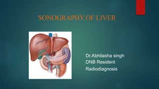
  • 1. SONOGRAPHY OF LIVER Dr.Abhilasha singh DNB Resident Radiodiagnosis
  • 2. Liver-Introduction • Also called ‘hepar’. • Largest/heaviest solid organ in the body. • Weighs about 1600 gm in males,1300 gm in females. • Occupies the right hypochondrium, epigastrium & left hypochondrium. • Most part of the liver is covered by ribs & costal cartilages. • It is covered by network of connective tissue (Glisson’s Capsule)
  • 3. Peritoneal ligaments These ligaments connect the liver to the undersurface of the diaphragm Falciform ligament = It is a double fold of peritoneum from umblicus to liver.Contains ligamentum teres,the remnant of umblical vein,which attaches to the left portal vein. Falciform ligaments split into coronary ligament ( which becomes the right triangular ligament) and left triangular ligament, between which lies the bare area of the liver.
  • 4. Lobes of Liver • Liver is divided into right • & left lobes by falciform ligament . • The right lobe , left lobe and caudate lobe.
  • 5. Liver surfaces • Divided into 2 anatomicalregions: 1.Diaphragmatic surface:  Smooth and dome-shaped surface  Inferior to diaphragm  Separated from diaphragm by subphrenic recessandfrom posterior organs {kidney and suprarenal glands} by hepatorenal recess  Covered by peritoneum except on the posterior surfaceof liver which is not invested in peritoneum and is known as the bare area of liver.
  • 7. 2. Visceralsurface  Covered by visceral peritoneum except porta hepatis andgall bladder bed. • Thevisceral surface is related to:  Right side of the stomach i.e. gastric and pyloric areas  Superior part of the duodenum i.e. duodenal area  Lesser omentum  Gall bladder  Right colic flexure and right transverse colon ;colic area  Right kidney and suprarenal gland; Renalarea
  • 8. Visceral relations :impression of neighbouring viscera
  • 9. Peritoneal relations of theLiver The Lesser omentum • Encloses the portal triad (bile duct, hepatic artery and portal vein ) • Passes from the liver to lesser curvature of the stomach + 2 cm of duodenum. • Thick free edge -- hepatoduodenal ligament • Sheet like remainder – hepatogastric ligament
  • 10. Development ofLiver Liver development requires two linked processes: 1. Differentiation of the various hepatic cell types from their embryonic progenitors. 2. The arrangement of those cells into structures that permit the distinctive and excretorycirculatory, metabolic functions of theliver. Mediated by many essential regulators which include signaling molecules, and transcription factors.
  • 11. Development of Liver: Stages • Specification • Liver bud formation andexpansion • Epithelial differentiation stage Endoderm cells adjacent to the cardiogenic mesoderm begin to differentiate into hepatoblasts. Hepatoblasts proliferate and penetrate the endoderm basement membrane to form the liver bud (25d). The liver bud then expands in size, intercalating into the adjacent septum transversummesenchyme. During the epithelialdifferentiation stage,hepatoblasts mature into hepatocytes or differentiate into cholangiocytes. 13/26
  • 12. Development of liver and biliary passages • The hepatic diverticulum enlarges rapidly and divides into two parts ie pars hepatica(cranial bud) and pars cystica(caudal bud) as it grows between the layers of the ventralmesentery. Superior Caudalbud Inferior Gallbladder, cysticduct Ventral pancreas
  • 13. Pars hepatica • It is thelarger cranial part of the hepatic diverticulum. • Givesrise to: 1. Hepatocytes 2. Hepatic sinusoids 3. Kupffer cells and hematopoietic tissue 4. Intrahepatic bile ducts • The liver grows rapidly to fill a large part of the abdominal cavity. • At first, the 2 lobes are of the same size but soon the right become larger.
  • 14. Pars cystica • Becomes the gall bladder and the stem of the diverticulum forms the cystic duct. • The stalk connecting the hepatic andthe cystic ducts to the duodenum becomes the common bileduct. • The right and the left branches of the pars hepatica canalized to form the right and the left hepaticducts. • Bile begins to flow at about the 12th week. 16/26
  • 15. Development of Liver Parscystica Endoderm Parshepatica Hepatocytes Hepatic sinusoids Kupffer cellsand hematopoietic tissue Intrahepatic biliarytree Gallbladder Extrahepatic bile ducts (cystic duct ,CBD) Ventralmesentery Mesoderm Visceralperitoneum of liver Falciform ligament SeptumTransversum Cardiogenicmesoderm Hepatoblast Hepatic diverticulum Kupffer cells derive from circulating monocytes and possibly yolk sacmacrophages.
  • 16. Formation of the capsule & ligaments of the liver As the septum transversum is penetrated by the growing pars hepatica: • The mesoderm of the septum transversum between the liver and the anterior abdominal wall becomes the FALCIFORM LIGAMENT. • The mesoderm of the septum transversum between the liver and the foregut (stomach and duodenum) forms the LESSER OMENTUM. • The mesoderm on the surface of the liver differentiates into CAPSULE AND PERITONEAL COVERING.
  • 17. Vascular Development related to Liver Extraembryonic Major Venoussystem Intraembryonic Vitelline veins CardinalVeinsUmbilical Veins Sinusvenosus
  • 18. • During early development, there are 3 major venous systems in the embryo—2 extraembryonic and 1 intraembryonic. • The extraembryonic venous systems are the omphalomesenteric (vitelline) and umbilical (placental) veins, and the intraembryonic system includes the cardinal veins that drain the venous blood of the embryo to theheart. • All of these systems converge into the sinus venosus, a cavity that is incorporated into the heart.
  • 19. Development of Vitelline & umbilicalveins A: 4th week B. 5th week. Note the plexus around the duodenum, formation of the hepatic sinusoids, and initiation of left to right shunt between the vitelline veins. Before entering the sinus venosus, the vitelline veins form a plexus around the duodenum and pass through the septum transversum. The liver cords growing into the septuminterrupt the course of the veins and an extensive vascular network, the hepatic sinusoids,forms. There is also initiation of the left to right shunting between the vitelline veins
  • 20. Development of Vitelline & umbilicalveins • Blood from the left side of the liver is rechanneled towards the right, resulting in an enlargement of the right vitelline vein (right hepatocardiac channel). • The right hepatocardiac channel forms the hepatocardiac portion of the inferior vena cava. • The proximal part of the left vitelline vein disappears. The anastomotic network around the duodenum develops into a single vessel, the portal vein. • The superior mesenteric vein derives from the right vitelline vein. • The distal portion of the left vitelline vein also disappears. • The superior segment of vitelline veins becomes the hepatic veins A. 2nd mnth B.3rd mnth. Note formation of the ductus venosus, portal vein , hepatic portion of IVC.The splenic & SMVenter thePV.
  • 21. Development of Vitelline & umbilical veins A. 2nd mnth B.3rd mnth. Note formation of the ductus venosus, portal vein , hepatic portion of IVC. The splenic & SMVenterthe PV. 22/26 • Theumbilical veins run from the placenta to the heart and during fetal life are the predominant afferentvessels that supply theliver. • Initially the umbilical veins passon eachside of the liver but someconnect to the hepaticsinusoids. • Theproximal part of both umbilical veins and the remainder of the right umbilical vein then disappear. Theleft vein is the only one to carry blood from the placenta to theliver. • With the increaseof the placental circulation,a direct communication forms between the left umbilicalvein and the right hepatocardiac channel, the ductus venosus. • Theductus venosusbypassesthe sinusoidal plexusof the liver. After birth the left umbilical vein and the ductus venosus are obliterated and form the ligamentum teres hepatic or the round ligament and ligamentum venosum respectively.
  • 22. Arterial supply ofliver The arterial supply of the liver begins as an off shoot from the celiac trunk at around the eighth week ofgestation. By the 10th week, the first arterial radicles are visible in the central portion of the liver, and by the fifteenth week, they reach the periphery of theliver.
  • 23. Congenital anomalies ofliver Riedel lobe is a tongue-like, inferior projection of the right lobe of the liver beyond the level of the most inferior costal cartilage . It is notconsidered a true accessory lobe of the liver but an anatomical variant of the right lobe of the liver. • Congenital solitary nonparasitic cysts of the liver • Congenital Hepatic Fibrosis • Congenital vascular malformation of the liver • Intrahepatic BiliaryAtresia • Mesenchymal Hamartoma • Accessoryand Ectopic Lobesof the Liver
  • 24. Congenital anomalies ofliver Anomalous supradiaphragmatic lobe
  • 25. Ductul platemalformations • Intrahepatic bile ducts (IHBDs) develop from bi-potential liver progenitor cells(hepatoblasts) in contact with the mesenchyme of the portal vein and thus form the “ductalplates.” • The ductal plates are remodeled into mature tubular ducts. Lack of remodeling results in “ductal platemalformation”. • Aproposal is that virtually all congenital diseases of IHBDs represent examples of DPM. • DPM are developmental anomalies considered to result from lack of ductal plate remodeling during bile ductmorphogenesis.
  • 26. Classification of DPM • Autosomal recessive polycystic kidney disease (hepatic ARPKD) (50% of children, 70% of families): DPM of interlobular bile ducts associated with tubular dilatation of collecting renal tubules • Caroli disease : DPM of the larger IHBDs • Caroli syndrome: Caroli disease + congenital hepatic fibrosis • Von Meyenburg complexes: DPM of smaller interlobular ducts (liver cysts in autosomal dominant polycystic kidney disease) • Mesenchymal hamartoma • Meckel syndrome • Non-syndromal ductal plate malformation
  • 27. Liver is now divided into segmentsas per Couinaud System. • Caudate lobe = Segment I. • Portal and hepatic veins used as landmarks to divide the remainder of the liver into eight segment. • Right hepatic vein divides the right lobe into anterior (segment V and VIII) and posterior segments (segment VI and VII). • Middle hepatic vein divides the liver into right and left lobes (or right and left hemiliver). This plane runs from the inferior vena cava to the gallbladder fossa. • Left hepatic vein divides the left lobe into a medial (segment IV and lateral part (segment II and III).
  • 29. BLOOD SUPPLY The liver has dual blood supply : hepatic artery and portal vein. • Hepatic artery :  Provides 15% of hepatic blood supply.  Branch of coelic artery  Common hepatic artery passes over the head of the pancreas and gives of right gastric artery,then gives off gastroduodenal artery at the epiploic forame to become the hepatic artery proper.  Hepatic artery continues in the free edge of the lesser omentum,anterior to the portal vein and to the left side of the common bide duct (CBD)  Divides into left and right branches at the porta hepatis.
  • 30. • Portal Vein:  Provides 85% of blood suply to liver.  Formed by the union of the splenic vein and superior mesentric vein behind the neck of the pancreas at L1/L2.  Runs at the posterior aspect of free edge of lesser omentum to the porta hepatis; it lies posterior to hepatic artery and CBD.
  • 31. • Venous Drainage: Majority of the the liver is via the hepatic veins which unite into to drain into the IVCat T9close to thediaphragmatic hiatus. Caudatelobe drains directly into the IVCand may therefore be spared incaseof hepaticvein thrombosis. • Nerve Supply Parasympathetic supply is by the preganglionic fibers of the vagus nerve. Sympathetic innervation is by the postganglionic fibers from the coeliac plexus. • LymphaticDrainage Lymphatics from upper surface drain into nodes in the posterior mediastinum. Lymphatics from lower surface drain into hepatic nodes and celiac nodes.
  • 32.
  • 33.
  • 34.
  • 35.
  • 36.
  • 37.
  • 38.
  • 39.
  • 40.
  • 42. Images obtained through the anterior axillary line showthe porta hepatis along the long axis. On the gray-scale image, the portal vein is easily identified. Often, the hepaticartery (arrows) is identified only with color Doppler imaging.
  • 43. HEPATIC ABNORMALITIES 1. CONGENITAL ABNORMALITIES 2. INFECTIOUS DISEASE 3. DISORDER OF METABOLISM 4. VASCULAR ABNORMALITIES 5. TRAUMA 6. HEPATIC MASSES
  • 45. 1.HEPATIC CYST DEFINITION: A liver cyst is defined as a fluid- filled space with an epithelial lining. Occurs in 2.5% of general population increasing to 7% in population older than 80 years ULTRASONOGRAPHY: *Anechoic with a well demarcated thin wall & posterior acoustic enhancement *If complicated with hemorrhage or infection –May contain internal echoes & septations , thickened wall or may appear solid *If thick septae or nodules are seen within the cyst - CT is recommended as biliary cystadenoma & cystic metastases must be considered in the differential possibilities for complex appearing liver cysts
  • 46.
  • 47.
  • 48. 2.PERIBILIARY CYST  Peribiliary cysts have been described in patients with severe liver disease.  These cysts are small, 0.2 to 2.5 cm, and are usually located centrally within the porta hepatis or at the junction of the main right and left hepatic ducts.  USG : Discrete, clustered cysts or as tubular-appearing structures with thin septae, paralleling the bile ducts and portal veins.
  • 49.
  • 50. 3.POLYCYSTIC LIVER DISEASE  Usually associated with polycystic kidney disease but may also occur as an isolated finding in a rarer genetically distinct disease  USG : *Massive hepatomegaly with innumerable ,predominantly simple cysts are present *Portal vein patency should be assessed ,compression of the main portal vein may result in portal hypertension as well as associated findings such as splenomegaly and ascites
  • 51.
  • 52. 4.BILIARY HAMARTOMAS (VON MEYENBURG COMPLEXES)  Bile duct hamartomas, 1rst described by von Meyenburg in 1918, are small, focal developmental lesions of the liver composed of groups of dilated intrahepatic bile ducts set within a dense collagenous stroma  USG : Single, multiple, or most often innumerable well- defined solid nodules usually less than 1 cm in diameter  Usually uniformly hypoechoic and less frequently hyperechoic  Often confused with metastatic cancer
  • 53.
  • 54.
  • 56. 1.VIRAL HEPATITIS Viral hepatitis is a common disease that occurs world- wide. It is responsible for millions of deaths secondary to acute hepatic necrosis or chronic hepatitis, which in turn may lead to portal hypertension, cirrhosis, and hepatocellular carcinoma (HCC). Hepatitis A : -Occurs throughout the world -Diagnosed using serosurveys with the antibody to hepatitis A virus (anti-HAV) as the marker. -The primary mode of spread is the fecal-oral route. -Hepatitis A is an acute infection leading to complete recovery or death from acute liver failure.
  • 57. Hepatitis B : -Transmitted parenterally (e.g., blood transfusions, needle punctures) as well as by non percutaneous exposure through sexual contact and at birth. -The two most useful markers for acute infection are hepatitis B surface antigen (HBsAg) and antibody to hepatitis B core antigen (anti-HBc). Hepatitis C (predominantly) and hepatitis E : -Formerly called non-A, non-B (NANB) hepatitis,. -Acutely infected patients have a much greater risk of chronic infection, with up to 85% progressing to chronic liver disease. -Chronic hepatitis C virus (HCV) infection is diagnosed by the presence of anti- body to HCV(anti-HCV) in the blood.
  • 58. Hepatitis D, or delta hepatitis : -Entirely dependent on HBV for its infectivity, requiring the HBsAg to provide an envelope coat for the hepatitis D virus. - Its geographic distribution is therefore similar to that of hepatitis B. - HDV is an uncommon infection in North America, occurring primarily in intravenous (IV) drug users.
  • 59.  Uncomplicated acute hepatitis : Clinical recovery within 4 months. It is the outcome of 99% of cases of hepatitis A.  Subfulminant and fulminant hepatic failure : Follows the onset of jaundice and includes worsening jaundice, coagulopathy, and hepatic encephalopathy.  Chronic hepatitis : Persistence of biochemical abnormalities beyond 6 months. It has many etiologies other than viral, including metabolic (e.g., Wilson’s disease, alpha-1 antitrypsin deficiency, hemochromatosis), autoimmune, and drug induced. The prognosis and treatment of chronic hepatitis depend on specific etiology.  Acute hepatitis : Diffuse swelling of the hepatocytes, proliferation of Kupffer cells lining the sinusoids, and infiltration of the portal areas by lymphocytes and monocytes.
  • 60. Radiographic Features : U/S -In most cases , the liver appears normal -The liver parenchyma may have a diffusely decreased echogenicity with accentuated brightness of the portal triads (starry sky) , periportal cuffing -Hepatomegaly & thickening of the G.B. wall
  • 61.
  • 62.
  • 63. 2.BACTERIAL DISEASES  Pyogenic bacteria reach the liver by several routes, the most common being 1. Direct extension from the biliary tract in patients with suppurative cholangitis and cholecystitis. 2.Through the portal venous system in patients with diverticulitis or appendicitis. 3. Through the hepatic artery in patients with osteomyelitis and subacute bacterial endocarditis. 4. Trauma , surgery
  • 64. On USG : -Cystic with the fluid ranging from echofree to highly echogenic -Occasionally , gas producing organisms give rise to echogenic foci Fluid-fluid interfaces , internal septations & debris have all been observed -The abscess wall can vary from well defined to irregular & thick -May be multiple
  • 65.
  • 66.  Differential diagnosis of pyogenic liver abscess includes : 1. Amebic or echinococcal infection 2. Simple cyst with hemorrhage, 3. Hematoma 4. Necrotic or cystic neoplasm.  Ultrasound-guided liver aspiration is an expeditious means to confirm the diagnosis.  Specimens should be sent for both aerobic and anaerobic culture.  Once the diagnosis of liver abscess is made by the identification of pus or a positive Gram stain and culture, the collection can be drained percutaneously using ultrasound or CT guidance.
  • 67. 3.FUNGAL DISEASES : Candiasis  In immunocompromised  The liver is frequently involved secondary to hematogeneous spread of mycotic infections in other organs, most often the lungs.  USG features :  “Wheel within a wheel”: peripheral hypoechoic zone with inner echogenic wheel and central hypoechoic nidus. The central nidus represents focal necrosis in which fungal elements are found. This is seen early in the disease.  Bull’s-eye: 1 to 4 cm lesion with hyperechoic center and hypoechoic rim. It is present when neutrophil counts return to normal. The echogenic center contains inflammatory cells.  Uniformly hypoechoic: most common, corresponding to progressive fibrosis  Echogenic: variable calcification, representing scar formation
  • 68.
  • 69.
  • 70. 4.PARASITIC DISEASE : a) Amebiasis  Hepatic infection by the parasite Entamoeba histolytica is the most common extraintestinal manifestation of amebiasis.  Transmission is by the fecal-oral route.  The most common presenting symptom, pain occurs in 99% of patients with amebic abscess. Approximately 15% of patients have diarrhea at diagnosis  Radiographic Features :  Round or oval-shaped lesion  Absence of a prominent abscess wall  Hypoechogenecity compared to normal liver  Internal septations , 30%  Distal sonic enhancement  Contiguity with the diaphragm , may be associated with pleural effusion & lung collapse  Most amebic liver abscess occur in right lobe of liver.
  • 71.
  • 72. b) Hydatid disease  Caused by infestation by the parasite Echinococcus granulosus  It is most prevalent in sheep- and cattle-raising countries  E. granulosus is a tapeworm 3 to 6 mm in length that lives in the intestine of the definitive host, usually the dog.  Its eggs are excreted in the dog’s feces and swallowed by the intermediate hosts—sheep, cattle, goats, or humans.  The embryos are freed in the duodenum and pass through the mucosa to reach the liver through the portal venous system.  Most of the embryos remain trapped in the liver, although the lungs, kidneys, spleen, central nervous system, and bone may become secondarily involved.
  • 73.  In the liver the right lobe is more frequently involved  Ectocyst : The cyst wall consists of an external membrane that is approxi- mately 1 mm thick, which may calcify  Pericyst : The host forms a dense connective tissue capsule around the cyst  Endocyst : The inner germinal layer gives rise to brood capsules that enlarge to form protoscolices.  Hydatid sand : The brood capsules may separate from the wall and form a fine sediment  When hydatid cysts within the organs of a herbivore are eaten, the scolices attach to the intestine and grow to adult tapeworms, thus completing the life cycle.
  • 74.
  • 75.  Sonographic features of hepatic hydatid following four groups for hydatid cysts: 1. Simple cysts containing no internal architecture except sand 2. Cysts with detached endocyst secondary to rupture 3. Cysts with daughter cyst matrix (echogenic material between daughter cysts) 4. Densely calcified masses
  • 76.
  • 77. Hepatic alveolar echinococcus :  Rare parasitic infestation by the larvae of E. multilocularis.  The fox is the main host.  Sonographic features : Echo- genic lesions, which may be single or multiple; necrotic, irregular lesions without a well-defined wall; clusters of calcification within lesions; and dilated bile ducts.
  • 78. c)Schistosomiasis  Schistosomiasis is one of the most common parasitic infections in humans, estimated to affect 200 million people worldwide.  Hepatic schistosomiasis is caused by Schistosoma mansoni, S. japonicum, S. mekongi, and S. intercalatum.  Hepatic involvement by S. mansoni is particularly severe.  The ova reach the liver through the portal vein and incite a chronic granulomatous reaction, described by Symmers in 1904 as “clay- pipestem fibrosis.
  • 79.  Sonographic features :  Widened echogenic portal tracts , sometimes reaching a thickness of 2 cm  The porta hepatis is the region most often affected  Initially the liver is enlarged , however as the peri-portal fibrosis progress , the liver is contracted & the features of portal hypertension prevails
  • 80.
  • 81. d)Pnemocystis carinii  Pneumocystis carinii is the most common organism causing opportunistic infection in patients with acquired immunodeficiency syndrome (AIDS).  Pneumocystis pneumonia is the most common cause of life-threatening infection in patients with human immunodeficiency virus (HIV).  Extrapulmonary P. carinii infection has been documented in the liver, spleen, renal cortex, thyroid gland, pancreas, and lymph nodes.  Sonographic findings : Involvement of the liver range from tiny, diffuse, non shadowing echogenic foci to extensive replacement of the normal hepatic parenchyma by echogenic clumps representing dense calcification
  • 82.
  • 84. 1.FATTY LIVER  Fatty liver is an acquired, reversible disorder of metabolism, resulting in an accumulation of triglycerides within the hepatocytes.  Causes : 1.Obesity (most common cause) 2.Excessive alcohol intake 3.Hyperlipidemia 4.Diabetes 5.Steroids , cushing's syndrome 6. Pregnancy, 7.Total parenteral hyperalimentation 8.Severe hepatitis 9.Glycogen storage disease 10.Jejunoileal bypass procedures for obesity, 11.Cystic fibrosis, 12.Congenital generalized lipodystrophy 13.Several chemotherapeutic agents, including methotrexate 14.Toxins such as carbon tetrachloride and yellow phosphorus
  • 85.
  • 86.
  • 87. Focal fatty change includes : Focal fatty infiltration and focal fatty sparing , both may mimic neoplastic involvement a) Focal fatty infiltration , regions of increased echogenicity are present within a background of normal liver parenchyma b) Focal fatty sparing , islands of normal liver parenchyma may appear as hypoechoic masses within a dense fatty infiltrated liver
  • 88. -Features of focal fatty change include : a) Focal fatty sparing and focal fatty liver , both most commonly involve the peripheral region of the medial segment of the left lobe (segment IV) b) Sparing also occurs commonly by the gall bladder fossa and along the liver margins c) Lack of mass effect , hepatic vessels as a rule are not displaced d) Geographic margins are present , although focal fat may appear round , nodular , or interdigitated with normal tissue e) Rapid change with time , fatty infiltration may resolve as early as within 6 days f) CT scan of the liver will demonstrate corresponding regions of low attenuation
  • 90.
  • 92.
  • 93.
  • 94.  Contrast-enhanced ultrasound (CEUS) is valuable in the differentiation of fatty change from neoplasia, because the fatty or spared regions will all appear isovascular in both the arterial and the portal venous phase of enhancement.  Chemical shift MRI techniques are useful in distinguishing diffuse from focal fatty infiltration.  Radionuclide liver and spleen scintigraphic examination will yield normal results, indicating adequate numbers of Kupffer cells within the fatty regions.
  • 95. 2.GLYCOGEN STORAGE DISEASE(Glycogenesis) Von Gierke :  Von Gierke 1rst recognized glycogen storage disease (GSD) affecting the kidneys and liver in 1929.  Type 1 GSD (von Gierke’s disease, glucose 6-phosphatase deficiency) is manifested in the neonatal period by hepatomegaly, nephromegaly, and hypoglycemic convulsions.  Because of the enzyme deficiency, large quantities of glycogen are deposited in the hepatocytes and proximal convoluted tubules of the kidney.  Sonographically, type 1 GSD appears indistinguishable from other causes of diffuse fatty infiltration.
  • 96. Definition :Glucocerebrosidase deficiency leads to accumulation of ceramide in cells of the RES Radiographic Features : 1-Liver :-Hepatomegaly 2-Spleen :-Splenomegaly (marked) -Focal lesions (infarcts) typically are hyperechoic (US) 3-Musculoskeletal :-Erlenmeyer flask deformity of femur -Generalized osteopenia -Multiple lytic bone lesions -Aseptic necrosis of femoral head Gaucher’s Disease :
  • 97. 3.CIRRHOSIS  Diffuse process characterized by fibrosis and the conversion of normal liver architecture into structurally abnormal nodules.  Pathology :3 pathologic mechanisms which in combination create cirrhosis :cell death , fibrosis & regeneration  Classification : 1-Micronodular (nodule 1 mm to 1 cm) 2-Macronodular (up to 5 cm)
  • 98.  Alcohol consumption is the most common cause of micronodular cirrhosis.  Chronic viral hepatitis is the most frequent cause of the macronodular form.  Other etiologies : Biliary cirrhosis (primary and secondary), Wilson’s disease, Primary sclerosing cholangitis Hemochromatosis.  The classic clinical presentation of cirrhosis is : Hepatomegaly Jaundice Ascites
  • 99. Radiographic Features : 1-Volume redistribution 2-Coarse echotexture 3-Nodular surface 4-Nodules 5-Portal hypertension
  • 100. 1.Volume redistribution : -Early stages , the liver is enlarged -Advanced stages , the liver is often small with relative enlargement of the caudate , left lobe or both in comparison with the right lobe 2.Coarse echotexture : -Increased echogenicity and coarse echotexture are frequent observations in diffuse liver disease
  • 101.
  • 102.
  • 103. Hepatic C/RL ratio line 1: Right lateral border of the portal vein line 2: Left lateral border of the caudate lobe line 3: Midway between the portal vein and the IVC extended to the right liver edge RL measurement: C measurement caudate-right lobe ratio: C/RL C/RL >0.65 = 96% likely to be cirrhotic//C/RL >0.73 = 99% likely to be cirrhotic : axial slice immediately below the bifurcation of the main portal vein
  • 104. 3.Nodular surface : -Irregularity of the liver surface corresponds to the presence of regenerating nodules and fibrosis 4.Nodules (regenerative & dysplastic) : -Regenerative nodules (RN) , represents regenerating hepatocytes surrounded by a fibrous septa (isoechoic or hypoechoic with a thin echogenic border) -Dysplastic nodules , considered premalignant , 1 cm , they contain well differentiated hepatocytes , a portal venous blood supply and also atypical or frankly malignant cells 5.Portal hypertension : Ascites , splenomegaly and varices
  • 105.
  • 106.
  • 107.
  • 108.  Patients with compensated cirrhosis (no portal hypertension), the Doppler wave- form is abnormal. Two abnormal patterns of HV have been described: a) Decreased amplitude of phasic oscillations with loss of reversed flow and b) Flattened waveform  As cirrhosis progresses, luminal narrowing of the hepatic veins may be associated with flow alterations visible on color and spectral Doppler ultrasound. High- velocity signals through an area of narrowing produce color aliasing and turbulence  In patients with cirrhosis and chronic liver disease, the normal increase in postprandial resistive index of hepatic artery is blunted.
  • 109.
  • 111. 1.PORTAL HYPERTENSION  Normal portal vein pressure is 5 to 10 mm Hg (14 cm H2O).  Portal hypertension is defined by : (1) wedge hepatic vein pressure or direct portal vein pressure more than 5 mm Hg greater than IVC pressure, (2) splenic vein pressure greater than 15 mm Hg, or (3) portal vein pressure (measured surgically) greater than 30 cm H2O
  • 112. Causes :  1.Presinusoidal portal hypertension Extrahepatic : -Thrombosis of the portal or splenic veins. Intrahepatic forms : -Schistosomiasis, -Primary biliary cirrhosis, -Congenital hepatic fibrosis -Toxic substances, such as polyvinyl chloride and methotrexate  2.Sinusoidal :Cirrhosis  3.Postsinusoidal : -Budd-Chiari syndrome -Congestive heart failure
  • 113. Radiographic Features :  PV diameter > 13 mm  Splenomegaly  Ascites  Portosystemic venous collaterals (Varices) : Gastro- oesophageal junction , para-umbilical vein , spleno-renal & gastro- renal intestinal & hemorrhoidal
  • 114.
  • 115. Portal vein diameter –>9-12 mm in quietrespiration >13 mm indicates portal hypertension (>17mm –Large varices) Portal veindiameter (1-2 cm proximal to bifurcation)
  • 116. Splenic vein and Superior mesenteric vein Splenic vein and superior mesenteric vein - Diameter >10mm increase of diameter <20%during inspiration Lossof respiratory variation in these vesselsis important sign
  • 117. Normal portal vein flow and velocity • Undulating hepatopetal flow. • Remain above baseline • Mean portal venous flow velocity - 12 to 18 cm/sec • Average portal flow – 500-900 ml/min Normal portal venous flow direction andwaveform.
  • 118. Normal portal vein flow May become accentuated in portal hypertension
  • 119. Portal hypertension • Portal vein loses its undulatory pattern and becomes monophasic. • Later flow becomes biphasic and finally hepatofugal • Flow velocity < 12cm /sec US image shows slow flow in the main portal vein
  • 120. Reversal of flow –Hepatofugal flow
  • 121. Interpretation of flow Normal flow Toand Froflow Reversedflow Portalhypertension Cardiac failure AdvancedPHTN
  • 122. Vascular indices Liver Vascular Index (LVI = PV Vel/ Hepatic Artery PI) < 12 cm/sec Congestive index : Cross-sectional area /mean flow velocity of the portal trunk > 0.13 cm/sec---Portal HTN
  • 123. Important indicators of portal hypertension. Left gastric Short gastric Paraumblical Splenorenal Splenoretroperitoneal Splenocaval Splenoportal Portosystemic Venous Collaterals
  • 124. • Left gastric (coronary vein) – Normally <4mm • > 7mm :Associate with esophageal and gastricvarices • Short gastric veins-- spleen and gastric wall. Left gastric (coronary vein)
  • 126. Paraumbilical vein • Ligamentum teres in the left lobe of liver • Recanalized visible as a channel greater than 3 mm in diameter • Hepatofugal flow • Recanalization of umbilical vein is a highly specific sign of portal hypertension
  • 127. Splenorenal shunts –Splenic hilar Splenoretroperitoneal Spenocaval Splenoportal
  • 128. Hepatic vein assessment -Doppler spectral traces from normal hepatic veins have a triphasic appearance two waves that represent atrial and ventricular diastole and a small wave that occurs in atrial systole -Altered hepatic vein waveforms -50% of patients with cirrhosis  flattening of the phasic oscillations
  • 129. Loss of pattern -Monophasic flow -Cirrhosis (Poor prognosis) Infiltrative liver disease Budd Chiari (Hepatic vein thrombosis) Pulsed Doppler waveform analysis shows lossof the normal triphasic hepatic vein pulsatility.
  • 130. Exaggerated phasicity 1.Congestive heart failure 2.Tricuspid incompetence
  • 131. Hepaticartery normal hepatic artery (HA) -- 4-6 mm -- 25 to 30% of blood to the liver. -- anterior to the portal vein systolic velocity - 30 to 40 cm/sec and diastolic velocity - 10-15 cm/sec Resistivity Index -0.55 to 0.81 Pulsatility index (PI)- 1.16 to1.24 Increased in chronic liverdisease
  • 132. 2-Portal Vein Thrombosis :  Causes: 1.Malignancy-HCC,Metastatic liver disease,Ca pancreas,1o leiomyosarcoma 2.Chronic pancreatitis 3.Hepatitis 4.Septicemia 5.Trauma 6.Splenectomy 7.Potocaval shunts 8.Hypercoagulable state-Pregnancy and neonates
  • 133. Ultrasonographic Features : -Echogenic thrombus within the lumen of the vein, portal vein collaterals, expansion of the caliber of the vein, and cavernous transformations -Acute thrombosis may be difficult to detect with grey-scale imaging alone as the thrombus will be hypoechoic , with time it becomes more echogenic and easier to detect -Colour Doppler will of course be able to demonstrate absent flow in the portal vein and even detect partial thrombosis
  • 134.
  • 135.
  • 136. -Cavernous Transformations of the PV :numerous wormlike vessels at the porta hepatis which represent periportal collateral circulation , this pattern is observed in long standing thrombosis requiring up to 12 months to occur , so it is more likely to develop with benign disease Acute thrombus may appear relatively anechoic and thus may be overlooked unless Doppler ultrasound interrogation is performed. Malignant thrombosis of the portal vein has a high association with HCC and is often expansive, as is malignant occlusion from other primary or secondary disease
  • 137.
  • 138.
  • 139.
  • 140. Causes : 1-Idiopathic : 50%-75% 2-Secondary : 25%-50% -Coagulation anomalies , clotting disorders ,polycythemia -Tumors : HCC , RCC -Trauma -Oral contraceptives , chemotherapy Definition -Occlusion of the lumina of the hepatic veins with or without occlusion of the lumen of the IVC Degree of occlusion and presence of collateral circulation predict the clinical course. 3.Budd-Chiari Syndrome (BCS) :
  • 141. Classic patient in North america is a young adult woman taking oral contraceptives who presents with an acute onset of ascites, right upper quadrant pain, hepatomegaly and lesser extent, splenomegaly Clinical Picture : 1-Ascites 2-Pain 3-Hepatomegaly 4-Splenomegaly
  • 142. Ultrasonographic Features : -Liver is typically large and bulbous in acute phase -Partial or complete inability to see the hepatic veins , stenosis with proximal dilatation , intra-luminal echogenicity , thickened walls ,thrombosis & extensive intra-hepatic collaterals -Hemorrhagic infarction appears hypoechoic by US -Caudate lobe is often spared (emissary veins drain directly into the IVC) and appears enlarged, small right lobe -Membranous “webs” may be identified as echogenic or focal obliteration of the lumen
  • 143.  Real-time ultrasonography, however, underestimates the presence of thrombosis and webs and may be inconclusive in a cirrhotic patient with hepatic veins that are difficult to image.  Intrahepatic collaterals, on gray- scale images, show as tubular vascular structures in an abnormal location and typically are seen extending from a hepatic vein to the liver surface, where they anastomose with systemic capsular vessels.  Duplex Doppler ultrasound and color Doppler flow imaging (CDFI) can help determine both the presence and the direction of hepatic venous flow in the evaluation of patients with suspected Budd-Chiari syndrome.  The middle and left hepatic veins are best scanned in the transverse plane at the level of the xiphoid process.  The right hepatic vein is best evaluated from a right intercostal approach
  • 144.  The normal blood flow in the IVC and hepatic veins is phasic in response to both the cardiac and respiratory cycles  In Budd-Chiari syndrome, flow in the IVC, hepatic veins, or both, changes from phasic to absent, reversed, turbulent, or continuous.  Continuous flow has been called the pseudoportal Doppler signal and appears to reflect either partial IVC obstruction or extrinsic IVC compression.  Associated reversal of flow in the portal vein and epigastric collaterals is also optimally assessed with this technique
  • 145.
  • 146.
  • 147.
  • 148. Differential Diagnosis : -Hepatic veno-occlusive disease which causes progressive occlusion of small vessels , is clinically indistinguishable from BCS Caused by : 1-Bone marrow transplantation 2-Chemotherapy 3-Jamaican bush tea
  • 149. 4.PORTAL VEIN ANEURYSM  Rare,their origin is either congenital or acquired secondary to portal hypertension  Proximally at the junction of the superior mesenteric and splenic veins and distally involving the portal venous radicles.  The sonographic appearance is that of an anechoic cystic mass, which connects with the portal venous system.  Pulsed Doppler sonographic examination demonstrates turbulent venous flow.
  • 150. 5.Hepatic Artery Aneurysm : -Hepatic artery is fourth most common site. -Decreasing order of frequency of abdominal aneurysms : aorta > iliac artery> splenic artery > hepatic artery -10% of patients with hepatic artery aneurysm have sudden atastrophic rupture into the peritoneum, biliary tree, gastrointestinal tract, or portal vein. -Hepatic pseudoaneurysm may occur secondary to pancreatitis. -The duplex Doppler sonographic examination revealed turbulent arterial flow within a sonolucent mass.
  • 151. Hepatic artery aneurysm, (a) Left anterior oblique digital subtraction angiogram obtained with the catheter in the distal CHA shows a right hepatic artery branch aneurysm (arrowhead), (b) Digital subtraction completion angiogram shows exclusion of the aneurysm with use of the sac-packing coil embolization technique (arrowhead)
  • 152. Definition : -Refers to abnormal shunt or fistulous connection between the portal venous system and a hepatic arterial system within the liver Types : 1-Tumorous Shunt 2-Non-Tumorous Shunt 6. Arterio-Portal Shunting in Liver :
  • 153. 1-Tumorous Shunt : -Occurs with hepatocellular carcinoma -Trans-tumoral shunt is due to abnormal communication between the feeding artery and draining vein of the tumor which results in increased vascularity around the tumor manifested as peritumoral transient hepaticattenuation differences (THAD) -The portal vein may show early enhancement in dynamic arterial scan without enhancement of its main tributaries the splenic and superior mesenteric veins -THAD refer to areas of parenchymal enhancement visible during the hepatic artery phase on helical CT , they are thought to be a physiological phenomenon caused by the dual hepatic blood supply , occasionally they may be associated with hepatic tumors such as HCC
  • 154. 2-Non-Tumorous Shunt : -Mainly due to liver biopsy and other hepatic Intervention -Also may occur due to liver cirrhosis -Patients typically are middle aged and present with hepatic encephalopathy. -Sonography demonstrates a tortuous tubular vessel or complex vascular channels, which connect a branch of the portal vein to a hepatic vein or the IVC. -The diagnosis is confirmed angiographically.
  • 155. 7. HEREDITARY HEMORRHAGIC TELANGIECTASIA  Hereditary hemorrhagic telangiectasia, or Osler-Weber- Rendu disease, is an autosomal dominant disorder that causes arteriovenous (AV) malformations in the liver, hepatic fibrosis, and cirrhosis.  Patients present with multiple telangiectasias and recurrent episodes of bleeding.  Sonographic findings : A large feeding common hepatic artery up to 10 mm, multiple dilated tubular structures representing AV malformations, and large draining hepatic veins secondary to AV shunting.
  • 156. 8. PELIOSIS HEPATIS  Peliosis hepatis is a rare liver disorder characterized by blood filled cavities ranging from less than a millimeter to many centimeters in diameter.  It can be distinguished from hemangioma by the presence of portal tracts within the fibrous stroma of the blood spaces.  Pathogenesis : Rupture of the reticulin fibers that support the sinusoidal walls, secondary to cell injury or nonspecific hepatocellular necrosis.  The diagnosis of peliosis can be made with certainty only by histologic examination.
  • 157.  On sonography, described lesions are nonspecific and have shown single or multiple masses of heterogeneous echogenicity.  Calcifications have been reported  CT scans show low-attenuation nodular lesions that may or may not enhance with contrast injection
  • 158.
  • 159. TRAUMA
  • 160. Incidence : -The liver is the most common intraabdominal site of injury , however ,one must inspect other organs (spleen ,bowel) for coexistent trauma. -The predominant site of hepatic injury in blunt trauma is the right lobe in particular the posterior segment.
  • 161. Types : a) Laceration (most common) b) Hematoma , subcapsular or intraparenchymal c) Active hemorrhage d) Major hepatic vein injury e) AV fistula
  • 163. Ultrasonographic Features : - < 24 hrs following injury , the fresh hemorrhage is echogenic - Within the 1 week , the hepatic laceration becomes more hypoechoic & distinct as a result of resorption of devitalized tissue & ingress of intestinal fluid - At 2 or 3 weeks later , the laceration becomes increasingly indistinct as a result of resorption of the fluid & filling of the spaces of the granulation tissue st
  • 164. < 24 hrs < 24 hrs
  • 165.
  • 166. 1 week
  • 167.