2. The Liver
• Largest gland of the body.
• 1500 grams and 2.5% of total body weight.
• Location:
- Right hypochondrium
- Epigastric region
- Left hypochondrium
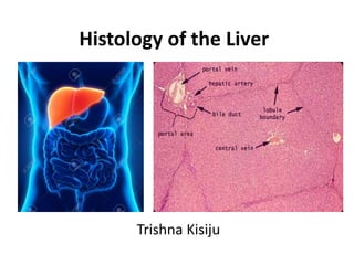
3. Vascular Supply of the Liver
• Receives dual vascular supply:
-Hepatic Portal Vein (75%)
- Hepatic Artery(25%)
• Both vessels enter the liver via Porta hepatis.
6. • Trabeculae: The Glisson’s capsule extends into the
interior of liver as numerous branching trabeculae and
septa.
7. • Reticular fibers:
- Supporting connective tissue of the liver.
- Line the sinusoids, support the endothelial cells, and
form a denser network of reticular fibers in the wall of
the central vein.
- Also merge with the collagen fibers in the interlobular
septum, where they surround the portal vein and the
bile duct.
8. The Liver Parenchyma
• Organized as thousands of small hepatic lobules.
• Hepatic Lobules: Structural units of Liver.
Roughly hexagonal arrangement of irregular plates or
cords of hepatocytes radiating outward from a central
vein.
9.
10. Concepts of Liver Lobules
• Classical Hepatic Lobule
• Portal Lobule
• Hepatic Acinus of Rappaport
11. Classical Hepatic Lobule
• Each lobule consists of a hexagonal mass of liver cells.
• Central axis occupied by:
Central vein.
• Is an independent venous unit.
• From the central vein, hepatocytes
radiate irregularly as plates known
as Hepatic lamina.
• Spaces between the hepatic
lamina are called Hepatic lacunae,
occupied by Hepatic sinusoids.
12.
13.
14. Classical hepatic lobule contd..
• Hepatic Sinusoids:
- Wide diameter capillaries.
- Their walls are fenestrated and made up of flattened
endothelial cells.
- Kupffer cells and pit cells are attached to the endothelial
surface.
15. Classical hepatic lobule contd..
• Hepatic sinusoids receive a mixture of blood from the
portal vein and the hepatic artery of adjacent portal area.
• They are interlaminar and centripetal in direction.
• The blood flows towards Central vein Sublobular
vein Hepatic vein Inferior Venacava.
16. The Portal Area
• Peripherally, each lobule has 3 to 6 portal areas with more
fibrous connective tissue, each of which contains
interlobular structures that comprise the portal triad. They
include:
• A venule branch of the portal vein, with blood rich in
nutrients but low in O2.
• An arteriole branch of the hepatic artery that supplies O2.
• One or two small bile ductules of cuboidal epithelium,
branches of the bile conducting system.
17.
18. Bile Canaliculi
• Formed by spaces present between plasma membranes of
adjacent liver cells.
• Form hexagonal networks around the liver cells.
• Borders around the canaliculi are sealed by tight junctions.
This forms the blood-bile barrier.
19. Bile Canaliculi
• The canaliculi pass to periphery of the hepatic lobules
where they form intralobular canal of Herring, that
finally drains into the interlobular duct of the portal
area.
• Bile canaliculi are intralaminar and centrifugal in
direction
20. Space of Disse / Perisinusoidal Space
• Potential space between the wall of sinusoids and
laminae of the liver cells.
• Filled with blood plasma and
chylomicrons that percolate
through the wall of sinusoids.
• Presence of Ito cells.
21.
22. Ito cells
• Irregular outline with numerous lipid vesicles.
• Function of Ito cells:
- Secrete collagenous matrix
- Provide growth factor for regeneration of
damaged liver cells.
- Store Vitamin A in their lipid vesicles.
23. Space of Mall
• Potential space, between the glisson’s capsule of portal
area and the hepatic plates of the cells.
• Lymphatics of liver
begin here.
24. The Portal Lobule
• Territory of liver tissue centered around a portal triad.
• Drawn by joining the central veins of three adjacent
lobules.
• Nutritional lobule of the liver.
25.
26. Hepatic acinus of Rappaport
• Diamond shaped area of liver parenchyma.
• Forms structural and metabolic functions of the liver.
• Numerous branches arise
at right angles from the
blood vessels of portal area,
these terminal vessels
form backbone of the liver
acinus.
27.
28. Hepatic acinus of Rappaport contd..
• The acinus can be divided into 3 zones based on the
gradient of blood supply:
Zone 1:
- Around the vascular backbone, is well oxygenated.
Zone 2:
- Intermediate zone, moderately oxygenated.
Zone 3:
- Close to the central vein and the least oxygenated; most
susceptible to anoxic injury.
29. The Hepatocytes
• Large cuboidal or polyhedral epithelial cells, with
large, round central nuclei and eosinophilic cytoplasm
rich in mitochondria.
31. References
• Dutta A.K., Essentials of Human Anatomy (Thorax and
abdomen) 9th ed. Kolkata: Current Books International.
• Mescher, A. L., & Junqueira, L. C. U. (2013). Junqueira's
basic histology: Text and atlas (Thirteenth edition.).
New York: McGraw Hill Medical.
• Eroschenko, V. P., & Fiore, M. S. H. d. (2000). Di Fiore's
atlas of histology with functional correlations (9th ed.).
Philadelphia: Lippincott Williams & Wilkins.