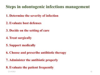
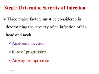
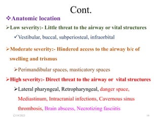
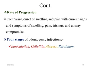
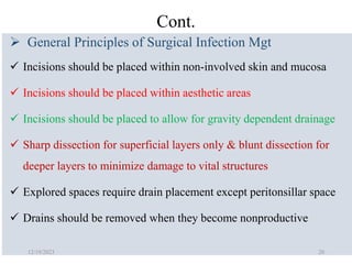
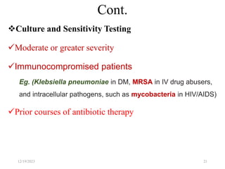
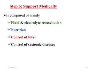
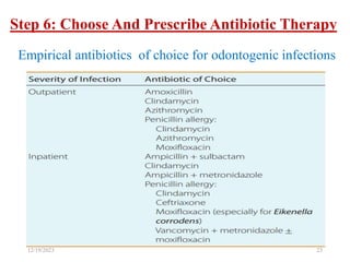
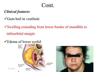
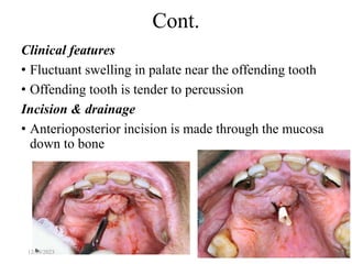
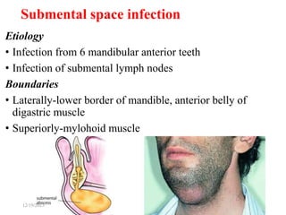
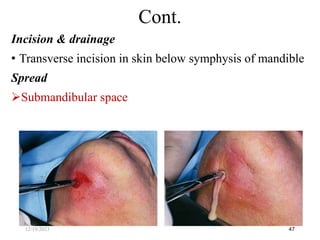
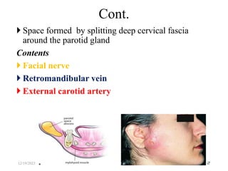
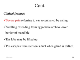
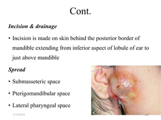
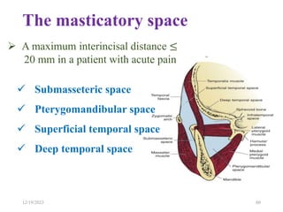
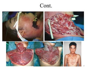
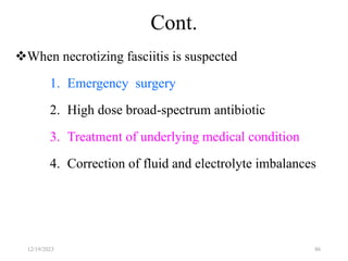
SlideShare a Scribd company logo
1 of 95
Head and neck space
infections
Dr. Nigusu G. ( DMD)
Outline
Introduction
 Etiology
 Microbiology
Principle of management
 Classification of fascial spaces
Possible untoward or life-threatening complications of
orofacial infection
12/19/2023 2
Introduction
Maxillofacial space infections particularly those of odontogenic
origin are the most common
It arises from periapical abscess to superficial and deep neck
infections
The infections generally spread by following the path of least
resistance through connective tissue and along fascial planes
Finally they spread to such an extent, distant from the site of
origin, causing considerable morbidity and occasional death
12/19/2023 3
Cont.
The palatine tonsils and peritonsilar area in children
and odontogenic structures in adults are the most
prevalent initial anatomic origins of deep neck space
infections
Almost all odontogenic infections are caused by
multiple bacteria with anaerobic-aerobic characteristic
12/19/2023 4
1-Odontogenic origin
Pulp disease, Periodontal disease, Secondarily infected
cyst & odontomes, Remaining root fragment, Pericoronal
infection
2-Trauma
3-Implant Surgery
4-Reconstructive Surgery
5-Contaminated Needle Puncture
6-Infections Of Maxillary Antrum
7-Infections of salivary glands
8-Secondary to oral malignancies
5
Etiology
12/19/2023
Invasion of dental pulp by bacteria after
decay of a tooth

Inflammation, edema & lack of collateral
blood supply

Venous congestion or avascular necrosis
(pulpal tissue death)

Reservoir of bacterial growth(anaerobic)

Periodic egress of bacteria into surrounding
alveolar bone 6
Pathways of odontogenic infections
12/19/2023
• Aerobic gram positive cocci bacteria(α-hemolytic
streptococci) or streptococci viridans that includes strept
milleri, strep. Sanguis, strep. Salivarius, strep. Mutans
• Anaerobic Cocci-peptostreptococcus
• Bacteriodes-porphyromonas, prevotella
Most odontogenic infections are caused by anaerobic and
aerobic bacteria
Aerobic bacteria-5%
Anaerobic bacteria-35%
Mixed infection-60% 7
Microbiology
12/19/2023
 Acute
Chronic
Acute stage:- 3 forms
Cellulitis
Abscess
Fulminating infection
8
Types of head and neck infection
12/19/2023
Cellulitis
It is spreading infection of loose connective tissues
It is a diffuse, erythematous, mucosal or cutaneous
infection
It is the result of streptococcal infection
Streptococcus produces streptokinase, hyaluronidase &
streptodornase which break down fibrin, connective
tissue ground substance & lyse cellular debris, which
facilitate rapid spread of bacteria
9
Cont.
12/19/2023
Abscess
It is a circumscribed collection of pus in a pathologic tissue space
 Infections are characterised by staphylococci and bacteroides
They produce coagulase, an enzyme, that may cause fibrin
deposition in citrated or oxalated blood
Fulminating Infections
In this the infection involves secondary spaces involving vital
structures along the pathway of least resistance
10
Cont.
12/19/2023
Chronic fistulous tract or sinus formation
• Abscesses neglected for a long time may discharge
intraorally or extra orally
Chronic osteomyelitis of Jaws
 Cervicofacial actinomycosis
11
Chronic stage
12/19/2023
Steps in odontogenic infections management
1. Determine the severity of infection
2. Evaluate host defenses
3. Decide on the setting of care
4. Treat surgically
5. Support medically
6. Choose and prescribe antibiotic therapy
7. Administer the antibiotic properly
8. Evaluate the patient frequently
12/19/2023 12
Step1: Determine Severity of Infection
Three major factors must be considered in
determining the severity of an infection of the
head and neck
Anatomic location
Rate of progression
Airway compromise
12/19/2023 13
Cont.
Anatomic location
Low severity:- Little threat to the airway or vital structures
Vestibular, buccal, subperiosteal, infraorbital
Moderate severity:- Hindered access to the airway b/c of
swelling and trismus
Perimandibular spaces, masticatory spaces
High severity:- Direct threat to the airway or vital structures
Lateral pharyngeal, Retropharyngeal, danger space,
Mediastinum, Intracranial infections, Cavernous sinus
thrombosis, Brain abscess, Necrotizing fasciitis
12/19/2023 14
Cont.
Rate of Progression
Comparing onset of swelling and pain with current signs
and symptoms of swelling, pain, trismus, and airway
compromise
Four stages of odontogenic infections:-
Innoculation, Cellulitis, Abscess, Resolution
12/19/2023 15
Cont.
Airway Compromise
The most frequent cause of death in odontogenic
infection is airway obstruction
Complete airway obstruction is a surgical emergency
Infection in the region of surgical airway access is not a
contraindication to an emergency cricothyroidotomy or
tracheotomy
12/19/2023 16
Step 2: Evaluate Host Defenses
12/19/2023 17
Factors associated with immune system compromise
DM, Steroid therapy, Organ transplants, Malignancy,
Chemotherapy, CKD, Malnutrition, Alcoholism, End-
stage AIDS
Step 3: Decide on Setting of Care
12/19/2023 18
Indications for Hospital Admission
Temperature > 101°F (38.3°C)
Dehydration
Threat to the airway or vital structures
Infection in moderate or high severity anatomic spaces
Need for general anesthesia
Need for inpatient control of systemic disease
Multiple space involvement
Step 4: Treat Surgically
Immediate establishment of airway security and early
aggressive surgical therapy
When to go to the operating room
To establish airway security
Moderate to high anatomic severity
Multiple space involvement
Rapidly progressing infection
Need for general anesthesia
12/19/2023 19
Cont.
 General Principles of Surgical Infection Mgt
 Incisions should be placed within non‐involved skin and mucosa
 Incisions should be placed within aesthetic areas
 Incisions should be placed to allow for gravity dependent drainage
 Sharp dissection for superficial layers only & blunt dissection for
deeper layers to minimize damage to vital structures
 Explored spaces require drain placement except peritonsillar space
 Drains should be removed when they become nonproductive
12/19/2023 20
Cont.
Culture and Sensitivity Testing
Moderate or greater severity
Immunocompromised patients
Eg. (Klebsiella pneumoniae in DM, MRSA in IV drug abusers,
and intracellular pathogens, such as mycobacteria in HIV/AIDS)
Prior courses of antibiotic therapy
12/19/2023 21
Step 5: Support Medically
Is composed of mainly
Fluid & electrolyte resuscitation
Nutrition
Control of fever
Control of systemic diseases
12/19/2023 22
Step 6: Choose And Prescribe Antibiotic Therapy
12/19/2023 23
Empirical antibiotics of choice for odontogenic infections
Step 7: Administer Antibiotic Properly
The tissue level of antibiotics determines their
effectiveness
PO antibiotics achieve much lower serum levels at a
slower rate than IV antibiotics
Some antibiotics are equally well absorbed intravenously
and orally
Eg. The fluoroquinolones( ciprofloxacin and
moxifloxacin)
12/19/2023 24
Step 8: Evaluate The Patient Frequently
In outpatient infections that have been treated by tooth
extraction and intraoral incision and drainage, the
appointment is usually at 2 days postoperatively
For odontogenic deep fascial space infections that are
serious enough for hospitalization, daily clinical
evaluation and wound care are recommended
12/19/2023 25
Cont.
By 2 to 3 postoperative days, the clinical signs of
improvement should be apparent like
Decreasing swelling
Defervescence
Cessation of wound drainage
Declining white blood cell count
Decreased malaise
Improvement in mouth opening
12/19/2023 26
Cont.
If the previous signs of clinical improvement are not
apparent, then it may be necessary to begin an
investigation for possible Rx failure
Causes of Treatment Failure
Inadequate surgery, Depressed host defenses, Foreign
body, Patient noncompliance, Drug not reaching site, Drug
dosage too low, Wrong bacterial diagnosis, Wrong
antibiotic
12/19/2023 27
Criteria for Changing Antibiotics
Allergy, toxic reaction, or intolerance
Culture and/or sensitivity test indicating resistance
Failure of clinical improvement
Removal of odontogenic cause
Adequate surgical drainage (suggest postoperative
imaging)
48–72 hr of the same antibiotic therapy
12/19/2023 28
Primary maxillary spaces
Canine
Buccal
Infratemporal
Primary mandibular spaces
Submental
Buccal
Submandibular
Sublingual
29
Classification of facial spaces
12/19/2023
Secondary facial spaces
Parotid space
Masseteric
Pterigomandibular
Superficial & deep temporal
Lateral pharyngeal
Retropharyngeal
Danger space
30
Cont.
12/19/2023
 Located between the levator anguli oris and the
levator labii superioris muscles
Etiology
 Infection of maxillary canine, premolars &
mesiobuccal root of 1st molar
31
Canine space infection
12/19/2023
Clinical features
• Swelling of cheek & upper lip
• Obliteration of nasolabial fold
• Drooping of angle of mouth
• Edema of lower eyelids
• Redness & marked tenderness of facial tissues
32
Cont.
12/19/2023
Treatment
Incision & drainage
• Through the mucosa of labial vestibule in the region of
lateral incisor & canine
• A curved mosquito artery forceps is inserted, pus is
evacuated & a drain is inserted & is secured with suture
33
Cont.
12/19/2023
Etiology
Infection of maxillary(mostly) & mandibular premolars &
molars
Pericoronitis of lower 3rd molar
Boundaries
• Anteromedially-buccinator muscle
• Posteromedially-masseter muscle
• Laterally-deep fascia from parotid capsule & platysma muscle
34
Buccal space infection
12/19/2023
• Inferiorly-deep fascia & depressor anguli oris
• Superiorly-zygomatic process of maxilla & zygomaticus
major & minor muscles
Contents
Buccal pad of fat
Buccal branch of CN Ⅶ
Stenson`s duct
Facial artery
35
Cont.
12/19/2023
Clinical features
Gum boil in vestibule
Swelling extending from lower border of mandible to
infraorbital margin
Edema of lower eyelid
36
Cont.
12/19/2023
Spread
To pterigomandibular space
Infratemporal space
Submasseteric space
Treatment
• Incision & drainage through mucosa of cheek in premolar
molar region
37
Cont.
12/19/2023
Also called retrozygomatic space because it is
situated behind the zygomatic bone
Regarded as a serious event
Etiology
Infection of buccal roots of maxillary 2nd & 3rd
molars
LA injection with contaminated needles in the area
of tuberosity
Spread from other spaces
38
Infratemporal space infection
12/19/2023
Boundaries
• Laterally - by ramus of mandible, temporalis muscle
• Medially - medial pterygoid plate, lateral pterygoid
muscle, medial pterygoid muscle, lower part of temporal
fossa of the skull & lateral wall of pharynx
• Superiorly - greater wing of sphenoid & by zygomatic
arch
39
Cont.
12/19/2023
• Inferiorly - lateral pterygoid muscle
• Anteriorly - infra temporal surface of maxilla
• Posteriorly- parotid gland
Contents
 Medial & lateral pterigoid muscle
Pterigoid venous plexus
Maxillary artery
Mandibular nerve and middle meningeal artery
40
Cont.
12/19/2023
Clinical features
Limitation of mouth opening
Swelling in front of ear on the affected side
Proptosis of eye
Swelling in the area of tuberosity
Fever
41
Cont.
12/19/2023
Incision & drainage
• Incision is given in buccal vestibule opposite the 2nd &
3rd molars
• In severe infection incision is made at the upper
posterior edge of temporalis muscle
• Sinus forceps is directed upwards & medially
42
Cont.
12/19/2023
Etiology
• Periodontal abscess from palatal pockets
• Apical abscess from palatal roots of posterior teeth
usually from the lateral incisor + 1st molar
43
Palatal abscess
12/19/2023
Clinical features
• Fluctuant swelling in palate near the offending tooth
• Offending tooth is tender to percussion
Incision & drainage
• Anterioposterior incision is made through the mucosa
down to bone
44
Cont.
12/19/2023
Etiology
• Infection from 6 mandibular anterior teeth
• Infection of submental lymph nodes
Boundaries
• Laterally-lower border of mandible, anterior belly of
digastric muscle
• Superiorly-mylohoid muscle
45
Submental space infection
12/19/2023
• Inferiorly-deep cervical fascia, platysma, superficial fascia, skin
Contents
Submental lymph nodes
Anterior jugular vein
Clinical features
Distinct, firm swelling in midline, beneath the chin
Skin overlying the swelling is board like & taut
Fluctuation of swelling
Nonvital, fractured or carious lower anterior teeth
Offending tooth is tender on percussion & sometimes
mobile
46
Cont.
12/19/2023
Incision & drainage
• Transverse incision in skin below symphysis of mandible
Spread
Submandibular space
47
Cont.
12/19/2023
Etiology
• Infection from mandibular molars
• Infection of submandibular salivary gland
• Infection from middle 1/3 of tongue, posterior part of
floor of mouth, maxillary teeth, cheek, maxillary sinus &
palate
48
Submandibular space infection
12/19/2023
Contents
• Submandibular salivary gland
• Submandibular lymph nodes
• Facial artery & vein
Clinical features
Firm swelling in submandibular region
Constitutional symptoms
49
Cont.
12/19/2023
Tenderness of swelling
Redness of overlying skin
Teeth are sensitive to percussion & mobile
Dysphagia
Moderate trismus
50
Cont.
12/19/2023
Incision & drainage
• Incision of 1.5 to 2cm length is made 2cm below the
lower border of mandible in the skin creases
• Skin & subcutaneous tissues are incised
51
Cont.
12/19/2023
Etiology
• Infection from mandibular incisors, canines, premolars & 1st
molars
Boundaries
• Inferiorly-mylohyoid muscle
• Laterally-medial side of mandible
• Medially-hyoglossus, genioglossus, geniohyoid muscles
• Posteriorly-hyoid bone
52
Sublingual space infection
12/19/2023
Contents
Geniohyoid, genioglossus, mylohyoid muscle
Deep part of submandibular salivary gland
Sublingual salivary gland
Lingual nerve
Hypoglossal nerve
53
Cont.
12/19/2023
Clinical features
Enlarged tender lymph nodes
Pain & discomfort on deglutition
Speech is affected
Painful swelling in floor of mouth
Tongue may be pushed superiorly
Incision & drainage
• Incision made close to lingual cortical plate
54
Cont.
12/19/2023
Spread
• Sublingual space of opposite side
• Submandibular space
• Pterigomandibular space
• Parapharyngeal space
• Submental & submandibular lymph nodes
55
Cont.
12/19/2023
Etiology
• Infection through stenson`s duct
• Blood borne infection
• Infection from submasseteric, pterigomandibular & lateral
pharyngeal space
Boundaries
• Inferiorly-stylomandibular ligament
• Anteriorly-masseteric space
56
Parotid space
12/19/2023
 Space formed by splitting deep cervical fascia
around the parotid gland
Contents
 Facial nerve
 Retromandibular vein
 External carotid artery
57
Cont.
12/19/2023
Clinical features
Severe pain referring to ear accentuated by eating
Swelling extending from zygomatic arch to lower
border of mandible
Ear lobe may be lifted up
Pus escapes from stenson`s duct when gland is milked
58
Cont.
12/19/2023
Incision & drainage
• Incision is made on skin behind the posterior border of
mandible extending from inferior aspect of lobule of ear to
just above mandible
Spread
• Submasseteric space
• Pterigomandibular space
• Lateral pharyngeal space
59
Cont.
12/19/2023
The masticatory space
60
12/19/2023
 Submasseteric space
 Pterygomandibular space
 Superficial temporal space
 Deep temporal space
 A maximum interincisal distance ≤
20 mm in a patient with acute pain
Etiology
• Spreading from buccal space
• Infection of lower 3rd Molar
Boundaries
• Anterior-anterior border of masseter & buccinator
muscle
• Posterior-parotid gland, posterior part of masseter
• Inferior- attachment of masseter to lower border of
mandible
• Medial-lateral surface of ramus of mandible
61
Submasseteric space infection
12/19/2023
• Lateral-medial surface of masseter muscle
Contents
• Masseteric nerve
• Superficial temporal artery
• Transverse facial artery
62
Cont.
12/19/2023
Clinical Features
• Moderate swelling extending from lower border of
mandible to zygomatic arch, anteriorly to anterior border
of masseter, posteriorly to posterior border of mandible
• Tenderness over angle of mandible
• Complete limitation of mouth opening
• Pyrexia & Malaise
63
Cont.
12/19/2023
Incision & drainage
• Intraoral-incision is made vertically over the lower
part of anterior border of ramus of mandible, deep
to bone
• Extraoral-incision is placed in skin behind the angle
of mandible
64
Cont.
12/19/2023
Etiology
• Pericoronitis related to the mandibular third molar
• Inferior alveolar nerve block using contaminated needle
• Infection form maxillary third molar
Boundaries
• Posterior - parotid gland
• Medial - lateral surface of medial pterygoid muscle
65
Pterigo-mandibular space infection
12/19/2023
• Lateral - medial surface of ramus of mandible
• Anterior -pterygomandibular raphae
• Superior - lateral pterygoid muscle
Contents
Lingual nerve
Mandibular nerve
Inferior alveolar artery
Mylohyoid muscle
66
Cont.
12/19/2023
Clinical features
Trismus
Tenderness & swelling medial to anterior border of
ramus of the mandible
Dysphagia
Difficulty in breathing
67
Cont.
12/19/2023
Incision & drainage .
• Intraoral – a vertical incision; approximately 1.5 cm in
length, is made on the anterior & medial aspect of the
ramus of mandible
• Extraoral - an incision is taken in the skin below the angle
of the mandible
Spread
Infra temporal space
Lateral pharyngeal space
Retropharygeal space
Submandibular space
68
Cont.
12/19/2023
Rarely becomes infected
Etiology
• Secondary to the involvement of infratemporal space
Boundaries
• Superficial temporal space-b/w temporal fascia &
temporalis muscle
• Deep temporal space-b/w temporalis muscle & skull
69
Temporal space
12/19/2023
Clinical features
Pain
Trismus
Swelling over temporal region
Incision & drainage
• Incision in temporal region in hairline 45 to
zygomatic arch
70
Cont.
12/19/2023
Deep cervical fascial space infection
Is uncommon occurrence
When it does happen may have serious life threating
sequele
They can compress, deviate or completely obstruct the
air way
Invade vital structure such as the major vessel and can
extend to mediastinum
71
12/19/2023
Etiology
Mandibular third molar area
Sublingual, submandibular & ptergomandibular space
infection
Boundaries
• Inferiorly - hyoid bone
• Anteriorly - pterygomandibular raphe
• Laterally - ascending ramus of mandibular
• Medially - pharyngeal wall
• Posteriorly - styloid muscle, upper part of carotid sheath,
prevertebral fascia
72
Lateral Pharyngeal Space
12/19/2023
Contents
• Anterior compartment - lymph nodes, facial artery,
loose areolar connective tissue
• Posterior compartment - carotid sheath, cranial
nerves IX, X and XII, cervical sympathetic trunk
73
Cont.
12/19/2023
Clinical signs
Truisms as a result of inflammation of medial pterygoid muscle
Lateral swelling of the neck between angle of mandible &
SCM
Swelling of lateral pharyngeal wall causing bulge to ward
midline
Difficulty swallowing
High temperature
74
Cont.
12/19/2023
Incision & drainage
• Extraoral - an incision is made along the anterior border
of sternocleidomastoid muscle, extending from below the
angle of the mandible, to the middle third of
submandibular gland
• Intraoral - a vertical incision is placed over the
pterygomandibular raphe
75
Cont.
12/19/2023
• Bounded anteriorly by the pharyngeal constrictor muscles
and retropharyngeal fascia and posteriorly by alar fascia
• It begins at the base of the skull and end inferiorly at
T4 vertebrae, where the alar fascia fuses anteriorly
with retropharyngeal fascia
• Contains only loose connective tissue and lymph nodes
• The major concern is that the infection can rupture the
alar fascia posteriorly to enter danger space
76
Retropharyngeal space
12/19/2023
Cont.
Etiology
• Infection from the lateral pharyngeal space
Clinical features
Painful deglutition, Snoring, Choking, Stertorous
breathing
Incision & drainage
• Same as lateral pharyngeal space
12/19/2023 77
Danger Space
• It lies between the alar fascia anteriorly and
prevertebral fascia posteriorly
• It extends from base of skull to the diaphragm
and it is continuous with posterior mediastinum
78
12/19/2023
Possible life threatening complications of orofacial
infection
1. Those related to the lower jaw
Ludwig‘s angina
Descending deep cellulitis of the neck, resulting in
mediastinitis
Carotid sheath invasion
2. Those related to the upper jaw
Intracranial complications, with possibilities of
cavernous sinus thrombosis, brain abscess, durral
meningitis and osteomyelitis of the skull
Retrobulbar cellulitis with possibility of blindness
12/19/2023 79
Ludwig`s angina
A massive, firm, brawny, cellulitis or induration & acute
toxic stage involving simultaneously submandibular,
sublingual & submental spaces bilaterally
Etiology
Odontogenic(90%)
Tonsillitis, Foreign bodies like fish bone, Oral soft tissue
lacerations, LA using contaminated needles, Trauma in orofacial
region, Osteomyelitis, Submandibular & sublingual sialadenitis,
Secondary infections of oral malignancies
80
12/19/2023
Cont.
Clinical features
Pyrexia, Anorexia, Chills, Malaise, Dysphagia
Hoarseness of voice
Firm, non pitting, non fluctuant,tender with ill defined
border of swelling in bilateral submandibular & submental
regions extending to the clavicles
Air way obstruction, ↑RR, Cyanosis
Trismus
Mouth remains open due to edema of sublingual tissues
Tongue is raised against palate
Drooling of saliva
81
12/19/2023
Cont.
82
12/19/2023
Ludwig’s angina, if untreated, can be fatal within 12– 24
hours; death due to asphyxia
Cont.
83
12/19/2023
Principles of Treatment of Ludwig’s Angina
Early diagnosis
Early aerobic and anaerobic antibiotics cover at aggressive
dosages
Urgent, skillful management of a patent, safe airway
Elimination of original focus (Exo of the offending teeth)
Incision and surgical drainage of fascial spaces involved to
decompress the involved areas and treat the cause
Stringent control of fluid management & adequate nutritional
Necrotizing fasciitis
Is an infection that spreads along the fascial planes,
causing subcutaneous tissue death with relative sparing of
skin and underlying muscles, characterized by rapid
progression, systemic toxicity, and even death
Diagnostic features of NF are high fever, hypotension,
prostration, and multiple organ failure. The condition is
known as streptococcal toxic shock syndrome
Gas in the tissues is considered a hallmark of NF
84
12/19/2023
Cont.
85
12/19/2023
Cont.
When necrotizing fasciitis is suspected
1. Emergency surgery
2. High dose broad-spectrum antibiotic
3. Treatment of underlying medical condition
4. Correction of fluid and electrolyte imbalances
86
12/19/2023
Osteomyelitis
It is defined as an inflammation of the bone
marrow with a tendency to progression
It involves adjacent cortical plates and often
periosteal tissues
The incidence of osteomyelitis is much higher
in the mandible
87
12/19/2023
Cont.
It has been associated
Systemic diseases including DM, autoimmune states,
malignancies, malnutrition, and AIDS
Medications (steroids, chemotherapeutic agents, and
bisphosphonates)
In the maxillofacial region, osteomyelitis primarily occurs
as a result of contiguous spread of odontogenic infections
or as a result of trauma
Primary hematogenous osteomyelitis is rare in the
maxillofacial region, generally occurring in the very
young
12/19/2023 88
Cont.
It is divided into acute or chronic forms based on the
presence of the disease for a 1-month duration
Clinical Presentation
Pain (deep and boring )
Swelling and erythema of overlying tissues
Adenopathy, Fever
Paresthesia of the inferior alveolar nerve (pressure on
the inferior alveolar nerve from the inflammatory
process within the medullary bone of the mandible )
Trismus, Malaise
Fistulas
12/19/2023 89
Cont.
Investigations
Laboratory work-up(leukocytosis, ESR, CRP)
Plain X-ray shows “moth-eaten” bone or sequestrum of
bone, which is the classic appearance of chronic
osteomyelitis
CT scanning, like plain films, requires 30 to 50%
demineralization of bone before changes can be seen
MRI may benefit in identifying the earlier stages of
osteomyelitis
Biopsy
12/19/2023 90
Cont.
Treatment
Medical
Complete bed rest, Supportive therapy, Control of pain,
Antimicrobial agents
Surgical
I&D, Exo offending teeth, Debridement, Decortication,
Sequestrectomy, Saucerization, Resection of jaw
12/19/2023 91
Actinomycosis
Usually caused by actinomyces israeli ( normal flora)
Uncommon disease because the bacteria has low degree
of virulence
If infection erodes through a cutaneous surface, which is
common with orofacial actinomycosis, multiple sinus
tract typically develop
92
12/19/2023
Cont.
Clinical presentation
Soft or firm tissue masses on the skin; which have
purplish, dark-red, oily areas with occasional small zones of
fluctuation
Spontaneous drainage of serous fluid containing granular
material. These granules are yellowish substances called
sulfur granules, and represent colonies of bacteria
93
12/19/2023
Cont.
Management
 I&D and excision of all sinus tracts
The antibiotics of choices in the non allergic patient is IV penicillin's,
followed by long term oral therapy
Alternative antibiotics:- doxycycline or clindamycin
94
12/19/2023
12/19/2023 95

More Related Content

Similar to Head and neck space infection which are common

Trachoma in Family Medicine
Trachoma in Family MedicineTrachoma in Family Medicine
Trachoma in Family Medicinemohammedlukman
 
Nosocomial infection
Nosocomial infectionNosocomial infection
Nosocomial infectionYoussef2000
 
SURGICAL SITE INFECTION (SSI)
SURGICAL SITE INFECTION (SSI)SURGICAL SITE INFECTION (SSI)
SURGICAL SITE INFECTION (SSI)Ahmed Azmy
 
Oral and Maxillofacial Infection
Oral and Maxillofacial InfectionOral and Maxillofacial Infection
Oral and Maxillofacial InfectionDr Chirag Ananth
 
Surgical site infection (ssi)
Surgical site infection (ssi)Surgical site infection (ssi)
Surgical site infection (ssi)tanjinaeva20
 
Infection prevention and control FULL COURSE(2).pdf
Infection prevention and control FULL COURSE(2).pdfInfection prevention and control FULL COURSE(2).pdf
Infection prevention and control FULL COURSE(2).pdfmsalahabd
 
Hospital acquired infections and their survival rate
Hospital acquired infections and their survival rateHospital acquired infections and their survival rate
Hospital acquired infections and their survival rateHafsaQasim1
 
Diphtheria_Pertussis_tetanus.ppt
Diphtheria_Pertussis_tetanus.pptDiphtheria_Pertussis_tetanus.ppt
Diphtheria_Pertussis_tetanus.pptMonishabasavaraj
 
Hernia mesh infection: a surgical disaster
Hernia mesh infection: a surgical disaster Hernia mesh infection: a surgical disaster
Hernia mesh infection: a surgical disaster KETAN VAGHOLKAR
 
Management of post operative wound infection
Management of post operative wound infectionManagement of post operative wound infection
Management of post operative wound infectionBashir BnYunus
 

Similar to Head and neck space infection which are common (20)

Trachoma in Family Medicine
Trachoma in Family MedicineTrachoma in Family Medicine
Trachoma in Family Medicine
 
Nosocomial infection
Nosocomial infectionNosocomial infection
Nosocomial infection
 
Antibiotics in endodontics
Antibiotics in endodontics Antibiotics in endodontics
Antibiotics in endodontics
 
SURGICAL SITE INFECTION (SSI)
SURGICAL SITE INFECTION (SSI)SURGICAL SITE INFECTION (SSI)
SURGICAL SITE INFECTION (SSI)
 
Pharmacotherapy in Endodontics
Pharmacotherapy in EndodonticsPharmacotherapy in Endodontics
Pharmacotherapy in Endodontics
 
LUDWIG’S ANGINA - DAVISpptx
LUDWIG’S ANGINA - DAVISpptxLUDWIG’S ANGINA - DAVISpptx
LUDWIG’S ANGINA - DAVISpptx
 
Oral and Maxillofacial Infection
Oral and Maxillofacial InfectionOral and Maxillofacial Infection
Oral and Maxillofacial Infection
 
INTENSIVE CARE UNIT (ICU) ASSOCIATED INFECTIONS Dr.T.V.Rao MD
INTENSIVE CARE UNIT (ICU)  ASSOCIATED INFECTIONS Dr.T.V.Rao MDINTENSIVE CARE UNIT (ICU)  ASSOCIATED INFECTIONS Dr.T.V.Rao MD
INTENSIVE CARE UNIT (ICU) ASSOCIATED INFECTIONS Dr.T.V.Rao MD
 
chapter28.pptx
chapter28.pptxchapter28.pptx
chapter28.pptx
 
Surgical site infection (ssi)
Surgical site infection (ssi)Surgical site infection (ssi)
Surgical site infection (ssi)
 
Infection okkkkkkkk
Infection okkkkkkkkInfection okkkkkkkk
Infection okkkkkkkk
 
-noscomial.pdf
-noscomial.pdf-noscomial.pdf
-noscomial.pdf
 
Acinetobater infection Updated Medical Microbiological View
Acinetobater infection Updated Medical Microbiological View Acinetobater infection Updated Medical Microbiological View
Acinetobater infection Updated Medical Microbiological View
 
FINAL MENINGITIS.pptx
FINAL MENINGITIS.pptxFINAL MENINGITIS.pptx
FINAL MENINGITIS.pptx
 
Infection prevention and control FULL COURSE(2).pdf
Infection prevention and control FULL COURSE(2).pdfInfection prevention and control FULL COURSE(2).pdf
Infection prevention and control FULL COURSE(2).pdf
 
Atypical mycobacteria
Atypical  mycobacteriaAtypical  mycobacteria
Atypical mycobacteria
 
Hospital acquired infections and their survival rate
Hospital acquired infections and their survival rateHospital acquired infections and their survival rate
Hospital acquired infections and their survival rate
 
Diphtheria_Pertussis_tetanus.ppt
Diphtheria_Pertussis_tetanus.pptDiphtheria_Pertussis_tetanus.ppt
Diphtheria_Pertussis_tetanus.ppt
 
Hernia mesh infection: a surgical disaster
Hernia mesh infection: a surgical disaster Hernia mesh infection: a surgical disaster
Hernia mesh infection: a surgical disaster
 
Management of post operative wound infection
Management of post operative wound infectionManagement of post operative wound infection
Management of post operative wound infection
 

Recently uploaded

ĐỀ THAM KHẢO KÌ THI TUYỂN SINH VÀO LỚP 10 MÔN TIẾNG ANH FORM 50 CÂU TRẮC NGHI...
ĐỀ THAM KHẢO KÌ THI TUYỂN SINH VÀO LỚP 10 MÔN TIẾNG ANH FORM 50 CÂU TRẮC NGHI...ĐỀ THAM KHẢO KÌ THI TUYỂN SINH VÀO LỚP 10 MÔN TIẾNG ANH FORM 50 CÂU TRẮC NGHI...
ĐỀ THAM KHẢO KÌ THI TUYỂN SINH VÀO LỚP 10 MÔN TIẾNG ANH FORM 50 CÂU TRẮC NGHI...Nguyen Thanh Tu Collection
 
The Liver & Gallbladder (Anatomy & Physiology).pptx
The Liver &  Gallbladder (Anatomy & Physiology).pptxThe Liver &  Gallbladder (Anatomy & Physiology).pptx
The Liver & Gallbladder (Anatomy & Physiology).pptxVishal Singh
 
Book Review of Run For Your Life Powerpoint
Book Review of Run For Your Life PowerpointBook Review of Run For Your Life Powerpoint
Book Review of Run For Your Life Powerpoint23600690
 
An overview of the various scriptures in Hinduism
An overview of the various scriptures in HinduismAn overview of the various scriptures in Hinduism
An overview of the various scriptures in HinduismDabee Kamal
 
24 ĐỀ THAM KHẢO KÌ THI TUYỂN SINH VÀO LỚP 10 MÔN TIẾNG ANH SỞ GIÁO DỤC HẢI DƯ...
24 ĐỀ THAM KHẢO KÌ THI TUYỂN SINH VÀO LỚP 10 MÔN TIẾNG ANH SỞ GIÁO DỤC HẢI DƯ...24 ĐỀ THAM KHẢO KÌ THI TUYỂN SINH VÀO LỚP 10 MÔN TIẾNG ANH SỞ GIÁO DỤC HẢI DƯ...
24 ĐỀ THAM KHẢO KÌ THI TUYỂN SINH VÀO LỚP 10 MÔN TIẾNG ANH SỞ GIÁO DỤC HẢI DƯ...Nguyen Thanh Tu Collection
 
8 Tips for Effective Working Capital Management
8 Tips for Effective Working Capital Management8 Tips for Effective Working Capital Management
8 Tips for Effective Working Capital ManagementMBA Assignment Experts
 
會考英文會考英文會考英文會考英文會考英文會考英文會考英文會考英文會考英文會考英文會考英文
會考英文會考英文會考英文會考英文會考英文會考英文會考英文會考英文會考英文會考英文會考英文會考英文會考英文會考英文會考英文會考英文會考英文會考英文會考英文會考英文會考英文會考英文
會考英文會考英文會考英文會考英文會考英文會考英文會考英文會考英文會考英文會考英文會考英文中 央社
 
Transparency, Recognition and the role of eSealing - Ildiko Mazar and Koen No...
Transparency, Recognition and the role of eSealing - Ildiko Mazar and Koen No...Transparency, Recognition and the role of eSealing - Ildiko Mazar and Koen No...
Transparency, Recognition and the role of eSealing - Ildiko Mazar and Koen No...EADTU
 
BỘ LUYỆN NGHE TIẾNG ANH 8 GLOBAL SUCCESS CẢ NĂM (GỒM 12 UNITS, MỖI UNIT GỒM 3...
BỘ LUYỆN NGHE TIẾNG ANH 8 GLOBAL SUCCESS CẢ NĂM (GỒM 12 UNITS, MỖI UNIT GỒM 3...BỘ LUYỆN NGHE TIẾNG ANH 8 GLOBAL SUCCESS CẢ NĂM (GỒM 12 UNITS, MỖI UNIT GỒM 3...
BỘ LUYỆN NGHE TIẾNG ANH 8 GLOBAL SUCCESS CẢ NĂM (GỒM 12 UNITS, MỖI UNIT GỒM 3...Nguyen Thanh Tu Collection
 
Sternal Fractures & Dislocations - EMGuidewire Radiology Reading Room
Sternal Fractures & Dislocations - EMGuidewire Radiology Reading RoomSternal Fractures & Dislocations - EMGuidewire Radiology Reading Room
Sternal Fractures & Dislocations - EMGuidewire Radiology Reading RoomSean M. Fox
 
The Story of Village Palampur Class 9 Free Study Material PDF
The Story of Village Palampur Class 9 Free Study Material PDFThe Story of Village Palampur Class 9 Free Study Material PDF
The Story of Village Palampur Class 9 Free Study Material PDFVivekanand Anglo Vedic Academy
 
An Overview of the Odoo 17 Knowledge App
An Overview of the Odoo 17 Knowledge AppAn Overview of the Odoo 17 Knowledge App
An Overview of the Odoo 17 Knowledge AppCeline George
 
Major project report on Tata Motors and its marketing strategies
Major project report on Tata Motors and its marketing strategiesMajor project report on Tata Motors and its marketing strategies
Major project report on Tata Motors and its marketing strategiesAmanpreetKaur157993
 
Stl Algorithms in C++ jjjjjjjjjjjjjjjjjj
Stl Algorithms in C++ jjjjjjjjjjjjjjjjjjStl Algorithms in C++ jjjjjjjjjjjjjjjjjj
Stl Algorithms in C++ jjjjjjjjjjjjjjjjjjMohammed Sikander
 
DEMONSTRATION LESSON IN ENGLISH 4 MATATAG CURRICULUM
DEMONSTRATION LESSON IN ENGLISH 4 MATATAG CURRICULUMDEMONSTRATION LESSON IN ENGLISH 4 MATATAG CURRICULUM
DEMONSTRATION LESSON IN ENGLISH 4 MATATAG CURRICULUMELOISARIVERA8
 
ANTI PARKISON DRUGS.pptx
ANTI         PARKISON          DRUGS.pptxANTI         PARKISON          DRUGS.pptx
ANTI PARKISON DRUGS.pptxPoojaSen20
 
e-Sealing at EADTU by Kamakshi Rajagopal
e-Sealing at EADTU by Kamakshi Rajagopale-Sealing at EADTU by Kamakshi Rajagopal
e-Sealing at EADTU by Kamakshi RajagopalEADTU
 
SPLICE Working Group: Reusable Code Examples
SPLICE Working Group:Reusable Code ExamplesSPLICE Working Group:Reusable Code Examples
SPLICE Working Group: Reusable Code ExamplesPeter Brusilovsky
 

Recently uploaded (20)

ĐỀ THAM KHẢO KÌ THI TUYỂN SINH VÀO LỚP 10 MÔN TIẾNG ANH FORM 50 CÂU TRẮC NGHI...
ĐỀ THAM KHẢO KÌ THI TUYỂN SINH VÀO LỚP 10 MÔN TIẾNG ANH FORM 50 CÂU TRẮC NGHI...ĐỀ THAM KHẢO KÌ THI TUYỂN SINH VÀO LỚP 10 MÔN TIẾNG ANH FORM 50 CÂU TRẮC NGHI...
ĐỀ THAM KHẢO KÌ THI TUYỂN SINH VÀO LỚP 10 MÔN TIẾNG ANH FORM 50 CÂU TRẮC NGHI...
 
The Liver & Gallbladder (Anatomy & Physiology).pptx
The Liver &  Gallbladder (Anatomy & Physiology).pptxThe Liver &  Gallbladder (Anatomy & Physiology).pptx
The Liver & Gallbladder (Anatomy & Physiology).pptx
 
Including Mental Health Support in Project Delivery, 14 May.pdf
Including Mental Health Support in Project Delivery, 14 May.pdfIncluding Mental Health Support in Project Delivery, 14 May.pdf
Including Mental Health Support in Project Delivery, 14 May.pdf
 
Book Review of Run For Your Life Powerpoint
Book Review of Run For Your Life PowerpointBook Review of Run For Your Life Powerpoint
Book Review of Run For Your Life Powerpoint
 
An overview of the various scriptures in Hinduism
An overview of the various scriptures in HinduismAn overview of the various scriptures in Hinduism
An overview of the various scriptures in Hinduism
 
24 ĐỀ THAM KHẢO KÌ THI TUYỂN SINH VÀO LỚP 10 MÔN TIẾNG ANH SỞ GIÁO DỤC HẢI DƯ...
24 ĐỀ THAM KHẢO KÌ THI TUYỂN SINH VÀO LỚP 10 MÔN TIẾNG ANH SỞ GIÁO DỤC HẢI DƯ...24 ĐỀ THAM KHẢO KÌ THI TUYỂN SINH VÀO LỚP 10 MÔN TIẾNG ANH SỞ GIÁO DỤC HẢI DƯ...
24 ĐỀ THAM KHẢO KÌ THI TUYỂN SINH VÀO LỚP 10 MÔN TIẾNG ANH SỞ GIÁO DỤC HẢI DƯ...
 
8 Tips for Effective Working Capital Management
8 Tips for Effective Working Capital Management8 Tips for Effective Working Capital Management
8 Tips for Effective Working Capital Management
 
Supporting Newcomer Multilingual Learners
Supporting Newcomer  Multilingual LearnersSupporting Newcomer  Multilingual Learners
Supporting Newcomer Multilingual Learners
 
會考英文會考英文會考英文會考英文會考英文會考英文會考英文會考英文會考英文會考英文會考英文
會考英文會考英文會考英文會考英文會考英文會考英文會考英文會考英文會考英文會考英文會考英文會考英文會考英文會考英文會考英文會考英文會考英文會考英文會考英文會考英文會考英文會考英文
會考英文會考英文會考英文會考英文會考英文會考英文會考英文會考英文會考英文會考英文會考英文
 
Transparency, Recognition and the role of eSealing - Ildiko Mazar and Koen No...
Transparency, Recognition and the role of eSealing - Ildiko Mazar and Koen No...Transparency, Recognition and the role of eSealing - Ildiko Mazar and Koen No...
Transparency, Recognition and the role of eSealing - Ildiko Mazar and Koen No...
 
BỘ LUYỆN NGHE TIẾNG ANH 8 GLOBAL SUCCESS CẢ NĂM (GỒM 12 UNITS, MỖI UNIT GỒM 3...
BỘ LUYỆN NGHE TIẾNG ANH 8 GLOBAL SUCCESS CẢ NĂM (GỒM 12 UNITS, MỖI UNIT GỒM 3...BỘ LUYỆN NGHE TIẾNG ANH 8 GLOBAL SUCCESS CẢ NĂM (GỒM 12 UNITS, MỖI UNIT GỒM 3...
BỘ LUYỆN NGHE TIẾNG ANH 8 GLOBAL SUCCESS CẢ NĂM (GỒM 12 UNITS, MỖI UNIT GỒM 3...
 
Sternal Fractures & Dislocations - EMGuidewire Radiology Reading Room
Sternal Fractures & Dislocations - EMGuidewire Radiology Reading RoomSternal Fractures & Dislocations - EMGuidewire Radiology Reading Room
Sternal Fractures & Dislocations - EMGuidewire Radiology Reading Room
 
The Story of Village Palampur Class 9 Free Study Material PDF
The Story of Village Palampur Class 9 Free Study Material PDFThe Story of Village Palampur Class 9 Free Study Material PDF
The Story of Village Palampur Class 9 Free Study Material PDF
 
An Overview of the Odoo 17 Knowledge App
An Overview of the Odoo 17 Knowledge AppAn Overview of the Odoo 17 Knowledge App
An Overview of the Odoo 17 Knowledge App
 
Major project report on Tata Motors and its marketing strategies
Major project report on Tata Motors and its marketing strategiesMajor project report on Tata Motors and its marketing strategies
Major project report on Tata Motors and its marketing strategies
 
Stl Algorithms in C++ jjjjjjjjjjjjjjjjjj
Stl Algorithms in C++ jjjjjjjjjjjjjjjjjjStl Algorithms in C++ jjjjjjjjjjjjjjjjjj
Stl Algorithms in C++ jjjjjjjjjjjjjjjjjj
 
DEMONSTRATION LESSON IN ENGLISH 4 MATATAG CURRICULUM
DEMONSTRATION LESSON IN ENGLISH 4 MATATAG CURRICULUMDEMONSTRATION LESSON IN ENGLISH 4 MATATAG CURRICULUM
DEMONSTRATION LESSON IN ENGLISH 4 MATATAG CURRICULUM
 
ANTI PARKISON DRUGS.pptx
ANTI         PARKISON          DRUGS.pptxANTI         PARKISON          DRUGS.pptx
ANTI PARKISON DRUGS.pptx
 
e-Sealing at EADTU by Kamakshi Rajagopal
e-Sealing at EADTU by Kamakshi Rajagopale-Sealing at EADTU by Kamakshi Rajagopal
e-Sealing at EADTU by Kamakshi Rajagopal
 
SPLICE Working Group: Reusable Code Examples
SPLICE Working Group:Reusable Code ExamplesSPLICE Working Group:Reusable Code Examples
SPLICE Working Group: Reusable Code Examples
 

Head and neck space infection which are common

  • 1. Head and neck space infections Dr. Nigusu G. ( DMD)
  • 2. Outline Introduction  Etiology  Microbiology Principle of management  Classification of fascial spaces Possible untoward or life-threatening complications of orofacial infection 12/19/2023 2
  • 3. Introduction Maxillofacial space infections particularly those of odontogenic origin are the most common It arises from periapical abscess to superficial and deep neck infections The infections generally spread by following the path of least resistance through connective tissue and along fascial planes Finally they spread to such an extent, distant from the site of origin, causing considerable morbidity and occasional death 12/19/2023 3
  • 4. Cont. The palatine tonsils and peritonsilar area in children and odontogenic structures in adults are the most prevalent initial anatomic origins of deep neck space infections Almost all odontogenic infections are caused by multiple bacteria with anaerobic-aerobic characteristic 12/19/2023 4
  • 5. 1-Odontogenic origin Pulp disease, Periodontal disease, Secondarily infected cyst & odontomes, Remaining root fragment, Pericoronal infection 2-Trauma 3-Implant Surgery 4-Reconstructive Surgery 5-Contaminated Needle Puncture 6-Infections Of Maxillary Antrum 7-Infections of salivary glands 8-Secondary to oral malignancies 5 Etiology 12/19/2023
  • 6. Invasion of dental pulp by bacteria after decay of a tooth  Inflammation, edema & lack of collateral blood supply  Venous congestion or avascular necrosis (pulpal tissue death)  Reservoir of bacterial growth(anaerobic)  Periodic egress of bacteria into surrounding alveolar bone 6 Pathways of odontogenic infections 12/19/2023
  • 7. • Aerobic gram positive cocci bacteria(α-hemolytic streptococci) or streptococci viridans that includes strept milleri, strep. Sanguis, strep. Salivarius, strep. Mutans • Anaerobic Cocci-peptostreptococcus • Bacteriodes-porphyromonas, prevotella Most odontogenic infections are caused by anaerobic and aerobic bacteria Aerobic bacteria-5% Anaerobic bacteria-35% Mixed infection-60% 7 Microbiology 12/19/2023
  • 8.  Acute Chronic Acute stage:- 3 forms Cellulitis Abscess Fulminating infection 8 Types of head and neck infection 12/19/2023
  • 9. Cellulitis It is spreading infection of loose connective tissues It is a diffuse, erythematous, mucosal or cutaneous infection It is the result of streptococcal infection Streptococcus produces streptokinase, hyaluronidase & streptodornase which break down fibrin, connective tissue ground substance & lyse cellular debris, which facilitate rapid spread of bacteria 9 Cont. 12/19/2023
  • 10. Abscess It is a circumscribed collection of pus in a pathologic tissue space  Infections are characterised by staphylococci and bacteroides They produce coagulase, an enzyme, that may cause fibrin deposition in citrated or oxalated blood Fulminating Infections In this the infection involves secondary spaces involving vital structures along the pathway of least resistance 10 Cont. 12/19/2023
  • 11. Chronic fistulous tract or sinus formation • Abscesses neglected for a long time may discharge intraorally or extra orally Chronic osteomyelitis of Jaws  Cervicofacial actinomycosis 11 Chronic stage 12/19/2023
  • 12. Steps in odontogenic infections management 1. Determine the severity of infection 2. Evaluate host defenses 3. Decide on the setting of care 4. Treat surgically 5. Support medically 6. Choose and prescribe antibiotic therapy 7. Administer the antibiotic properly 8. Evaluate the patient frequently 12/19/2023 12
  • 13. Step1: Determine Severity of Infection Three major factors must be considered in determining the severity of an infection of the head and neck Anatomic location Rate of progression Airway compromise 12/19/2023 13
  • 14. Cont. Anatomic location Low severity:- Little threat to the airway or vital structures Vestibular, buccal, subperiosteal, infraorbital Moderate severity:- Hindered access to the airway b/c of swelling and trismus Perimandibular spaces, masticatory spaces High severity:- Direct threat to the airway or vital structures Lateral pharyngeal, Retropharyngeal, danger space, Mediastinum, Intracranial infections, Cavernous sinus thrombosis, Brain abscess, Necrotizing fasciitis 12/19/2023 14
  • 15. Cont. Rate of Progression Comparing onset of swelling and pain with current signs and symptoms of swelling, pain, trismus, and airway compromise Four stages of odontogenic infections:- Innoculation, Cellulitis, Abscess, Resolution 12/19/2023 15
  • 16. Cont. Airway Compromise The most frequent cause of death in odontogenic infection is airway obstruction Complete airway obstruction is a surgical emergency Infection in the region of surgical airway access is not a contraindication to an emergency cricothyroidotomy or tracheotomy 12/19/2023 16
  • 17. Step 2: Evaluate Host Defenses 12/19/2023 17 Factors associated with immune system compromise DM, Steroid therapy, Organ transplants, Malignancy, Chemotherapy, CKD, Malnutrition, Alcoholism, End- stage AIDS
  • 18. Step 3: Decide on Setting of Care 12/19/2023 18 Indications for Hospital Admission Temperature > 101°F (38.3°C) Dehydration Threat to the airway or vital structures Infection in moderate or high severity anatomic spaces Need for general anesthesia Need for inpatient control of systemic disease Multiple space involvement
  • 19. Step 4: Treat Surgically Immediate establishment of airway security and early aggressive surgical therapy When to go to the operating room To establish airway security Moderate to high anatomic severity Multiple space involvement Rapidly progressing infection Need for general anesthesia 12/19/2023 19
  • 20. Cont.  General Principles of Surgical Infection Mgt  Incisions should be placed within non‐involved skin and mucosa  Incisions should be placed within aesthetic areas  Incisions should be placed to allow for gravity dependent drainage  Sharp dissection for superficial layers only & blunt dissection for deeper layers to minimize damage to vital structures  Explored spaces require drain placement except peritonsillar space  Drains should be removed when they become nonproductive 12/19/2023 20
  • 21. Cont. Culture and Sensitivity Testing Moderate or greater severity Immunocompromised patients Eg. (Klebsiella pneumoniae in DM, MRSA in IV drug abusers, and intracellular pathogens, such as mycobacteria in HIV/AIDS) Prior courses of antibiotic therapy 12/19/2023 21
  • 22. Step 5: Support Medically Is composed of mainly Fluid & electrolyte resuscitation Nutrition Control of fever Control of systemic diseases 12/19/2023 22
  • 23. Step 6: Choose And Prescribe Antibiotic Therapy 12/19/2023 23 Empirical antibiotics of choice for odontogenic infections
  • 24. Step 7: Administer Antibiotic Properly The tissue level of antibiotics determines their effectiveness PO antibiotics achieve much lower serum levels at a slower rate than IV antibiotics Some antibiotics are equally well absorbed intravenously and orally Eg. The fluoroquinolones( ciprofloxacin and moxifloxacin) 12/19/2023 24
  • 25. Step 8: Evaluate The Patient Frequently In outpatient infections that have been treated by tooth extraction and intraoral incision and drainage, the appointment is usually at 2 days postoperatively For odontogenic deep fascial space infections that are serious enough for hospitalization, daily clinical evaluation and wound care are recommended 12/19/2023 25
  • 26. Cont. By 2 to 3 postoperative days, the clinical signs of improvement should be apparent like Decreasing swelling Defervescence Cessation of wound drainage Declining white blood cell count Decreased malaise Improvement in mouth opening 12/19/2023 26
  • 27. Cont. If the previous signs of clinical improvement are not apparent, then it may be necessary to begin an investigation for possible Rx failure Causes of Treatment Failure Inadequate surgery, Depressed host defenses, Foreign body, Patient noncompliance, Drug not reaching site, Drug dosage too low, Wrong bacterial diagnosis, Wrong antibiotic 12/19/2023 27
  • 28. Criteria for Changing Antibiotics Allergy, toxic reaction, or intolerance Culture and/or sensitivity test indicating resistance Failure of clinical improvement Removal of odontogenic cause Adequate surgical drainage (suggest postoperative imaging) 48–72 hr of the same antibiotic therapy 12/19/2023 28
  • 29. Primary maxillary spaces Canine Buccal Infratemporal Primary mandibular spaces Submental Buccal Submandibular Sublingual 29 Classification of facial spaces 12/19/2023
  • 30. Secondary facial spaces Parotid space Masseteric Pterigomandibular Superficial & deep temporal Lateral pharyngeal Retropharyngeal Danger space 30 Cont. 12/19/2023
  • 31.  Located between the levator anguli oris and the levator labii superioris muscles Etiology  Infection of maxillary canine, premolars & mesiobuccal root of 1st molar 31 Canine space infection 12/19/2023
  • 32. Clinical features • Swelling of cheek & upper lip • Obliteration of nasolabial fold • Drooping of angle of mouth • Edema of lower eyelids • Redness & marked tenderness of facial tissues 32 Cont. 12/19/2023
  • 33. Treatment Incision & drainage • Through the mucosa of labial vestibule in the region of lateral incisor & canine • A curved mosquito artery forceps is inserted, pus is evacuated & a drain is inserted & is secured with suture 33 Cont. 12/19/2023
  • 34. Etiology Infection of maxillary(mostly) & mandibular premolars & molars Pericoronitis of lower 3rd molar Boundaries • Anteromedially-buccinator muscle • Posteromedially-masseter muscle • Laterally-deep fascia from parotid capsule & platysma muscle 34 Buccal space infection 12/19/2023
  • 35. • Inferiorly-deep fascia & depressor anguli oris • Superiorly-zygomatic process of maxilla & zygomaticus major & minor muscles Contents Buccal pad of fat Buccal branch of CN Ⅶ Stenson`s duct Facial artery 35 Cont. 12/19/2023
  • 36. Clinical features Gum boil in vestibule Swelling extending from lower border of mandible to infraorbital margin Edema of lower eyelid 36 Cont. 12/19/2023
  • 37. Spread To pterigomandibular space Infratemporal space Submasseteric space Treatment • Incision & drainage through mucosa of cheek in premolar molar region 37 Cont. 12/19/2023
  • 38. Also called retrozygomatic space because it is situated behind the zygomatic bone Regarded as a serious event Etiology Infection of buccal roots of maxillary 2nd & 3rd molars LA injection with contaminated needles in the area of tuberosity Spread from other spaces 38 Infratemporal space infection 12/19/2023
  • 39. Boundaries • Laterally - by ramus of mandible, temporalis muscle • Medially - medial pterygoid plate, lateral pterygoid muscle, medial pterygoid muscle, lower part of temporal fossa of the skull & lateral wall of pharynx • Superiorly - greater wing of sphenoid & by zygomatic arch 39 Cont. 12/19/2023
  • 40. • Inferiorly - lateral pterygoid muscle • Anteriorly - infra temporal surface of maxilla • Posteriorly- parotid gland Contents  Medial & lateral pterigoid muscle Pterigoid venous plexus Maxillary artery Mandibular nerve and middle meningeal artery 40 Cont. 12/19/2023
  • 41. Clinical features Limitation of mouth opening Swelling in front of ear on the affected side Proptosis of eye Swelling in the area of tuberosity Fever 41 Cont. 12/19/2023
  • 42. Incision & drainage • Incision is given in buccal vestibule opposite the 2nd & 3rd molars • In severe infection incision is made at the upper posterior edge of temporalis muscle • Sinus forceps is directed upwards & medially 42 Cont. 12/19/2023
  • 43. Etiology • Periodontal abscess from palatal pockets • Apical abscess from palatal roots of posterior teeth usually from the lateral incisor + 1st molar 43 Palatal abscess 12/19/2023
  • 44. Clinical features • Fluctuant swelling in palate near the offending tooth • Offending tooth is tender to percussion Incision & drainage • Anterioposterior incision is made through the mucosa down to bone 44 Cont. 12/19/2023
  • 45. Etiology • Infection from 6 mandibular anterior teeth • Infection of submental lymph nodes Boundaries • Laterally-lower border of mandible, anterior belly of digastric muscle • Superiorly-mylohoid muscle 45 Submental space infection 12/19/2023
  • 46. • Inferiorly-deep cervical fascia, platysma, superficial fascia, skin Contents Submental lymph nodes Anterior jugular vein Clinical features Distinct, firm swelling in midline, beneath the chin Skin overlying the swelling is board like & taut Fluctuation of swelling Nonvital, fractured or carious lower anterior teeth Offending tooth is tender on percussion & sometimes mobile 46 Cont. 12/19/2023
  • 47. Incision & drainage • Transverse incision in skin below symphysis of mandible Spread Submandibular space 47 Cont. 12/19/2023
  • 48. Etiology • Infection from mandibular molars • Infection of submandibular salivary gland • Infection from middle 1/3 of tongue, posterior part of floor of mouth, maxillary teeth, cheek, maxillary sinus & palate 48 Submandibular space infection 12/19/2023
  • 49. Contents • Submandibular salivary gland • Submandibular lymph nodes • Facial artery & vein Clinical features Firm swelling in submandibular region Constitutional symptoms 49 Cont. 12/19/2023
  • 50. Tenderness of swelling Redness of overlying skin Teeth are sensitive to percussion & mobile Dysphagia Moderate trismus 50 Cont. 12/19/2023
  • 51. Incision & drainage • Incision of 1.5 to 2cm length is made 2cm below the lower border of mandible in the skin creases • Skin & subcutaneous tissues are incised 51 Cont. 12/19/2023
  • 52. Etiology • Infection from mandibular incisors, canines, premolars & 1st molars Boundaries • Inferiorly-mylohyoid muscle • Laterally-medial side of mandible • Medially-hyoglossus, genioglossus, geniohyoid muscles • Posteriorly-hyoid bone 52 Sublingual space infection 12/19/2023
  • 53. Contents Geniohyoid, genioglossus, mylohyoid muscle Deep part of submandibular salivary gland Sublingual salivary gland Lingual nerve Hypoglossal nerve 53 Cont. 12/19/2023
  • 54. Clinical features Enlarged tender lymph nodes Pain & discomfort on deglutition Speech is affected Painful swelling in floor of mouth Tongue may be pushed superiorly Incision & drainage • Incision made close to lingual cortical plate 54 Cont. 12/19/2023
  • 55. Spread • Sublingual space of opposite side • Submandibular space • Pterigomandibular space • Parapharyngeal space • Submental & submandibular lymph nodes 55 Cont. 12/19/2023
  • 56. Etiology • Infection through stenson`s duct • Blood borne infection • Infection from submasseteric, pterigomandibular & lateral pharyngeal space Boundaries • Inferiorly-stylomandibular ligament • Anteriorly-masseteric space 56 Parotid space 12/19/2023
  • 57.  Space formed by splitting deep cervical fascia around the parotid gland Contents  Facial nerve  Retromandibular vein  External carotid artery 57 Cont. 12/19/2023
  • 58. Clinical features Severe pain referring to ear accentuated by eating Swelling extending from zygomatic arch to lower border of mandible Ear lobe may be lifted up Pus escapes from stenson`s duct when gland is milked 58 Cont. 12/19/2023
  • 59. Incision & drainage • Incision is made on skin behind the posterior border of mandible extending from inferior aspect of lobule of ear to just above mandible Spread • Submasseteric space • Pterigomandibular space • Lateral pharyngeal space 59 Cont. 12/19/2023
  • 60. The masticatory space 60 12/19/2023  Submasseteric space  Pterygomandibular space  Superficial temporal space  Deep temporal space  A maximum interincisal distance ≤ 20 mm in a patient with acute pain
  • 61. Etiology • Spreading from buccal space • Infection of lower 3rd Molar Boundaries • Anterior-anterior border of masseter & buccinator muscle • Posterior-parotid gland, posterior part of masseter • Inferior- attachment of masseter to lower border of mandible • Medial-lateral surface of ramus of mandible 61 Submasseteric space infection 12/19/2023
  • 62. • Lateral-medial surface of masseter muscle Contents • Masseteric nerve • Superficial temporal artery • Transverse facial artery 62 Cont. 12/19/2023
  • 63. Clinical Features • Moderate swelling extending from lower border of mandible to zygomatic arch, anteriorly to anterior border of masseter, posteriorly to posterior border of mandible • Tenderness over angle of mandible • Complete limitation of mouth opening • Pyrexia & Malaise 63 Cont. 12/19/2023
  • 64. Incision & drainage • Intraoral-incision is made vertically over the lower part of anterior border of ramus of mandible, deep to bone • Extraoral-incision is placed in skin behind the angle of mandible 64 Cont. 12/19/2023
  • 65. Etiology • Pericoronitis related to the mandibular third molar • Inferior alveolar nerve block using contaminated needle • Infection form maxillary third molar Boundaries • Posterior - parotid gland • Medial - lateral surface of medial pterygoid muscle 65 Pterigo-mandibular space infection 12/19/2023
  • 66. • Lateral - medial surface of ramus of mandible • Anterior -pterygomandibular raphae • Superior - lateral pterygoid muscle Contents Lingual nerve Mandibular nerve Inferior alveolar artery Mylohyoid muscle 66 Cont. 12/19/2023
  • 67. Clinical features Trismus Tenderness & swelling medial to anterior border of ramus of the mandible Dysphagia Difficulty in breathing 67 Cont. 12/19/2023
  • 68. Incision & drainage . • Intraoral – a vertical incision; approximately 1.5 cm in length, is made on the anterior & medial aspect of the ramus of mandible • Extraoral - an incision is taken in the skin below the angle of the mandible Spread Infra temporal space Lateral pharyngeal space Retropharygeal space Submandibular space 68 Cont. 12/19/2023
  • 69. Rarely becomes infected Etiology • Secondary to the involvement of infratemporal space Boundaries • Superficial temporal space-b/w temporal fascia & temporalis muscle • Deep temporal space-b/w temporalis muscle & skull 69 Temporal space 12/19/2023
  • 70. Clinical features Pain Trismus Swelling over temporal region Incision & drainage • Incision in temporal region in hairline 45 to zygomatic arch 70 Cont. 12/19/2023
  • 71. Deep cervical fascial space infection Is uncommon occurrence When it does happen may have serious life threating sequele They can compress, deviate or completely obstruct the air way Invade vital structure such as the major vessel and can extend to mediastinum 71 12/19/2023
  • 72. Etiology Mandibular third molar area Sublingual, submandibular & ptergomandibular space infection Boundaries • Inferiorly - hyoid bone • Anteriorly - pterygomandibular raphe • Laterally - ascending ramus of mandibular • Medially - pharyngeal wall • Posteriorly - styloid muscle, upper part of carotid sheath, prevertebral fascia 72 Lateral Pharyngeal Space 12/19/2023
  • 73. Contents • Anterior compartment - lymph nodes, facial artery, loose areolar connective tissue • Posterior compartment - carotid sheath, cranial nerves IX, X and XII, cervical sympathetic trunk 73 Cont. 12/19/2023
  • 74. Clinical signs Truisms as a result of inflammation of medial pterygoid muscle Lateral swelling of the neck between angle of mandible & SCM Swelling of lateral pharyngeal wall causing bulge to ward midline Difficulty swallowing High temperature 74 Cont. 12/19/2023
  • 75. Incision & drainage • Extraoral - an incision is made along the anterior border of sternocleidomastoid muscle, extending from below the angle of the mandible, to the middle third of submandibular gland • Intraoral - a vertical incision is placed over the pterygomandibular raphe 75 Cont. 12/19/2023
  • 76. • Bounded anteriorly by the pharyngeal constrictor muscles and retropharyngeal fascia and posteriorly by alar fascia • It begins at the base of the skull and end inferiorly at T4 vertebrae, where the alar fascia fuses anteriorly with retropharyngeal fascia • Contains only loose connective tissue and lymph nodes • The major concern is that the infection can rupture the alar fascia posteriorly to enter danger space 76 Retropharyngeal space 12/19/2023
  • 77. Cont. Etiology • Infection from the lateral pharyngeal space Clinical features Painful deglutition, Snoring, Choking, Stertorous breathing Incision & drainage • Same as lateral pharyngeal space 12/19/2023 77
  • 78. Danger Space • It lies between the alar fascia anteriorly and prevertebral fascia posteriorly • It extends from base of skull to the diaphragm and it is continuous with posterior mediastinum 78 12/19/2023
  • 79. Possible life threatening complications of orofacial infection 1. Those related to the lower jaw Ludwig‘s angina Descending deep cellulitis of the neck, resulting in mediastinitis Carotid sheath invasion 2. Those related to the upper jaw Intracranial complications, with possibilities of cavernous sinus thrombosis, brain abscess, durral meningitis and osteomyelitis of the skull Retrobulbar cellulitis with possibility of blindness 12/19/2023 79
  • 80. Ludwig`s angina A massive, firm, brawny, cellulitis or induration & acute toxic stage involving simultaneously submandibular, sublingual & submental spaces bilaterally Etiology Odontogenic(90%) Tonsillitis, Foreign bodies like fish bone, Oral soft tissue lacerations, LA using contaminated needles, Trauma in orofacial region, Osteomyelitis, Submandibular & sublingual sialadenitis, Secondary infections of oral malignancies 80 12/19/2023
  • 81. Cont. Clinical features Pyrexia, Anorexia, Chills, Malaise, Dysphagia Hoarseness of voice Firm, non pitting, non fluctuant,tender with ill defined border of swelling in bilateral submandibular & submental regions extending to the clavicles Air way obstruction, ↑RR, Cyanosis Trismus Mouth remains open due to edema of sublingual tissues Tongue is raised against palate Drooling of saliva 81 12/19/2023
  • 82. Cont. 82 12/19/2023 Ludwig’s angina, if untreated, can be fatal within 12– 24 hours; death due to asphyxia
  • 83. Cont. 83 12/19/2023 Principles of Treatment of Ludwig’s Angina Early diagnosis Early aerobic and anaerobic antibiotics cover at aggressive dosages Urgent, skillful management of a patent, safe airway Elimination of original focus (Exo of the offending teeth) Incision and surgical drainage of fascial spaces involved to decompress the involved areas and treat the cause Stringent control of fluid management & adequate nutritional
  • 84. Necrotizing fasciitis Is an infection that spreads along the fascial planes, causing subcutaneous tissue death with relative sparing of skin and underlying muscles, characterized by rapid progression, systemic toxicity, and even death Diagnostic features of NF are high fever, hypotension, prostration, and multiple organ failure. The condition is known as streptococcal toxic shock syndrome Gas in the tissues is considered a hallmark of NF 84 12/19/2023
  • 86. Cont. When necrotizing fasciitis is suspected 1. Emergency surgery 2. High dose broad-spectrum antibiotic 3. Treatment of underlying medical condition 4. Correction of fluid and electrolyte imbalances 86 12/19/2023
  • 87. Osteomyelitis It is defined as an inflammation of the bone marrow with a tendency to progression It involves adjacent cortical plates and often periosteal tissues The incidence of osteomyelitis is much higher in the mandible 87 12/19/2023
  • 88. Cont. It has been associated Systemic diseases including DM, autoimmune states, malignancies, malnutrition, and AIDS Medications (steroids, chemotherapeutic agents, and bisphosphonates) In the maxillofacial region, osteomyelitis primarily occurs as a result of contiguous spread of odontogenic infections or as a result of trauma Primary hematogenous osteomyelitis is rare in the maxillofacial region, generally occurring in the very young 12/19/2023 88
  • 89. Cont. It is divided into acute or chronic forms based on the presence of the disease for a 1-month duration Clinical Presentation Pain (deep and boring ) Swelling and erythema of overlying tissues Adenopathy, Fever Paresthesia of the inferior alveolar nerve (pressure on the inferior alveolar nerve from the inflammatory process within the medullary bone of the mandible ) Trismus, Malaise Fistulas 12/19/2023 89
  • 90. Cont. Investigations Laboratory work-up(leukocytosis, ESR, CRP) Plain X-ray shows “moth-eaten” bone or sequestrum of bone, which is the classic appearance of chronic osteomyelitis CT scanning, like plain films, requires 30 to 50% demineralization of bone before changes can be seen MRI may benefit in identifying the earlier stages of osteomyelitis Biopsy 12/19/2023 90
  • 91. Cont. Treatment Medical Complete bed rest, Supportive therapy, Control of pain, Antimicrobial agents Surgical I&D, Exo offending teeth, Debridement, Decortication, Sequestrectomy, Saucerization, Resection of jaw 12/19/2023 91
  • 92. Actinomycosis Usually caused by actinomyces israeli ( normal flora) Uncommon disease because the bacteria has low degree of virulence If infection erodes through a cutaneous surface, which is common with orofacial actinomycosis, multiple sinus tract typically develop 92 12/19/2023
  • 93. Cont. Clinical presentation Soft or firm tissue masses on the skin; which have purplish, dark-red, oily areas with occasional small zones of fluctuation Spontaneous drainage of serous fluid containing granular material. These granules are yellowish substances called sulfur granules, and represent colonies of bacteria 93 12/19/2023
  • 94. Cont. Management  I&D and excision of all sinus tracts The antibiotics of choices in the non allergic patient is IV penicillin's, followed by long term oral therapy Alternative antibiotics:- doxycycline or clindamycin 94 12/19/2023

Editor's Notes

  1. The most common causative organisms are streptococci, which comprise about 90% of the aerobic bacterial species that cause odontogenic infections. Staphylococci account for about 5% of the aerobic bacteria, and many miscellaneous bacteria contribute 1% or less. Rarely found bacteria include group D Streptococcus organisms, Neisseria spp., Corynebacterium spp., and Haemophilus spp
  2. The bacteria that cause odontogenic infections are part of the normal oral flora: those that comprise the bacteria of plaque, those found on mucosal surfaces, and those found in the gingival sulcus They are causing- dental caries, gingivitis and periodontitis through which they will get access to deeper underlying tissues odontogenic infection The anaerobic gram positive cocci and gram negative anaerobic rods play a more important pathogenic role The aerobic bacteria found in odontogenic infection are primarily gram-positive cocci, most are viridans streptococci species(80% ), include Streptococcus milleri, S. sanguis, S. salivarius, S. mutans Staphylococci found are about 6% Almost all odontogenic infections are caused multiple bacteria ( lab test can identify average of five species)
  3. The anatomic spaces of the head and neck can be graded in severity by the level to which they threaten the airway or vital structures Ultrasound offers the advantages of portability and avoiding radiation; however, ultrasound for deep neck structures is a sensitive technique, requiring a level of expertise that is not as widely available as that for CECT. The dentoalveolar complex is composed of the teeth and surrounding gingival and bony tissues The mandibular incisors and canines often show labial extension in the vestibule. Lingual extension involving the incisors can occur. The mandibular premolars and molars often show buccal extension The maxillary incisors, canines, and buccal root of the first premolars often show labial extension. Infections of the premolar palatal root more often spread to the palate The maxillary second premolar and molars frequently show a buccal extension with the exception of infections involving the palatal root of the molars.
  4. In highly skilled hands, one brief attempt at endotracheal intubation may be made, but a direct surgical approach to the airway by cricothyroidotomy or tracheotomy is more predictably successful In partial airway obstruction, abnormal breath sounds will be evident, consisting of stridor or coarse airway sounds suggestive of fluid in the upper airways
  5. Diabetes is listed first because it is the most common immune-compromising disease such as Klebsiella pneumoniae in diabetes, intracellular pathogens in HIV infection, methicillin-resistant Staphylococcus aureus (MRSA) with drug abuse, and fungi in iatrogenic or oncologic immunodeficiency. Diabetics have the combination of a white blood cell migration defect, which inhibits successful chemotaxis of white blood cells to the infected site from the blood stream A vascular defect that impairs blood flow to small vessel tissue beds, especially in end organs such as the foot. Orally, diabetics have an increased susceptibility to periodontal infections. Dehydration can be assessed in the presence of normal serum creatinine by an elevated urine specific gravity (>1.030) or an elevated blood urea nitrogen (BUN) with a normal serum creatinine, which indicates prerenal azotemia. Evaluation of the anatomic extent and source of the infection, medical compromise, and possibly C-reactive protein are important in determining the severity of infection as well as in estimating the length of hospital stay The host response to severe infection can place a damaging physiologic load on the body The statistically significant predictors of the length of stay, based on multivariate analysis, were indicators of the severity of infection, such as number of infected anatomic spaces, time in the operating room, and complications, such as therapeutic antibiotic failure, and the need for reoperation. In other studies,number of involved spaces,diabetes,Neck swelling, lower facial swelling, C-reactive protein greater than 100, and trismus also have been associated with an increased hospital stay. Fever can increase sensible and insensible fluid losses and caloric requirements shifting the body metabolism to a catabolic state A prolonged fever may cause dehydration, which can, therefore, decrease cardiovascular reserves and deplete glycogen stores Clinical signs of dry skin, chapped lips, loss of skin turgor,and dry mucous membranes
  6. The inpatient setting affords the patient with continual professional monitoring, supportive medical care, the availability of radiologic and medical consultative services, and, most important, a team that can rapidly secure the airway should it become compromised The involvement of moderate- or high severity anatomic spaces generally necessitates a definitive airway management procedure, as well as surgical intervention in anatomic locations that are not amenable to profound local anesthesia
  7. Guide line of surgical drainage By using appropriate anatomic landmarks to use small incisions and blunt dissection without direct exposure and visualization of the entire infected anatomic space It is crucial to insert the instrument closed, then open it at the depth of penetration, and then withdraw the instrument in the open position
  8. Incisions placed within necrotic or inflamed tissues result in delayed healing and unaesthetic scars Sharp dissection for superficial layers only (skin, subcutaneous tissues, and mucosa) & blunt dissection for deeper layers to minimize damage to vital structures Pus usually stops flowing from surgically drained abscesses in 24 to 72 hours, but this process may take somewhat longer when only cellulitis has been encountered Identification of an abscess is not required before surgical intervention Early incision and drainage aborts the spread of infection into deeper and more critical anatomic spaces, even when it is in the cellulites stage
  9. Daily sensible fluid loss, consisting primarily of sweat, is increased by 250 mL per degree of fever. Insensible fluid loss, consisting mainly of evaporation from lungs and skin, is increased by 50 to 75 mL per degree of fever per day. Therefore, a 70-kg patient with a fever of 102.2°F would have a daily fluid requirement of about 3100 mL. This would translate to a required intravenous infusion rate of approximately 130 mL/hr, assuming no oral intake and no other extraordinary fluid losses Therefore, it may be necessary to supplement the infected patient’s oral intake, which is likely to be significantly inhibited by the local effects of the infection and surgery, by using supplementary feedings or even enteral nutrition via a feeding tube
  10. Ampicillin-sulbactam is now the antibiotic of choice in odontogenic infections that are serious enough to warrant hospital admission. And also penicillin + metronidazole Erythromycin and clarithromycin, for example, are metabolized in the liver by the cytochrome P-450 CYP3A4, which is responsible for 50% of drug interactions. While azithromycin is metabolized in different way although in vitro antibiotic sensitivity testing has recently shown that the macrolide antibiotics are generally ineffective against oral anaerobes and even streptococci the fact that azithromycin is concentrated in phagocytic macrophages by 10 to 15 times the serum concentration may explain its clinical effectiveness for oral infections when combined with appropriate surgical therapy Clindamycin, conversely, does not cross the blood-brain barrier. Therefore, it is appropriate to use penicillin plus metronidazole or ampicillin-sulbactam when there is a risk of an odontogenic infection entering the cranial cavity. Metronidazole has a disulfiram-like reaction with alcohol; it should be avoided in patients that may have difficulty abstaining from alcohol. Because ceftriaxone can cause pseudocholelithiasis owing to sludging of bile salts, it should be used with caution or avoided in patients with hepatobiliary disease
  11. One of the best methods of reevaluation is the postoperative CT. that have been treated by tooth extraction and intraoral incision and drainage
  12. 1.
  13. Shapiro defined fascial spaces as potential spaces between the layers of fascia. These spaces are normally filled with loose connective tissues and various anatomical structures like veins, arteries, glands, lymph nodes
  14. Problem that should be considered When this space is involved, the OI is severe and may be progressing at rapid rate. Direct effect on the contents that include thrombosis of internal jugular vein, erosion of carotid artery or its branch and interference with function of cranial nerves. Can progress to retropharyngeal space or beyond.
  15. Referred to by laypersons as “flesh eating bacteria” Necrosis of the overlying platysma, subcutaneous tissue and skin occurs because of thrombosis and occlusion of the arterioles that pass through the muscle to provide blood supply to overlying tissue The distinguishing features are fast progression, pain, systemic toxicity, and presence of subdermal gas
  16. necrosis and liquefaction of the fat and fascia lead to arterial thrombosis, wet gangrene, and finally ischemic death of the skin
  17. The incidence of osteomyelitis is much higher in the mandible due to the dense poorly vascularized cortical plates and the blood supply primarily from the inferior alveolar artery. It is much less common in the maxilla due to the excellent blood supply from multiple nutrient feeder vessels. In addition the maxillary bone is much less dense than the mandible.
  18. The clinician must be aware that malignancies can mimic the presentation of osteomyelitis and must be kept in the differential diagnosis until ruled out by tissue histopathology