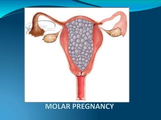
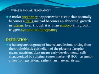
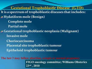
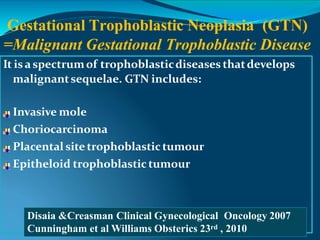
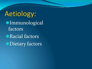
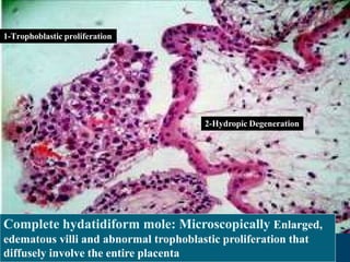
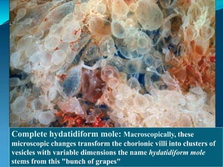
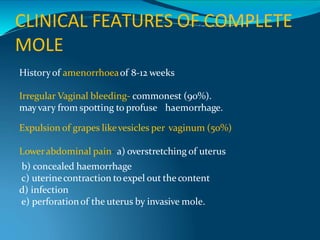
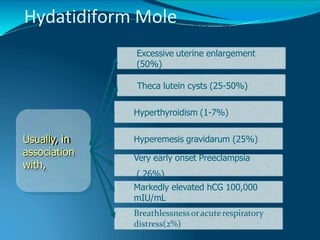
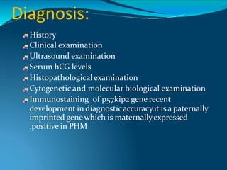
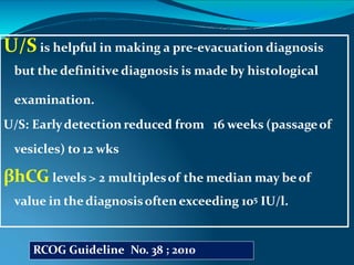
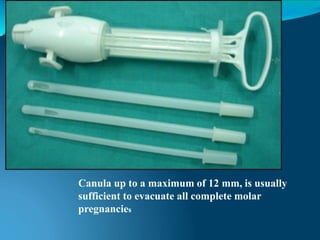
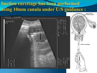
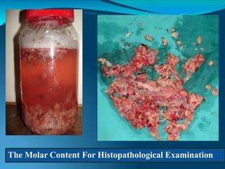
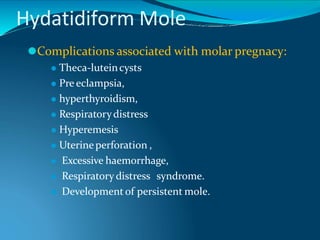
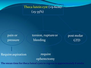
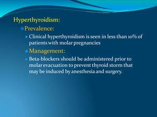
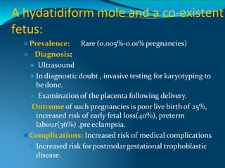
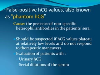
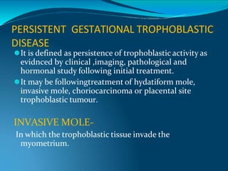
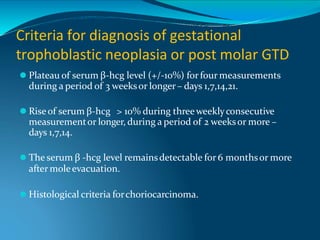
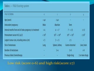
SlideShare a Scribd company logo
MOLAR
PREGNANCY
DR. YOGESH PATEL
MBBS, DGO
DIPLOMA IN LAPAROSCOPY (D. MAS)
FELLOWSHIP IN LAPAROSCOPY (F. MAS)
FELLOWSHIP IN INFERTILITY (F. ART)
PG DIPLOMA IN ULTRASONOGRAPHY (D. USG)
EMERGENCY MEDICINE SPECIALIST
FORMER CONSULTANT AIIMS NEW DELHI
MEMBER OF THE WORLD ASSOCIATION OF LAPROSCOPIC SURGEONS
WHAT IS MOLAR PREGNANCY?
⚫A molarpregnancy happenswhen tissue that normally
becomes a fetus instead becomes an abnormal growth
in uterus. Even though it isn't an embryo, this growth
triggers symptomsof pregnancy
DEFINATION:
⚫A heterogeneous group of interrelated lesions arising from
the trophoblastic epithelium of the placenta ,(tropho
means nutrition, blast means early developmental cells)
characterized by adistinct tumor marker –β HCG as tumor
arises from gestational rather than maternal tissue.
What is the classification of
gestational trophoblastic
disease?
Gestational Trophoblastic Disease (GTD)
It is aspectrumof trophoblasticdiseases that includes:
Hydatiform mole (Benign)
Complete mole
Partial mole
Gestational trophoblastic neoplasia (Malignant)
Invasive mole
Choriocarcinoma
Placental site trophoblastictumour
Epitheloid trophoblastic tumour
The last 3 may follow abortion, ectopic or normal pregnancy
FIGO oncology committee; Williams Obsterics
23rd , 2010
It is aspectrumof trophoblasticdiseases thatdevelops
malignantsequelae. GTN includes:
Invasive mole
Choriocarcinoma
Placental site trophoblastictumour
Epitheloid trophoblastic tumour
Gestational Trophoblastic Neoplasia (GTN)
=Malignant Gestational Trophoblastic Disease
Disaia &Creasman Clinical Gynecological Oncology 2007
Cunningham et al Williams Obsterics 23rd , 2010
MOLAR PREGNANCY ?
Aetiology:
⚫Immunological
factors
⚫Racial factors
⚫Dietary factors
Risk Factors
Age: extremes of age <15 yr and >40 yr
Reproductive history:
Prior Molar Pregnancy
Previous spontaneous abortion: double the incidence
Second molar: 1% - Third molar : 20%!
Diet: increase incidence in high carbohydrate diet, low protein and Vit.A or
carotene diet (complete mole)
Malnutrition and debilitated condition.
Repetitive H. moles in women with different partners
Maternal Blood GroupAB andA
High gamma globin in absence of hepatic disesase
WHAT IS THE INCIDENCE?
In the United States,
1 in 1,500 -2,000
pregnancies
In Asian countries,
•The rate is 10 times
higher than in Europeand
North America
In Saudi Arabia;,
•1.48 in 1000 live births
(hospital-based study;
Felemban AA, etal; 1969)
1 in 200-300 pregnancies in
south eastasia.
1-2 in 1,000 pregnancies in
Chinaand Japan.
Incidence
highest in
philippines i,e
1 in 80
WHAT IS THE CHROMOSOMAL
BASIS OF DEVELOPMENT OF
MOLE?
Pathogenesis and Cytogenetics of HM
Genetic
Constitution
Diploid Triploid/ teraploid
Patho-genesis
4%
Fertilization
of an empty
ovum by two
sperms
“Diandric
dispermy”
90%
Triploid
fertilization of
a normal
ovum by two
sperms
“Dispermic
triploidy”
96%
Fertilization
of an empty
ovum by one
sperms that
undergoes
duplication
“Diandric
diploidy”
10%
Tetraploid
fertilization of
a normal
ovum by
three sperms
“Dispermic
triploidy”
Karyotype
46XX
69XXX
69YXX
69YYX
46XX
46XY
Complete Partial
Complete Mole, Pathogenesis
Duplication 46XX
Empty ovum
23X
Diandric diploidy
Androgenesis M:F 2:0
Paternal
chromosomes only
Complete Mole, Pathogenesis
Empty ovum
23X
Dispermic diploidy
Paternal
chromosomes only
23X 23X
46XX
23X
Partial Mole, Pathogenesis
69XXY
Normal ovum
23X
Dispermic triploidy M:F 2:1
Paternal extra set
23Y 23X
23Y 23X
23X
Familial biparental hydatidiform
mole
⚫Familial biparental hydatidiform mole (FBHM) is
inherited in an autosomal recessive pattern .
⚫Extensive mapping studies had demonstrated a
defective locus at 19q13.4. thisabnormality have been
localised toa singlegene- NALP7.
⚫This is the first causativegenedefect identified in H.
MOLE.
-
Describe Complete
Hydatidiform Mole
Feature
Karyotype
Partial mole
Most commonly
69, XXX or - XXY
Complete mole
Most commonly
46, XX or -,XY
Often present
Usually present
Variable, focal
Focal, slight-moderate
Absent
Absent
Diffuse
Diffuse, slight-severe
Pathology
Fetus
Amnion, fetal RBC
Villous edema
Trophoblastic proliferation
Clinical presentation
Diagnosis
Uterine size
Theca lutein cysts
Medical complications
Postmolar GTN
Missed abortion
Small for dates
Rare
Rare
1-5%
Molar gestation
50% large for dates
25-30%
10-25%
15-20%
Disaia &Creasman Clinical Gynecological Oncology 2007
Cunningham et al Williams Obsterics 23rd , 2010
What is the pathological features of
complete hydatidiform mole?
Complete H. Mole
Macroscopically, these microscopic changes transform the
chorionic villi into clusters of vesicles with variable
dimensions “ like bunch of grapes“.
No fetal or embryonic tissue are produced
Uterine enlargement in excess of gestational age .
Theca-lutein cyst associated in 30%
Microscopically Enlarged, edematous villi and abnormal
trophoblastic proliferation that diffusely involve the
entire villi.
No fetal tissue, RBCs or amnion are produced
1-Trophoblastic proliferation
2-Hydropic Degeneration
Complete hydatidiform mole: Microscopically Enlarged,
edematous villi and abnormal trophoblastic proliferation that
diffusely involve the entire placenta
Complete hydatidiform mole: Macroscopically, these
microscopic changes transform the chorionic villi into clusters of
vesicles with variable dimensions the name hydatidiform mole
stems from this "bunch of grapes"
Complete Hydatiform Mole
Uterine wall
CLINICAL FEATURES OF COMPLETE
MOLE
Historyof amenorrhoeaof 8-12 weeks
Irregular Vaginal bleeding- commonest (90%).
mayvary from spotting to profuse haemorrhage.
Expulsion of grapes likevesicles per vaginum (50%)
Lowerabdominal pain- a) overstretching of uterus
b) concealed haemorrhage
c) uterinecontraction toexpel out the content
d) infection
e) perforationof the uterus by invasive mole.
Hydatidiform Mole
Usually, in
association
with,
Theca lutein cysts (25-50%)
Very early onset Preeclampsia
( 26%)
Markedly elevated hCG 100,000
mIU/mL
Breathlessnessoracute respiratory
distress(2%)
Hyperemesis gravidarum (25%)
Hyperthyroidism (1-7%)
Excessive uterine enlargement
(50%)
Points to be noted during
examination
GENERAL EXAMINATION:
⚫Patient looks more ill and Pallor isoutof proportionof
bleeding.
⚫PR-tachycardia, RR- tachypnea /dyspnoea
⚫Features of preeclampsia present .usually there is early
onset of preeclampsia.
P/A-
⚫Uterine enlargement> than expected GA.
⚫The uterus is soft and doughydue toabsence of the
amniotic fluid sac.
⚫Fetal parts not felt
⚫FHS cannot be detected.
P/S-
⚫Cervical os may beopen orclose.
⚫Vuval orvaginal metastasis mayappearas purple
nodule.
P/V-
⚫Note the uterinesize.
⚫Internal ballotmentcannot beelicited.
⚫Unilateral or bilateral theca luteincystof ovary
palpable in 25-35% of cases.
PATHOLOGY OF PARTIAL
H. MOLE
Partial H. Mole
Microscopically: The enlarged, edematous villi and
abnormal trophoblastic proliferation are slight and
focal and did not involve the entire villi.
There is a scalloping of chorionic villi with undulating
border.
Fetal or embryonic or fetal RBCs
Macroscopically: The molar pattern did not involve
the entire placenta.
Uterine enlargement in excess of gestational age is
uncommon.
Theca-lutein cysts are rare
Fetal or embryonic tissue or amnion
Scalloping of chorionic villi
Partial Hydatidiform Mole
Trophoblastic proliferation are slight and focal
Partial Hydatiform Mole
Vesicles
Maternal side
Fetal hand demonstrating syndactyly. The fetus had a triploid
karyotype, and the chorionic tissues were a partial mole
Partial H. mole.
CLINICAL FEATURES OF PARTIAL
MOLE
History:
⚫Vaginal bleeding
⚫Usuallydiagnosed as missed or incomplete
abortion(91%).
Physical :
⚫A uteruscorrespondsorsmall forgestation age
⚫Excessiveuterinesize noted(4%)
⚫Toxemiaof pregnancy(4%)
Diagnosis:
History
Clinical examination
Ultrasound examination
Serum hCG levels
Histopathological examination
Cytogenetic and molecular biological examination
Immunostaining of p57kip2 gene recent
development in diagnosticaccuracy.it is a paternally
imprinted genewhich is maternallyexpressed
.positive in PHM
U/S is helpful in making a pre-evacuation diagnosis
but the definitive diagnosis is made by histological
examination.
U/S: Earlydetection reduced from 16 weeks (passageof
vesicles) to 12 wks
βhCG levels > 2 multiplesof the median may beof
value in thediagnosisoften exceeding 105 IU/l.
RCOG Guideline No. 38 ; 2010
Guideline to hCG Levels During Pregnancy
hCG levels in weeks from LMP (gestational age)* :
3 weeks LMP: 5 – 50 mIU/ml
4 weeks LMP: 5 – 426 mIU/ml
5 weeks LMP: 18 – 7,340 mIU/ml
6 weeks LMP: 1,080 – 56,500 mIU/ml
7 – 8 weeks LMP: 7, 650 – 229,000 mIU/ml
9 – 12 weeks LMP: 25,700 – 288,000 mIU/ml
13 – 16 weeks LMP: 13,300 – 254,000 mIU/ml
17 – 24 weeks LMP: 4,060 – 165,400 mIU/ml
25 – 40 weeks LMP: 3,640 – 117,000 mIU/ml
Non-pregnant females: <5.0 mIU/ml
Postmenopausal females: <9.5 mIU/ml
* These numbers are just a GUIDELINE– every woman’s level of
hCG can rise differently. It is not necessarily the level that
matters but rather the change in the level.
Complete Molar Pregnancy
Complete hydatidiform mole. The classic "snowstorm"
appearance is created by the multiple placental vesicles.
In most patients with a partial mole,
the clinical and U/S diagnosis is
Usually missed or incompleteabortion.
Thisemphasizes the need fora
thorough histopathologicevaluationof
all missed or incompleteabortions
How Is Partial H .Mole Diagnosed?
RCOG Guideline No. 38 ; 2010
Classically: A thickened, hydropicplacentawith
fetal orembryonic tissue
Multiplesoft markers, including:
Cysticspaces in the placentaand
Transverseto AP dimension a ratio of the
gestationsacof > 1.5, is required forthe reliable
diagnosisof a partial molarpregnancy βhCG ≥
105 U/L
RCOG Guideline No. 38 ; 2010
How Is Partial H .Mole Diagnosed?
egnancies
Hydatidiform Mole
⚫Diagnosis:
⚫Histopathological
examination:
⚫It should always be doneas faras
possible .
There are 2 important basic lines :
1 Evacuationof the mole
2Regular follow-up to detect
persistent trophoblasticdisease
If both basic lines are done
appropriately, mortalityratescan be
reduced to zero.
What Is The Plan of Management?
The Management Of
Gestational Trophoblastic
Disease
RCOG Guideline No. 38 ; 2010
Management:
Investigations:
Laboratory:
 Pre-evacuation hCG
 Complete blood count
 Electrolytes, BUN, creatinine
 Liver function tests
 Thyroid function tests
 HIV
Imaging:
 Pelvic ultrasound
 Chestx-ray
Management:
The patientshould be stabilized hemodynamically 
Medical care:
Correction of:
i.
ii.
iii.
Anemia
Dehydration
Hyperthyroidism
iv. Hypertension
Surgical care
⚫Suction Evacuation is method of choice –
can be done by conventional suction
curretageaswell as by MVA
⚫Two MVA setshould beavailable
⚫Cervical preparation with prostaglandinsor
misoprostol , should be avoided to reduce
the risk of embolisation
RCOG Guideline No. 38 ; 2010
What Is The Best Method Of
Evacuating A Molar Pregnancy?
For Partial mole: Itdepends on the fetal parts
Small fetal parts :Suction curettage
Large fetal parts: Medical (oxytocics)
In partial mole the oxytocics is safe ,as the hazard to
embolise and disseminate trophoblastic tissue is
very low
Also, the needing forchemotherapy is 0.1- 0.5%.
RCOG Guideline No. 38 ; 2010
Is That The Same For Partial Mole?
Canula up to a maximum of 12 mm, is usually
sufficient to evacuate all complete molar
pregnancies
Suction curettage has been performed
using 10mm canula under U/S guidance :
E
El
l SH
SHE
ER
RBIN
BINY
Y
H
O
S
H
O
S
P
P
Canula
Garner UpToDate 2010
Suction Curettage
⚫Supervision of seniorsurgeon
⚫Blood should becross matched and
keptavailable
⚫Maintain two i/v line
⚫Procedure to be carried out in OT
⚫Cervical os to bedilated up to 12mm
sizesuction cannula.
⚫Deep insertion of suction cannula
avoided
⚫Gentle curettage is performed after
evacuation is complete and tissue
sent for histopathology.
⚫ Intraop USG , if available help to
ensure thecomplitionof procedure.
• The useof oxytocin infusion priorto
completion of evacuation is not
recommended
The Molar Content For Histopathological Examination
When Anti-D Is Required?
It is required in Partial due to the presenceof
fetal RBCs
⚫In Complete mole because of poor
vascularisation of thechorionicvilli and
absence of the anti-D antigen, anti-D
prophylaxis is not required.
⚫Although ACOG recommend togive Anti-D
in all cases.
RCOG Guideline No. 38 ; 2010
Barrier methods – most preffered method.
Oral contraceptive method-Once βhCG level have
normalized: COC pill may be used.( as it mayactsas
growth factorfortrophoblastic tissue)
Lowdose OCP is preffered.
If oral COCwasstarted before thediagnosisof GTD
,COCcan becontinueas its potential to increase
risk of GTN isvery low
IUCD should not be used until β hCG levelsare
normal to reduceuterineperforation.
Permanentsterilisation- prefered in thosecouples
whose family has been completed.
What Is Safe Contraception Following GTD?
RCOG Guideline No. 38 ; 2010
Hysterectomy may be preferred to suction
curettage at age ≥ 40 years with no desire for
further pregnancies especially with other risk
factors for GTN as :
 Large theca lutein cysts( >6 cm)
 Significant uterine enlargement
 Pretreatment βhCG ≥ 105 U/L.
Although hysterectomy does not eliminate
possibility of GTN, it markedly reduces its
likelihood. Post hyst. GTN is observed in 3-5%
of cases.
Soper. Obstet Gynecol 108:176, 2006 Garner UpToDate 2010
Cunningham et al,Williams Obstetrics,23rd ,2010
Theca-lutein cyst associated with a complete H. mole in >30%
Second Uterine Evacuation : not required
routinely RCOG Guideline No. 38 ; 2010
Prophylactic Chemotherapy: The long-term
prognosis for women with a H. mole is not
improved with prophylactic chemotherapy. Because
toxicity—including death—may be significant, it is
not recommended routinely *
It may be useful in the high-risk cases when
follow-up are unavailable or unreliable. * *
American College of Obstetricians and Gynecologists, 2004*
Prophylactic chemotherapy after
molar pregnancy
⚫The controversial practice of prophylactic
chemotherapy in GTN in women with H.mole is not
recommended ( According to recent cochrane data
based review 2012).
⚫ Overall PC reduces the risk of GTN however
researcherconsider thisevidence to be lowquality.
⚫When GTN did occurthe time todiagnosed in women
received PC is longer and these women require more
coursesare require tocure GTN.
⚫Unnecessary exposure to toxicadverseeffect.
⚫Prophylactic Chemotherapy:
⚫In one randomized clinical trial, a single course of
methotrexate(0.4mg/ kg/day ) and folinicacid reduced
the incidence of postmolar trophoblastic disease from
47.4% to 14.3% in patients with high-risk moles:
Age >40 yrs
previous history of molar pregnancy
βhCG levels greater than 100,000 mIU/mL,
Uterine size greater than gestational age,
Ovarian sizegreater than 6 cm
All associated factors like PE, hyperthyroidism,
hyperemesis. Acute respiratorydistress.
Post evacuation uterine haemorrhage/subinvolution
⚫Still thegold standard is careful follow up
of each and every patient and serial
estimation of β HCG.
Post-evacuation Surveillance
Why?
Todeterminewhen pregnancy
can beallowed
Todetect persistent trophoblastic
disease (i.e. GTN)
A baseline serum β -hCG level is obtained within 48
hoursafterevacuation.
Levelsare monitored everyweek till a normal value is
achieved. Level usually becomes normal by 8-10
weeks.
Monitor HCG every month forfurther 6 months from
the date of evacuation if HCG has to return to normal
within 56 daysafterpregnancyevent.
>56 daysof the pregnancyevent :Follow up is 6 months
from normalization of the hCG level.
These levelsshould progressively fall toan undetectable
level (<5 mu/ml).
RCOG Guideline No. 38 ; 2010
The Post-evacuation Surveillance. How?
In resource poor setting – UPT and USG.
Monthly USG 1 month after evacuation.
UPT should be perfomed once monthly starting from 3rd to
4th month untill 1 year after evacuation.
The normal time for βhCG to normalise is 99 days in
complete moles and 59 days in partial mole.
Pelvic examination:
⚫ Duration: while hCG is elevated to monitor the
involution of pelvic structures and to aid in the
identification of vaginal metastasis
Hydatidiform Mole
⚫Complications associated with molar pregnacy:
⚫ Theca-luteincysts
⚫ Pre eclampsia,
⚫ hyperthyroidism,
⚫ Respiratorydistress
⚫ Hyperemesis
⚫ Uterineperforation ,
⚫ Excessive haemorrhage,
⚫ Respiratory distress syndrome.
⚫ Development of persistent mole.
Theca lutein cyst (>5-6cm)
(25-35%)
pain or
pressure
torsion, ruptureor
bleeding
post molar
GTD
Requireaspiration require
opherectomy
The mean time fortheca luteal cysts toregress isapproximately 8 weeks
RESPIRATORY DISTRESS SYNDROME
(rare event)
Pathophysiology:
⚫embolisation of trophoblastic tissue , pulmonary
metastasis
⚫ Risk factors :
⚫uterinesize > 14-16 weeks
⚫ high HCG level
⚫Contributing factors- anaemia, thyrotoxicosis
It should resolvewithin 24 to 48 hoursafter molarevacuation
Hyperthyroidism:
⚫Prevalence:
⚫ Clinical hyperthyroidism is seen in less than 10% of
patientswith molarpregnancies
⚫ Management:
⚫ Beta-blockers should be administered prior to
molarevacuation to prevent thyroid storm that
may be induced byanesthesiaand surgery.
When can women whose last pregnancy was a
complete or partial hydatidiform molar
pregnancy try to conceive in the future ?
⚫Womenshould beadvised not toconceiveuntil
their follow-up is complete.
⚫Womenwho undergochemotherapyareadvised
not toconceive for 1 yearaftercompletionof
treatment.
⚫Patientwith metastatic GTN – 2 years
Pregnancy after Hydatidiform Mole:
⚫Risk of another molarpregnancy:
(1–2% incidence)
⚫Current recommendations for
managementof subsequent pregnancies:
⚫P/V in first trimesterand ultrasound toconfirm
normal gestational developmentand dates
⚫Examination of the placentaorproductsof
conception histologicallyat the timeof delivery
orevacuation .
⚫An hCG level should be obtained 6 weeks
and 10 weeks post evacuation or delivery
to confirm normalization.
A hydatidiform mole and a co-existent
fetus:
⚫Prevalence: Rare (0.005%-0.01% pregnancies)
⚫ Diagnosis:
⚫ Ultrasound
⚫ In diagnosticdoubt , invasive testing for karyotyping to
be done.
⚫ Examinationof the placenta following delivery.
Outcome of such pregnancies is poor live birth of 25%,
increased risk of early fetal loss(40%), preterm
labour(36%) ,pre eclampsia.
⚫Complications: Increased risk of medical complications
⚫ Increased risk forpostmolargestational trophoblastic
disease.
False-positive hCG values, also known
as “phantom hCG”
Cause: the presence of non-specific
heterophil antibodies in the patients’ sera.
⚫Should be suspected if hCG values plateau
at relatively low levels and do not respond
to therapeutic maneuvers
⚫Evaluation of patients with :
• Urinary hCG
• Serial dilutionsof the serum
Prognosis:
⚫Post-molar gestational trophoblastic disease:
⚫ Risk:
⚫ Following complete mole: 20%
⚫ Following partial mole: 5%
⚫ Type:
⚫ 70% to 90% are persistentor invasive moles
⚫ 10% to 30% are choriocarcinomas.
Recurrence-
Risk of recurrencwith prior molar pregnancy is
1-4%
PERSISTENT GESTATIONAL TROPHOBLASTIC
DISEASE
⚫It is defined as persistence of trophoblastic activity as
evidnced by clinical ,imaging, pathological and
hormonal study following initial treatment.
⚫It may be followingtreatment of hydatiform mole,
invasive mole, choriocarcinoma or placental site
trophoblastic tumour.
INVASIVE MOLE-
In which the trophoblastic tissue invade the
myometrium.
Criteria for diagnosis of gestational
trophoblastic neoplasia or post molar GTD
⚫ Plateau of serum β-hcg level (+/-10%) forfour measurements
during a period of 3 weeksor longer– days 1,7,14,21.
⚫ Riseof serum β-hcg > 10% during threeweeklyconsecutive
measurementor longer, during a period of 2 weeksor more –
days 1,7,14.
⚫ The serum β -hcg level remainsdetectable for 6 monthsor more
after moleevacuation.
⚫ Histological criteria forchoriocarcinoma.
Low risk (score 0-6) and high risk(score ≥7)
Staging
International staging of WHO may be summarized as
follows:
Ⅰ : lesion localized in uterus, no metastasis;
Ⅱ: lesion extends beyond uterus, but still confined to
internal genitalias;
Ⅲ
: pulmonary lesion
Ⅳ: metastasis tootherdistantsites.
Indication for therapy
Indication for therapyafterevacuation-
⚫An abnormal hcg regression pattern (10% or> rise in hcg
level or plateauing hcg or 3 stablesvalueover 2 weeks)
⚫An hcg rebound.
⚫Histological diagnosis of choriocarcinoma or placental site
trophoblastic tumour.
⚫The presenceof metastases.
⚫High hcg levels(.20,000miu/ml more than 4 weeks
postevacuation)
⚫Persistentlyelevated hcg levels 6 months post evacuation.
Hydatiform mole
Evacuation
Serial hcg level resolution
GTN
FIGO scoring
Low risk(≤6) high risk(>6)
Single agent (MTX)
chemotherapy
combination(MAC/EMACO)
chemotherapy
Serial hcg level resolution(life–long
follow up)
Relapse /resistantdisease
Second –linechemotherapy ±surgical debulking
THANK YOU ! ! !

More Related Content

Similar to fgtfgjjg.pptx

23. gestational trophoblastic diseases
23. gestational trophoblastic diseases23. gestational trophoblastic diseases
23. gestational trophoblastic diseases
Chifuniro
 
Hydatid form mole
Hydatid form moleHydatid form mole
Hydatid form mole
ELIZEBETH RANI V
 
hmolesrd-190102152314.pptx
hmolesrd-190102152314.pptxhmolesrd-190102152314.pptx
hmolesrd-190102152314.pptx
kajalgupta681731
 
HYDATIDIFORM MOLE: APPROACH AND MANAGEMENT
HYDATIDIFORM MOLE: APPROACH AND MANAGEMENTHYDATIDIFORM MOLE: APPROACH AND MANAGEMENT
HYDATIDIFORM MOLE: APPROACH AND MANAGEMENT
Sharad Dahal
 
Gestational Trophoblastic disease
Gestational Trophoblastic diseaseGestational Trophoblastic disease
Hydatidiform mole
Hydatidiform moleHydatidiform mole
Hydatidiform mole
SusheelaSubedi
 
Gestational trophoblastic disease ( Molar pregnancy )
Gestational trophoblastic disease ( Molar pregnancy )Gestational trophoblastic disease ( Molar pregnancy )
Gestational trophoblastic disease ( Molar pregnancy )
Mohammed Barznji
 
Gestational Trophoblastic Disease - www.jinekolojivegebelik.com
Gestational Trophoblastic Disease - www.jinekolojivegebelik.comGestational Trophoblastic Disease - www.jinekolojivegebelik.com
Gestational Trophoblastic Disease - www.jinekolojivegebelik.com
jinekolojivegebelik.com
 
2. ECTOPIC & MOLAR PREGNANCY.pptx
2. ECTOPIC & MOLAR PREGNANCY.pptx2. ECTOPIC & MOLAR PREGNANCY.pptx
2. ECTOPIC & MOLAR PREGNANCY.pptx
MaximilianTUNGARAZA
 
Gestational trophoblastic disease (gtd.version gao)
Gestational trophoblastic disease (gtd.version gao)Gestational trophoblastic disease (gtd.version gao)
Gestational trophoblastic disease (gtd.version gao)
Ayub Medical College
 
Gestational Trophoblastic Disease - Obstetrics
Gestational Trophoblastic Disease - ObstetricsGestational Trophoblastic Disease - Obstetrics
Gestational Trophoblastic Disease - Obstetrics
NasreenSultana53
 
gestationaltrophoblasticneoplasia-120504132331-phpapp01 (2).pdf
gestationaltrophoblasticneoplasia-120504132331-phpapp01 (2).pdfgestationaltrophoblasticneoplasia-120504132331-phpapp01 (2).pdf
gestationaltrophoblasticneoplasia-120504132331-phpapp01 (2).pdf
DrSaniaAli2
 
GTN.pdf
GTN.pdfGTN.pdf
GTN.pdf
VidushRatan1
 
GESTATIONAL TROPHOBLASTIC DISEASES 27.02.2020.ppt
GESTATIONAL TROPHOBLASTIC DISEASES 27.02.2020.pptGESTATIONAL TROPHOBLASTIC DISEASES 27.02.2020.ppt
GESTATIONAL TROPHOBLASTIC DISEASES 27.02.2020.ppt
FatimaMalazai1
 
screening for down syndrome
screening for down syndromescreening for down syndrome
screening for down syndrome
Sadaf Khan
 
Gestational Trophoblastic Disease By Prof. Dr. Rafia Baloch
Gestational Trophoblastic Disease By Prof. Dr. Rafia BalochGestational Trophoblastic Disease By Prof. Dr. Rafia Baloch
Gestational Trophoblastic Disease By Prof. Dr. Rafia Balochrafiabaloch
 
Gynecology 5th year, 5th & 6th lectures (Dr. Muhabat Salih Saeid)
Gynecology 5th year, 5th & 6th lectures (Dr. Muhabat Salih Saeid)Gynecology 5th year, 5th & 6th lectures (Dr. Muhabat Salih Saeid)
Gynecology 5th year, 5th & 6th lectures (Dr. Muhabat Salih Saeid)
College of Medicine, Sulaymaniyah
 
Gestational Trophoblastic Disease - www.jinekolojivegebelik.com
Gestational Trophoblastic Disease - www.jinekolojivegebelik.comGestational Trophoblastic Disease - www.jinekolojivegebelik.com
Gestational Trophoblastic Disease - www.jinekolojivegebelik.com
jinekolojivegebelik.com
 
Gestational trophoblastic neoplasia (2).ppt44444
Gestational trophoblastic neoplasia (2).ppt44444Gestational trophoblastic neoplasia (2).ppt44444
Gestational trophoblastic neoplasia (2).ppt44444Farrukh Masood
 

Similar to fgtfgjjg.pptx (20)

23. gestational trophoblastic diseases
23. gestational trophoblastic diseases23. gestational trophoblastic diseases
23. gestational trophoblastic diseases
 
Hydatid form mole
Hydatid form moleHydatid form mole
Hydatid form mole
 
Molar pregnancy
Molar pregnancyMolar pregnancy
Molar pregnancy
 
hmolesrd-190102152314.pptx
hmolesrd-190102152314.pptxhmolesrd-190102152314.pptx
hmolesrd-190102152314.pptx
 
HYDATIDIFORM MOLE: APPROACH AND MANAGEMENT
HYDATIDIFORM MOLE: APPROACH AND MANAGEMENTHYDATIDIFORM MOLE: APPROACH AND MANAGEMENT
HYDATIDIFORM MOLE: APPROACH AND MANAGEMENT
 
Gestational Trophoblastic disease
Gestational Trophoblastic diseaseGestational Trophoblastic disease
Gestational Trophoblastic disease
 
Hydatidiform mole
Hydatidiform moleHydatidiform mole
Hydatidiform mole
 
Gestational trophoblastic disease ( Molar pregnancy )
Gestational trophoblastic disease ( Molar pregnancy )Gestational trophoblastic disease ( Molar pregnancy )
Gestational trophoblastic disease ( Molar pregnancy )
 
Gestational Trophoblastic Disease - www.jinekolojivegebelik.com
Gestational Trophoblastic Disease - www.jinekolojivegebelik.comGestational Trophoblastic Disease - www.jinekolojivegebelik.com
Gestational Trophoblastic Disease - www.jinekolojivegebelik.com
 
2. ECTOPIC & MOLAR PREGNANCY.pptx
2. ECTOPIC & MOLAR PREGNANCY.pptx2. ECTOPIC & MOLAR PREGNANCY.pptx
2. ECTOPIC & MOLAR PREGNANCY.pptx
 
Gestational trophoblastic disease (gtd.version gao)
Gestational trophoblastic disease (gtd.version gao)Gestational trophoblastic disease (gtd.version gao)
Gestational trophoblastic disease (gtd.version gao)
 
Gestational Trophoblastic Disease - Obstetrics
Gestational Trophoblastic Disease - ObstetricsGestational Trophoblastic Disease - Obstetrics
Gestational Trophoblastic Disease - Obstetrics
 
gestationaltrophoblasticneoplasia-120504132331-phpapp01 (2).pdf
gestationaltrophoblasticneoplasia-120504132331-phpapp01 (2).pdfgestationaltrophoblasticneoplasia-120504132331-phpapp01 (2).pdf
gestationaltrophoblasticneoplasia-120504132331-phpapp01 (2).pdf
 
GTN.pdf
GTN.pdfGTN.pdf
GTN.pdf
 
GESTATIONAL TROPHOBLASTIC DISEASES 27.02.2020.ppt
GESTATIONAL TROPHOBLASTIC DISEASES 27.02.2020.pptGESTATIONAL TROPHOBLASTIC DISEASES 27.02.2020.ppt
GESTATIONAL TROPHOBLASTIC DISEASES 27.02.2020.ppt
 
screening for down syndrome
screening for down syndromescreening for down syndrome
screening for down syndrome
 
Gestational Trophoblastic Disease By Prof. Dr. Rafia Baloch
Gestational Trophoblastic Disease By Prof. Dr. Rafia BalochGestational Trophoblastic Disease By Prof. Dr. Rafia Baloch
Gestational Trophoblastic Disease By Prof. Dr. Rafia Baloch
 
Gynecology 5th year, 5th & 6th lectures (Dr. Muhabat Salih Saeid)
Gynecology 5th year, 5th & 6th lectures (Dr. Muhabat Salih Saeid)Gynecology 5th year, 5th & 6th lectures (Dr. Muhabat Salih Saeid)
Gynecology 5th year, 5th & 6th lectures (Dr. Muhabat Salih Saeid)
 
Gestational Trophoblastic Disease - www.jinekolojivegebelik.com
Gestational Trophoblastic Disease - www.jinekolojivegebelik.comGestational Trophoblastic Disease - www.jinekolojivegebelik.com
Gestational Trophoblastic Disease - www.jinekolojivegebelik.com
 
Gestational trophoblastic neoplasia (2).ppt44444
Gestational trophoblastic neoplasia (2).ppt44444Gestational trophoblastic neoplasia (2).ppt44444
Gestational trophoblastic neoplasia (2).ppt44444
 

More from FatimaMalazai1

Design Inspiration for College by Slidesgo.pptx
Design Inspiration for College by Slidesgo.pptxDesign Inspiration for College by Slidesgo.pptx
Design Inspiration for College by Slidesgo.pptx
FatimaMalazai1
 
health-pptx
health-pptxhealth-pptx
health-pptx
FatimaMalazai1
 
Copy.pptx
 Copy.pptx Copy.pptx
Copy.pptx
FatimaMalazai1
 
DrCopy.pptx
DrCopy.pptxDrCopy.pptx
DrCopy.pptx
FatimaMalazai1
 
Lecture2.pptx
Lecture2.pptxLecture2.pptx
Lecture2.pptx
FatimaMalazai1
 
Lecture3.ppt
Lecture3.pptLecture3.ppt
Lecture3.ppt
FatimaMalazai1
 
Lecture4.pptx
Lecture4.pptxLecture4.pptx
Lecture4.pptx
FatimaMalazai1
 
Lecture5.pptx
Lecture5.pptxLecture5.pptx
Lecture5.pptx
FatimaMalazai1
 
molarpregnancy-190830175936 (1).pptx
molarpregnancy-190830175936 (1).pptxmolarpregnancy-190830175936 (1).pptx
molarpregnancy-190830175936 (1).pptx
FatimaMalazai1
 

More from FatimaMalazai1 (10)

Design Inspiration for College by Slidesgo.pptx
Design Inspiration for College by Slidesgo.pptxDesign Inspiration for College by Slidesgo.pptx
Design Inspiration for College by Slidesgo.pptx
 
health-pptx
health-pptxhealth-pptx
health-pptx
 
Copy.pptx
 Copy.pptx Copy.pptx
Copy.pptx
 
DrCopy.pptx
DrCopy.pptxDrCopy.pptx
DrCopy.pptx
 
Lecture_CFD.ppt
Lecture_CFD.pptLecture_CFD.ppt
Lecture_CFD.ppt
 
Lecture2.pptx
Lecture2.pptxLecture2.pptx
Lecture2.pptx
 
Lecture3.ppt
Lecture3.pptLecture3.ppt
Lecture3.ppt
 
Lecture4.pptx
Lecture4.pptxLecture4.pptx
Lecture4.pptx
 
Lecture5.pptx
Lecture5.pptxLecture5.pptx
Lecture5.pptx
 
molarpregnancy-190830175936 (1).pptx
molarpregnancy-190830175936 (1).pptxmolarpregnancy-190830175936 (1).pptx
molarpregnancy-190830175936 (1).pptx
 

Recently uploaded

A Classical Text Review on Basavarajeeyam
A Classical Text Review on BasavarajeeyamA Classical Text Review on Basavarajeeyam
A Classical Text Review on Basavarajeeyam
Dr. Jyothirmai Paindla
 
Physiology of Special Chemical Sensation of Taste
Physiology of Special Chemical Sensation of TastePhysiology of Special Chemical Sensation of Taste
Physiology of Special Chemical Sensation of Taste
MedicoseAcademics
 
Evaluation of antidepressant activity of clitoris ternatea in animals
Evaluation of antidepressant activity of clitoris ternatea in animalsEvaluation of antidepressant activity of clitoris ternatea in animals
Evaluation of antidepressant activity of clitoris ternatea in animals
Shweta
 
Gram Stain introduction, principle, Procedure
Gram Stain introduction, principle, ProcedureGram Stain introduction, principle, Procedure
Gram Stain introduction, principle, Procedure
Suraj Goswami
 
Pharma Pcd Franchise in Jharkhand - Yodley Lifesciences
Pharma Pcd Franchise in Jharkhand - Yodley LifesciencesPharma Pcd Franchise in Jharkhand - Yodley Lifesciences
Pharma Pcd Franchise in Jharkhand - Yodley Lifesciences
Yodley Lifesciences
 
Non-respiratory Functions of the Lungs.pdf
Non-respiratory Functions of the Lungs.pdfNon-respiratory Functions of the Lungs.pdf
Non-respiratory Functions of the Lungs.pdf
MedicoseAcademics
 
Colonic and anorectal physiology with surgical implications
Colonic and anorectal physiology with surgical implicationsColonic and anorectal physiology with surgical implications
Colonic and anorectal physiology with surgical implications
Dr Maria Tamanna
 
KDIGO 2024 guidelines for diabetologists
KDIGO 2024 guidelines for diabetologistsKDIGO 2024 guidelines for diabetologists
KDIGO 2024 guidelines for diabetologists
د.محمود نجيب
 
Ophthalmology Clinical Tests for OSCE exam
Ophthalmology Clinical Tests for OSCE examOphthalmology Clinical Tests for OSCE exam
Ophthalmology Clinical Tests for OSCE exam
KafrELShiekh University
 
Vision-1.pptx, Eye structure, basics of optics
Vision-1.pptx, Eye structure, basics of opticsVision-1.pptx, Eye structure, basics of optics
Vision-1.pptx, Eye structure, basics of optics
Sai Sailesh Kumar Goothy
 
planning for change nursing Management ppt
planning for change nursing Management pptplanning for change nursing Management ppt
planning for change nursing Management ppt
Thangamjayarani
 
Dehradun #ℂall #gIRLS Oyo Hotel 9719300533 #ℂall #gIRL in Dehradun
Dehradun #ℂall #gIRLS Oyo Hotel 9719300533 #ℂall #gIRL in DehradunDehradun #ℂall #gIRLS Oyo Hotel 9719300533 #ℂall #gIRL in Dehradun
Dehradun #ℂall #gIRLS Oyo Hotel 9719300533 #ℂall #gIRL in Dehradun
chandankumarsmartiso
 
Maxilla, Mandible & Hyoid Bone & Clinical Correlations by Dr. RIG.pptx
Maxilla, Mandible & Hyoid Bone & Clinical Correlations by Dr. RIG.pptxMaxilla, Mandible & Hyoid Bone & Clinical Correlations by Dr. RIG.pptx
Maxilla, Mandible & Hyoid Bone & Clinical Correlations by Dr. RIG.pptx
Dr. Rabia Inam Gandapore
 
ARTIFICIAL INTELLIGENCE IN HEALTHCARE.pdf
ARTIFICIAL INTELLIGENCE IN  HEALTHCARE.pdfARTIFICIAL INTELLIGENCE IN  HEALTHCARE.pdf
ARTIFICIAL INTELLIGENCE IN HEALTHCARE.pdf
Anujkumaranit
 
Hemodialysis: Chapter 3, Dialysis Water Unit - Dr.Gawad
Hemodialysis: Chapter 3, Dialysis Water Unit - Dr.GawadHemodialysis: Chapter 3, Dialysis Water Unit - Dr.Gawad
Hemodialysis: Chapter 3, Dialysis Water Unit - Dr.Gawad
NephroTube - Dr.Gawad
 
Pictures of Superficial & Deep Fascia.ppt.pdf
Pictures of Superficial & Deep Fascia.ppt.pdfPictures of Superficial & Deep Fascia.ppt.pdf
Pictures of Superficial & Deep Fascia.ppt.pdf
Dr. Rabia Inam Gandapore
 
Novas diretrizes da OMS para os cuidados perinatais de mais qualidade
Novas diretrizes da OMS para os cuidados perinatais de mais qualidadeNovas diretrizes da OMS para os cuidados perinatais de mais qualidade
Novas diretrizes da OMS para os cuidados perinatais de mais qualidade
Prof. Marcus Renato de Carvalho
 
NVBDCP.pptx Nation vector borne disease control program
NVBDCP.pptx Nation vector borne disease control programNVBDCP.pptx Nation vector borne disease control program
NVBDCP.pptx Nation vector borne disease control program
Sapna Thakur
 
How to Give Better Lectures: Some Tips for Doctors
How to Give Better Lectures: Some Tips for DoctorsHow to Give Better Lectures: Some Tips for Doctors
How to Give Better Lectures: Some Tips for Doctors
LanceCatedral
 
Physiology of Chemical Sensation of smell.pdf
Physiology of Chemical Sensation of smell.pdfPhysiology of Chemical Sensation of smell.pdf
Physiology of Chemical Sensation of smell.pdf
MedicoseAcademics
 

Recently uploaded (20)

A Classical Text Review on Basavarajeeyam
A Classical Text Review on BasavarajeeyamA Classical Text Review on Basavarajeeyam
A Classical Text Review on Basavarajeeyam
 
Physiology of Special Chemical Sensation of Taste
Physiology of Special Chemical Sensation of TastePhysiology of Special Chemical Sensation of Taste
Physiology of Special Chemical Sensation of Taste
 
Evaluation of antidepressant activity of clitoris ternatea in animals
Evaluation of antidepressant activity of clitoris ternatea in animalsEvaluation of antidepressant activity of clitoris ternatea in animals
Evaluation of antidepressant activity of clitoris ternatea in animals
 
Gram Stain introduction, principle, Procedure
Gram Stain introduction, principle, ProcedureGram Stain introduction, principle, Procedure
Gram Stain introduction, principle, Procedure
 
Pharma Pcd Franchise in Jharkhand - Yodley Lifesciences
Pharma Pcd Franchise in Jharkhand - Yodley LifesciencesPharma Pcd Franchise in Jharkhand - Yodley Lifesciences
Pharma Pcd Franchise in Jharkhand - Yodley Lifesciences
 
Non-respiratory Functions of the Lungs.pdf
Non-respiratory Functions of the Lungs.pdfNon-respiratory Functions of the Lungs.pdf
Non-respiratory Functions of the Lungs.pdf
 
Colonic and anorectal physiology with surgical implications
Colonic and anorectal physiology with surgical implicationsColonic and anorectal physiology with surgical implications
Colonic and anorectal physiology with surgical implications
 
KDIGO 2024 guidelines for diabetologists
KDIGO 2024 guidelines for diabetologistsKDIGO 2024 guidelines for diabetologists
KDIGO 2024 guidelines for diabetologists
 
Ophthalmology Clinical Tests for OSCE exam
Ophthalmology Clinical Tests for OSCE examOphthalmology Clinical Tests for OSCE exam
Ophthalmology Clinical Tests for OSCE exam
 
Vision-1.pptx, Eye structure, basics of optics
Vision-1.pptx, Eye structure, basics of opticsVision-1.pptx, Eye structure, basics of optics
Vision-1.pptx, Eye structure, basics of optics
 
planning for change nursing Management ppt
planning for change nursing Management pptplanning for change nursing Management ppt
planning for change nursing Management ppt
 
Dehradun #ℂall #gIRLS Oyo Hotel 9719300533 #ℂall #gIRL in Dehradun
Dehradun #ℂall #gIRLS Oyo Hotel 9719300533 #ℂall #gIRL in DehradunDehradun #ℂall #gIRLS Oyo Hotel 9719300533 #ℂall #gIRL in Dehradun
Dehradun #ℂall #gIRLS Oyo Hotel 9719300533 #ℂall #gIRL in Dehradun
 
Maxilla, Mandible & Hyoid Bone & Clinical Correlations by Dr. RIG.pptx
Maxilla, Mandible & Hyoid Bone & Clinical Correlations by Dr. RIG.pptxMaxilla, Mandible & Hyoid Bone & Clinical Correlations by Dr. RIG.pptx
Maxilla, Mandible & Hyoid Bone & Clinical Correlations by Dr. RIG.pptx
 
ARTIFICIAL INTELLIGENCE IN HEALTHCARE.pdf
ARTIFICIAL INTELLIGENCE IN  HEALTHCARE.pdfARTIFICIAL INTELLIGENCE IN  HEALTHCARE.pdf
ARTIFICIAL INTELLIGENCE IN HEALTHCARE.pdf
 
Hemodialysis: Chapter 3, Dialysis Water Unit - Dr.Gawad
Hemodialysis: Chapter 3, Dialysis Water Unit - Dr.GawadHemodialysis: Chapter 3, Dialysis Water Unit - Dr.Gawad
Hemodialysis: Chapter 3, Dialysis Water Unit - Dr.Gawad
 
Pictures of Superficial & Deep Fascia.ppt.pdf
Pictures of Superficial & Deep Fascia.ppt.pdfPictures of Superficial & Deep Fascia.ppt.pdf
Pictures of Superficial & Deep Fascia.ppt.pdf
 
Novas diretrizes da OMS para os cuidados perinatais de mais qualidade
Novas diretrizes da OMS para os cuidados perinatais de mais qualidadeNovas diretrizes da OMS para os cuidados perinatais de mais qualidade
Novas diretrizes da OMS para os cuidados perinatais de mais qualidade
 
NVBDCP.pptx Nation vector borne disease control program
NVBDCP.pptx Nation vector borne disease control programNVBDCP.pptx Nation vector borne disease control program
NVBDCP.pptx Nation vector borne disease control program
 
How to Give Better Lectures: Some Tips for Doctors
How to Give Better Lectures: Some Tips for DoctorsHow to Give Better Lectures: Some Tips for Doctors
How to Give Better Lectures: Some Tips for Doctors
 
Physiology of Chemical Sensation of smell.pdf
Physiology of Chemical Sensation of smell.pdfPhysiology of Chemical Sensation of smell.pdf
Physiology of Chemical Sensation of smell.pdf
 

fgtfgjjg.pptx

  • 1. MOLAR PREGNANCY DR. YOGESH PATEL MBBS, DGO DIPLOMA IN LAPAROSCOPY (D. MAS) FELLOWSHIP IN LAPAROSCOPY (F. MAS) FELLOWSHIP IN INFERTILITY (F. ART) PG DIPLOMA IN ULTRASONOGRAPHY (D. USG) EMERGENCY MEDICINE SPECIALIST FORMER CONSULTANT AIIMS NEW DELHI MEMBER OF THE WORLD ASSOCIATION OF LAPROSCOPIC SURGEONS
  • 2.
  • 3. WHAT IS MOLAR PREGNANCY? ⚫A molarpregnancy happenswhen tissue that normally becomes a fetus instead becomes an abnormal growth in uterus. Even though it isn't an embryo, this growth triggers symptomsof pregnancy DEFINATION: ⚫A heterogeneous group of interrelated lesions arising from the trophoblastic epithelium of the placenta ,(tropho means nutrition, blast means early developmental cells) characterized by adistinct tumor marker –β HCG as tumor arises from gestational rather than maternal tissue.
  • 4. What is the classification of gestational trophoblastic disease?
  • 5. Gestational Trophoblastic Disease (GTD) It is aspectrumof trophoblasticdiseases that includes: Hydatiform mole (Benign) Complete mole Partial mole Gestational trophoblastic neoplasia (Malignant) Invasive mole Choriocarcinoma Placental site trophoblastictumour Epitheloid trophoblastic tumour The last 3 may follow abortion, ectopic or normal pregnancy FIGO oncology committee; Williams Obsterics 23rd , 2010
  • 6. It is aspectrumof trophoblasticdiseases thatdevelops malignantsequelae. GTN includes: Invasive mole Choriocarcinoma Placental site trophoblastictumour Epitheloid trophoblastic tumour Gestational Trophoblastic Neoplasia (GTN) =Malignant Gestational Trophoblastic Disease Disaia &Creasman Clinical Gynecological Oncology 2007 Cunningham et al Williams Obsterics 23rd , 2010
  • 9. Risk Factors Age: extremes of age <15 yr and >40 yr Reproductive history: Prior Molar Pregnancy Previous spontaneous abortion: double the incidence Second molar: 1% - Third molar : 20%! Diet: increase incidence in high carbohydrate diet, low protein and Vit.A or carotene diet (complete mole) Malnutrition and debilitated condition. Repetitive H. moles in women with different partners Maternal Blood GroupAB andA High gamma globin in absence of hepatic disesase
  • 10. WHAT IS THE INCIDENCE?
  • 11. In the United States, 1 in 1,500 -2,000 pregnancies In Asian countries, •The rate is 10 times higher than in Europeand North America In Saudi Arabia;, •1.48 in 1000 live births (hospital-based study; Felemban AA, etal; 1969) 1 in 200-300 pregnancies in south eastasia. 1-2 in 1,000 pregnancies in Chinaand Japan. Incidence highest in philippines i,e 1 in 80
  • 12. WHAT IS THE CHROMOSOMAL BASIS OF DEVELOPMENT OF MOLE?
  • 13. Pathogenesis and Cytogenetics of HM Genetic Constitution Diploid Triploid/ teraploid Patho-genesis 4% Fertilization of an empty ovum by two sperms “Diandric dispermy” 90% Triploid fertilization of a normal ovum by two sperms “Dispermic triploidy” 96% Fertilization of an empty ovum by one sperms that undergoes duplication “Diandric diploidy” 10% Tetraploid fertilization of a normal ovum by three sperms “Dispermic triploidy” Karyotype 46XX 69XXX 69YXX 69YYX 46XX 46XY Complete Partial
  • 14. Complete Mole, Pathogenesis Duplication 46XX Empty ovum 23X Diandric diploidy Androgenesis M:F 2:0 Paternal chromosomes only
  • 15. Complete Mole, Pathogenesis Empty ovum 23X Dispermic diploidy Paternal chromosomes only 23X 23X 46XX 23X
  • 16. Partial Mole, Pathogenesis 69XXY Normal ovum 23X Dispermic triploidy M:F 2:1 Paternal extra set 23Y 23X 23Y 23X 23X
  • 17. Familial biparental hydatidiform mole ⚫Familial biparental hydatidiform mole (FBHM) is inherited in an autosomal recessive pattern . ⚫Extensive mapping studies had demonstrated a defective locus at 19q13.4. thisabnormality have been localised toa singlegene- NALP7. ⚫This is the first causativegenedefect identified in H. MOLE.
  • 19. Feature Karyotype Partial mole Most commonly 69, XXX or - XXY Complete mole Most commonly 46, XX or -,XY Often present Usually present Variable, focal Focal, slight-moderate Absent Absent Diffuse Diffuse, slight-severe Pathology Fetus Amnion, fetal RBC Villous edema Trophoblastic proliferation Clinical presentation Diagnosis Uterine size Theca lutein cysts Medical complications Postmolar GTN Missed abortion Small for dates Rare Rare 1-5% Molar gestation 50% large for dates 25-30% 10-25% 15-20% Disaia &Creasman Clinical Gynecological Oncology 2007 Cunningham et al Williams Obsterics 23rd , 2010
  • 20. What is the pathological features of complete hydatidiform mole?
  • 21. Complete H. Mole Macroscopically, these microscopic changes transform the chorionic villi into clusters of vesicles with variable dimensions “ like bunch of grapes“. No fetal or embryonic tissue are produced Uterine enlargement in excess of gestational age . Theca-lutein cyst associated in 30% Microscopically Enlarged, edematous villi and abnormal trophoblastic proliferation that diffusely involve the entire villi. No fetal tissue, RBCs or amnion are produced
  • 22. 1-Trophoblastic proliferation 2-Hydropic Degeneration Complete hydatidiform mole: Microscopically Enlarged, edematous villi and abnormal trophoblastic proliferation that diffusely involve the entire placenta
  • 23. Complete hydatidiform mole: Macroscopically, these microscopic changes transform the chorionic villi into clusters of vesicles with variable dimensions the name hydatidiform mole stems from this "bunch of grapes"
  • 25. CLINICAL FEATURES OF COMPLETE MOLE Historyof amenorrhoeaof 8-12 weeks Irregular Vaginal bleeding- commonest (90%). mayvary from spotting to profuse haemorrhage. Expulsion of grapes likevesicles per vaginum (50%) Lowerabdominal pain- a) overstretching of uterus b) concealed haemorrhage c) uterinecontraction toexpel out the content d) infection e) perforationof the uterus by invasive mole.
  • 26. Hydatidiform Mole Usually, in association with, Theca lutein cysts (25-50%) Very early onset Preeclampsia ( 26%) Markedly elevated hCG 100,000 mIU/mL Breathlessnessoracute respiratory distress(2%) Hyperemesis gravidarum (25%) Hyperthyroidism (1-7%) Excessive uterine enlargement (50%)
  • 27. Points to be noted during examination GENERAL EXAMINATION: ⚫Patient looks more ill and Pallor isoutof proportionof bleeding. ⚫PR-tachycardia, RR- tachypnea /dyspnoea ⚫Features of preeclampsia present .usually there is early onset of preeclampsia. P/A- ⚫Uterine enlargement> than expected GA. ⚫The uterus is soft and doughydue toabsence of the amniotic fluid sac. ⚫Fetal parts not felt ⚫FHS cannot be detected.
  • 28. P/S- ⚫Cervical os may beopen orclose. ⚫Vuval orvaginal metastasis mayappearas purple nodule. P/V- ⚫Note the uterinesize. ⚫Internal ballotmentcannot beelicited. ⚫Unilateral or bilateral theca luteincystof ovary palpable in 25-35% of cases.
  • 30. Partial H. Mole Microscopically: The enlarged, edematous villi and abnormal trophoblastic proliferation are slight and focal and did not involve the entire villi. There is a scalloping of chorionic villi with undulating border. Fetal or embryonic or fetal RBCs Macroscopically: The molar pattern did not involve the entire placenta. Uterine enlargement in excess of gestational age is uncommon. Theca-lutein cysts are rare Fetal or embryonic tissue or amnion
  • 31. Scalloping of chorionic villi Partial Hydatidiform Mole Trophoblastic proliferation are slight and focal
  • 33. Fetal hand demonstrating syndactyly. The fetus had a triploid karyotype, and the chorionic tissues were a partial mole
  • 35. CLINICAL FEATURES OF PARTIAL MOLE History: ⚫Vaginal bleeding ⚫Usuallydiagnosed as missed or incomplete abortion(91%). Physical : ⚫A uteruscorrespondsorsmall forgestation age ⚫Excessiveuterinesize noted(4%) ⚫Toxemiaof pregnancy(4%)
  • 36. Diagnosis: History Clinical examination Ultrasound examination Serum hCG levels Histopathological examination Cytogenetic and molecular biological examination Immunostaining of p57kip2 gene recent development in diagnosticaccuracy.it is a paternally imprinted genewhich is maternallyexpressed .positive in PHM
  • 37. U/S is helpful in making a pre-evacuation diagnosis but the definitive diagnosis is made by histological examination. U/S: Earlydetection reduced from 16 weeks (passageof vesicles) to 12 wks βhCG levels > 2 multiplesof the median may beof value in thediagnosisoften exceeding 105 IU/l. RCOG Guideline No. 38 ; 2010
  • 38. Guideline to hCG Levels During Pregnancy hCG levels in weeks from LMP (gestational age)* : 3 weeks LMP: 5 – 50 mIU/ml 4 weeks LMP: 5 – 426 mIU/ml 5 weeks LMP: 18 – 7,340 mIU/ml 6 weeks LMP: 1,080 – 56,500 mIU/ml 7 – 8 weeks LMP: 7, 650 – 229,000 mIU/ml 9 – 12 weeks LMP: 25,700 – 288,000 mIU/ml 13 – 16 weeks LMP: 13,300 – 254,000 mIU/ml 17 – 24 weeks LMP: 4,060 – 165,400 mIU/ml 25 – 40 weeks LMP: 3,640 – 117,000 mIU/ml Non-pregnant females: <5.0 mIU/ml Postmenopausal females: <9.5 mIU/ml * These numbers are just a GUIDELINE– every woman’s level of hCG can rise differently. It is not necessarily the level that matters but rather the change in the level.
  • 40. Complete hydatidiform mole. The classic "snowstorm" appearance is created by the multiple placental vesicles.
  • 41. In most patients with a partial mole, the clinical and U/S diagnosis is Usually missed or incompleteabortion. Thisemphasizes the need fora thorough histopathologicevaluationof all missed or incompleteabortions How Is Partial H .Mole Diagnosed? RCOG Guideline No. 38 ; 2010
  • 42. Classically: A thickened, hydropicplacentawith fetal orembryonic tissue Multiplesoft markers, including: Cysticspaces in the placentaand Transverseto AP dimension a ratio of the gestationsacof > 1.5, is required forthe reliable diagnosisof a partial molarpregnancy βhCG ≥ 105 U/L RCOG Guideline No. 38 ; 2010 How Is Partial H .Mole Diagnosed?
  • 45. There are 2 important basic lines : 1 Evacuationof the mole 2Regular follow-up to detect persistent trophoblasticdisease If both basic lines are done appropriately, mortalityratescan be reduced to zero. What Is The Plan of Management?
  • 46. The Management Of Gestational Trophoblastic Disease RCOG Guideline No. 38 ; 2010
  • 47. Management: Investigations: Laboratory:  Pre-evacuation hCG  Complete blood count  Electrolytes, BUN, creatinine  Liver function tests  Thyroid function tests  HIV Imaging:  Pelvic ultrasound  Chestx-ray
  • 48. Management: The patientshould be stabilized hemodynamically  Medical care: Correction of: i. ii. iii. Anemia Dehydration Hyperthyroidism iv. Hypertension Surgical care
  • 49. ⚫Suction Evacuation is method of choice – can be done by conventional suction curretageaswell as by MVA ⚫Two MVA setshould beavailable ⚫Cervical preparation with prostaglandinsor misoprostol , should be avoided to reduce the risk of embolisation RCOG Guideline No. 38 ; 2010 What Is The Best Method Of Evacuating A Molar Pregnancy?
  • 50. For Partial mole: Itdepends on the fetal parts Small fetal parts :Suction curettage Large fetal parts: Medical (oxytocics) In partial mole the oxytocics is safe ,as the hazard to embolise and disseminate trophoblastic tissue is very low Also, the needing forchemotherapy is 0.1- 0.5%. RCOG Guideline No. 38 ; 2010 Is That The Same For Partial Mole?
  • 51. Canula up to a maximum of 12 mm, is usually sufficient to evacuate all complete molar pregnancies
  • 52. Suction curettage has been performed using 10mm canula under U/S guidance : E El l SH SHE ER RBIN BINY Y H O S H O S P P Canula
  • 53. Garner UpToDate 2010 Suction Curettage ⚫Supervision of seniorsurgeon ⚫Blood should becross matched and keptavailable ⚫Maintain two i/v line ⚫Procedure to be carried out in OT ⚫Cervical os to bedilated up to 12mm sizesuction cannula. ⚫Deep insertion of suction cannula avoided ⚫Gentle curettage is performed after evacuation is complete and tissue sent for histopathology. ⚫ Intraop USG , if available help to ensure thecomplitionof procedure. • The useof oxytocin infusion priorto completion of evacuation is not recommended
  • 54. The Molar Content For Histopathological Examination
  • 55. When Anti-D Is Required? It is required in Partial due to the presenceof fetal RBCs ⚫In Complete mole because of poor vascularisation of thechorionicvilli and absence of the anti-D antigen, anti-D prophylaxis is not required. ⚫Although ACOG recommend togive Anti-D in all cases. RCOG Guideline No. 38 ; 2010
  • 56. Barrier methods – most preffered method. Oral contraceptive method-Once βhCG level have normalized: COC pill may be used.( as it mayactsas growth factorfortrophoblastic tissue) Lowdose OCP is preffered. If oral COCwasstarted before thediagnosisof GTD ,COCcan becontinueas its potential to increase risk of GTN isvery low IUCD should not be used until β hCG levelsare normal to reduceuterineperforation. Permanentsterilisation- prefered in thosecouples whose family has been completed. What Is Safe Contraception Following GTD? RCOG Guideline No. 38 ; 2010
  • 57. Hysterectomy may be preferred to suction curettage at age ≥ 40 years with no desire for further pregnancies especially with other risk factors for GTN as :  Large theca lutein cysts( >6 cm)  Significant uterine enlargement  Pretreatment βhCG ≥ 105 U/L. Although hysterectomy does not eliminate possibility of GTN, it markedly reduces its likelihood. Post hyst. GTN is observed in 3-5% of cases. Soper. Obstet Gynecol 108:176, 2006 Garner UpToDate 2010 Cunningham et al,Williams Obstetrics,23rd ,2010
  • 58. Theca-lutein cyst associated with a complete H. mole in >30%
  • 59. Second Uterine Evacuation : not required routinely RCOG Guideline No. 38 ; 2010 Prophylactic Chemotherapy: The long-term prognosis for women with a H. mole is not improved with prophylactic chemotherapy. Because toxicity—including death—may be significant, it is not recommended routinely * It may be useful in the high-risk cases when follow-up are unavailable or unreliable. * * American College of Obstetricians and Gynecologists, 2004*
  • 60. Prophylactic chemotherapy after molar pregnancy ⚫The controversial practice of prophylactic chemotherapy in GTN in women with H.mole is not recommended ( According to recent cochrane data based review 2012). ⚫ Overall PC reduces the risk of GTN however researcherconsider thisevidence to be lowquality. ⚫When GTN did occurthe time todiagnosed in women received PC is longer and these women require more coursesare require tocure GTN. ⚫Unnecessary exposure to toxicadverseeffect.
  • 61. ⚫Prophylactic Chemotherapy: ⚫In one randomized clinical trial, a single course of methotrexate(0.4mg/ kg/day ) and folinicacid reduced the incidence of postmolar trophoblastic disease from 47.4% to 14.3% in patients with high-risk moles: Age >40 yrs previous history of molar pregnancy βhCG levels greater than 100,000 mIU/mL, Uterine size greater than gestational age, Ovarian sizegreater than 6 cm All associated factors like PE, hyperthyroidism, hyperemesis. Acute respiratorydistress. Post evacuation uterine haemorrhage/subinvolution ⚫Still thegold standard is careful follow up of each and every patient and serial estimation of β HCG.
  • 62. Post-evacuation Surveillance Why? Todeterminewhen pregnancy can beallowed Todetect persistent trophoblastic disease (i.e. GTN)
  • 63. A baseline serum β -hCG level is obtained within 48 hoursafterevacuation. Levelsare monitored everyweek till a normal value is achieved. Level usually becomes normal by 8-10 weeks. Monitor HCG every month forfurther 6 months from the date of evacuation if HCG has to return to normal within 56 daysafterpregnancyevent. >56 daysof the pregnancyevent :Follow up is 6 months from normalization of the hCG level. These levelsshould progressively fall toan undetectable level (<5 mu/ml). RCOG Guideline No. 38 ; 2010 The Post-evacuation Surveillance. How?
  • 64. In resource poor setting – UPT and USG. Monthly USG 1 month after evacuation. UPT should be perfomed once monthly starting from 3rd to 4th month untill 1 year after evacuation. The normal time for βhCG to normalise is 99 days in complete moles and 59 days in partial mole. Pelvic examination: ⚫ Duration: while hCG is elevated to monitor the involution of pelvic structures and to aid in the identification of vaginal metastasis
  • 65. Hydatidiform Mole ⚫Complications associated with molar pregnacy: ⚫ Theca-luteincysts ⚫ Pre eclampsia, ⚫ hyperthyroidism, ⚫ Respiratorydistress ⚫ Hyperemesis ⚫ Uterineperforation , ⚫ Excessive haemorrhage, ⚫ Respiratory distress syndrome. ⚫ Development of persistent mole.
  • 66. Theca lutein cyst (>5-6cm) (25-35%) pain or pressure torsion, ruptureor bleeding post molar GTD Requireaspiration require opherectomy The mean time fortheca luteal cysts toregress isapproximately 8 weeks
  • 67. RESPIRATORY DISTRESS SYNDROME (rare event) Pathophysiology: ⚫embolisation of trophoblastic tissue , pulmonary metastasis ⚫ Risk factors : ⚫uterinesize > 14-16 weeks ⚫ high HCG level ⚫Contributing factors- anaemia, thyrotoxicosis It should resolvewithin 24 to 48 hoursafter molarevacuation
  • 68. Hyperthyroidism: ⚫Prevalence: ⚫ Clinical hyperthyroidism is seen in less than 10% of patientswith molarpregnancies ⚫ Management: ⚫ Beta-blockers should be administered prior to molarevacuation to prevent thyroid storm that may be induced byanesthesiaand surgery.
  • 69. When can women whose last pregnancy was a complete or partial hydatidiform molar pregnancy try to conceive in the future ? ⚫Womenshould beadvised not toconceiveuntil their follow-up is complete. ⚫Womenwho undergochemotherapyareadvised not toconceive for 1 yearaftercompletionof treatment. ⚫Patientwith metastatic GTN – 2 years
  • 70. Pregnancy after Hydatidiform Mole: ⚫Risk of another molarpregnancy: (1–2% incidence) ⚫Current recommendations for managementof subsequent pregnancies: ⚫P/V in first trimesterand ultrasound toconfirm normal gestational developmentand dates ⚫Examination of the placentaorproductsof conception histologicallyat the timeof delivery orevacuation . ⚫An hCG level should be obtained 6 weeks and 10 weeks post evacuation or delivery to confirm normalization.
  • 71. A hydatidiform mole and a co-existent fetus: ⚫Prevalence: Rare (0.005%-0.01% pregnancies) ⚫ Diagnosis: ⚫ Ultrasound ⚫ In diagnosticdoubt , invasive testing for karyotyping to be done. ⚫ Examinationof the placenta following delivery. Outcome of such pregnancies is poor live birth of 25%, increased risk of early fetal loss(40%), preterm labour(36%) ,pre eclampsia. ⚫Complications: Increased risk of medical complications ⚫ Increased risk forpostmolargestational trophoblastic disease.
  • 72. False-positive hCG values, also known as “phantom hCG” Cause: the presence of non-specific heterophil antibodies in the patients’ sera. ⚫Should be suspected if hCG values plateau at relatively low levels and do not respond to therapeutic maneuvers ⚫Evaluation of patients with : • Urinary hCG • Serial dilutionsof the serum
  • 73. Prognosis: ⚫Post-molar gestational trophoblastic disease: ⚫ Risk: ⚫ Following complete mole: 20% ⚫ Following partial mole: 5% ⚫ Type: ⚫ 70% to 90% are persistentor invasive moles ⚫ 10% to 30% are choriocarcinomas. Recurrence- Risk of recurrencwith prior molar pregnancy is 1-4%
  • 74. PERSISTENT GESTATIONAL TROPHOBLASTIC DISEASE ⚫It is defined as persistence of trophoblastic activity as evidnced by clinical ,imaging, pathological and hormonal study following initial treatment. ⚫It may be followingtreatment of hydatiform mole, invasive mole, choriocarcinoma or placental site trophoblastic tumour. INVASIVE MOLE- In which the trophoblastic tissue invade the myometrium.
  • 75. Criteria for diagnosis of gestational trophoblastic neoplasia or post molar GTD ⚫ Plateau of serum β-hcg level (+/-10%) forfour measurements during a period of 3 weeksor longer– days 1,7,14,21. ⚫ Riseof serum β-hcg > 10% during threeweeklyconsecutive measurementor longer, during a period of 2 weeksor more – days 1,7,14. ⚫ The serum β -hcg level remainsdetectable for 6 monthsor more after moleevacuation. ⚫ Histological criteria forchoriocarcinoma.
  • 76. Low risk (score 0-6) and high risk(score ≥7)
  • 77. Staging International staging of WHO may be summarized as follows: Ⅰ : lesion localized in uterus, no metastasis; Ⅱ: lesion extends beyond uterus, but still confined to internal genitalias; Ⅲ : pulmonary lesion Ⅳ: metastasis tootherdistantsites.
  • 78. Indication for therapy Indication for therapyafterevacuation- ⚫An abnormal hcg regression pattern (10% or> rise in hcg level or plateauing hcg or 3 stablesvalueover 2 weeks) ⚫An hcg rebound. ⚫Histological diagnosis of choriocarcinoma or placental site trophoblastic tumour. ⚫The presenceof metastases. ⚫High hcg levels(.20,000miu/ml more than 4 weeks postevacuation) ⚫Persistentlyelevated hcg levels 6 months post evacuation.
  • 79. Hydatiform mole Evacuation Serial hcg level resolution GTN FIGO scoring Low risk(≤6) high risk(>6) Single agent (MTX) chemotherapy combination(MAC/EMACO) chemotherapy Serial hcg level resolution(life–long follow up) Relapse /resistantdisease Second –linechemotherapy ±surgical debulking
  • 80. THANK YOU ! ! !