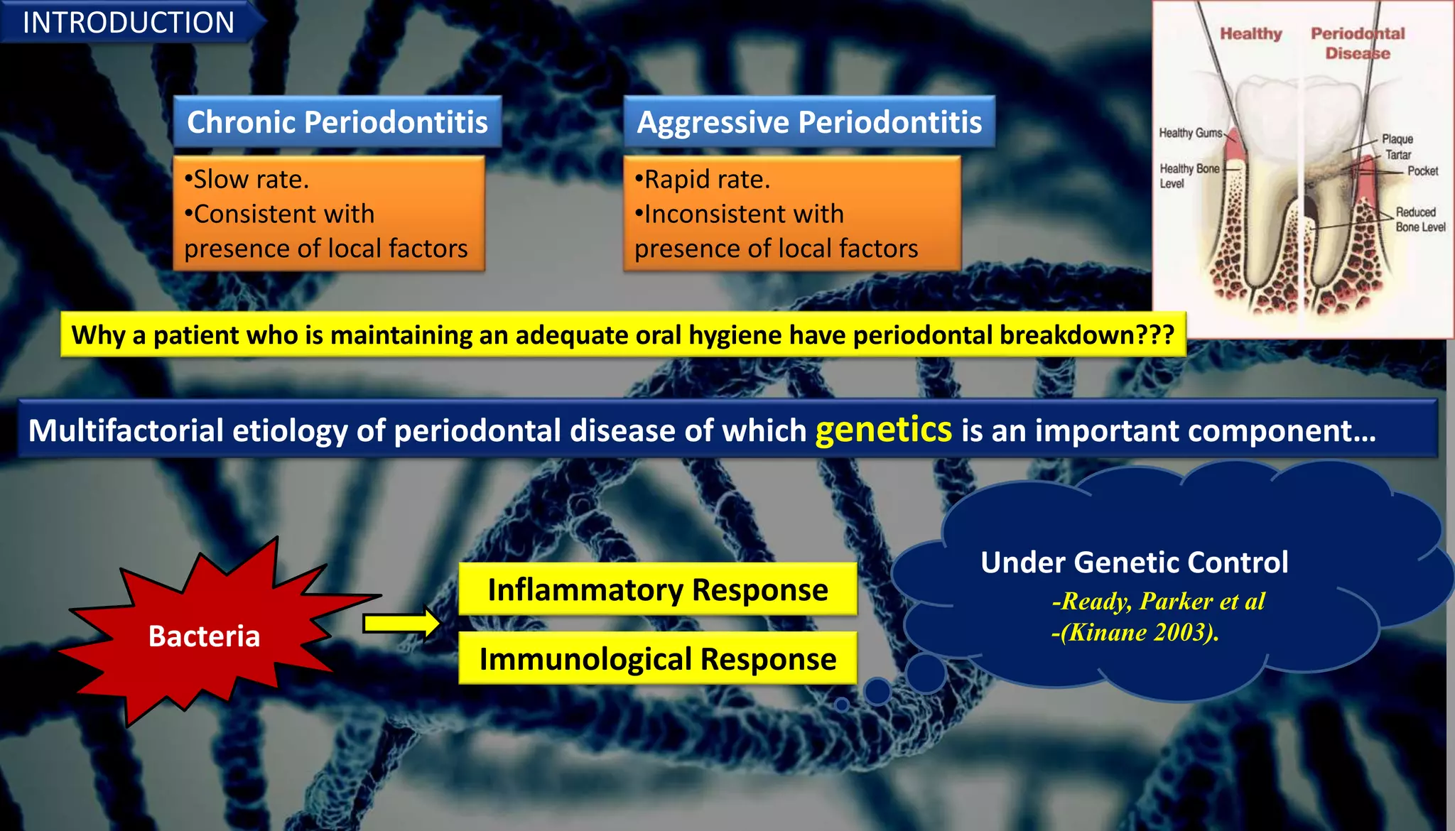
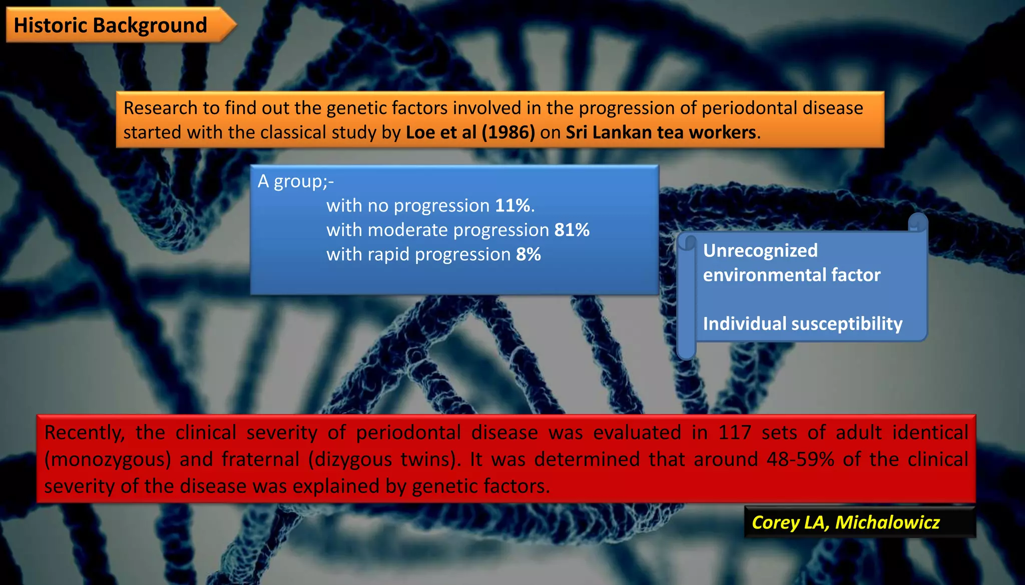
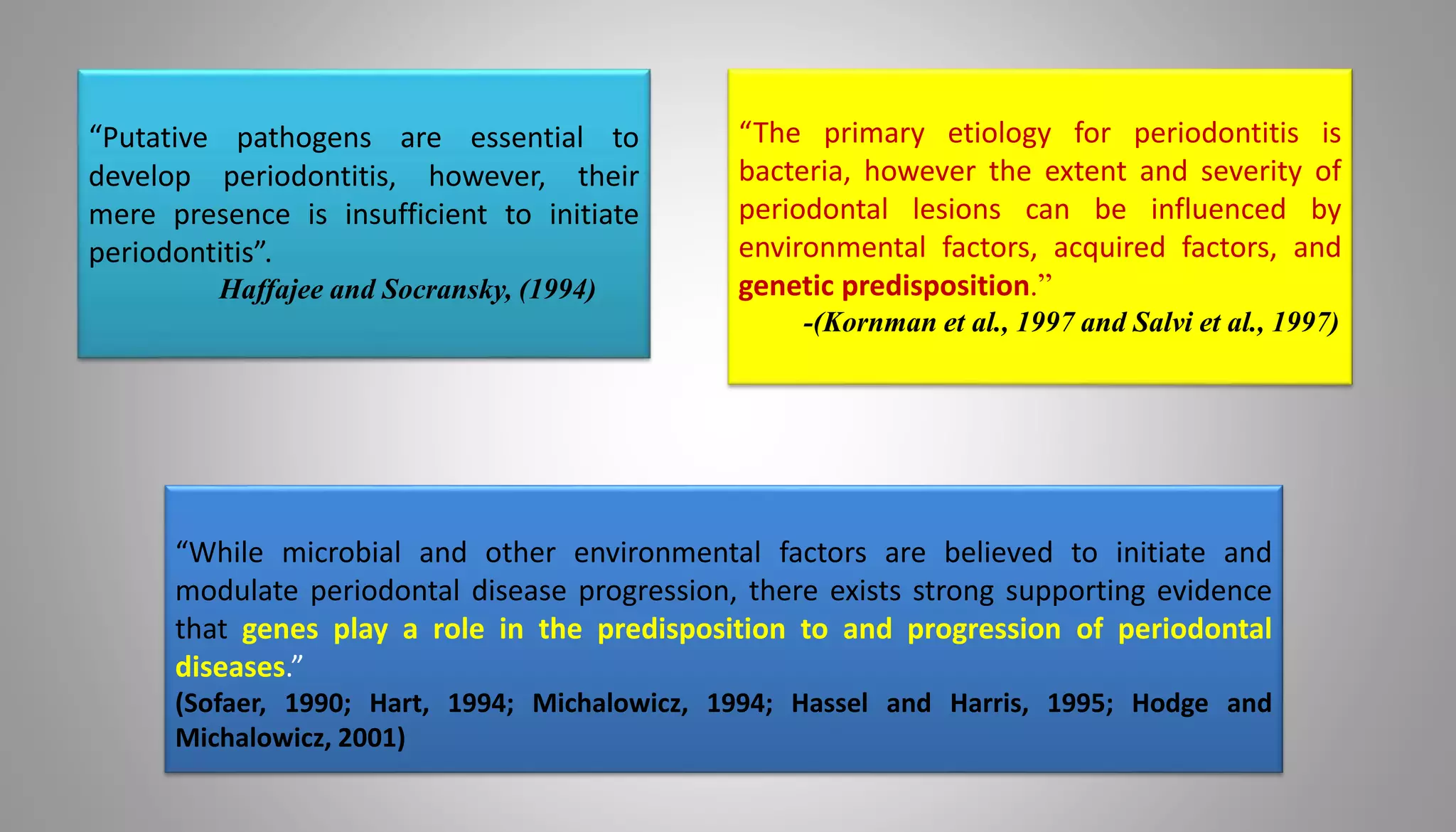
GENETICS IN PERIODONTAL
DISEASES.
Under the esteemed guidance of ;-
Prof. Dr. SUHAIL MAJID JAN (HOD)
Dr. ROOBAL BEHAL (Associate Professor)
By:-
MUMTAZ ALI PG (2nd Year)
‘’DEPARTMENTOF PERIODONTICSANDORAL IMPLANTOLOGY’’
CONTENTS
Introduction.
An insight to genetics.
Genetic basis of disease.
Methods of genetic analysis.
Evidence for the role of genetic variants in Periodontitis.
Genetic and Inherited Disorders associated with Aggressive Periodontitis.
Genetic polymorphism.
Gene therapy in Periodontics.
A futuristic approach to the application of genetic profiles.
Conclusion.
References.
INTRODUCTION
Chronic Periodontitis Aggressive Periodontitis
•Slow rate.
•Consistent with
presence of local factors
•Rapid rate.
•Inconsistent with
presence of local factors
Why a patient who is maintaining an adequate oral hygiene have periodontal breakdown???
Multifactorial etiology of periodontal disease of which genetics is an important component…
Inflammatory Response
Immunological Response
Bacteria
Under Genetic Control
-Ready, Parker et al
-(Kinane 2003).
Bacteria cause periodontal disease, but other factors determine how severe that disease will
become and how specific a patient responds to the therapy.
Environmental
Genetic
Acquired
Page RC &
Kornman KS.
Historic Background
Research to find out the genetic factors involved in the progression of periodontal disease
started with the classical study by Loe et al (1986) on Sri Lankan tea workers.
A group;-
with no progression 11%.
with moderate progression 81%
with rapid progression 8%
Recently, the clinical severity of periodontal disease was evaluated in 117 sets of adult identical
(monozygous) and fraternal (dizygous twins). It was determined that around 48-59% of the clinical
severity of the disease was explained by genetic factors.
Corey LA, Michalowicz
Unrecognized
environmental factor
Individual susceptibility
“While microbial and other environmental factors are believed to initiate and
modulate periodontal disease progression, there exists strong supporting evidence
that genes play a role in the predisposition to and progression of periodontal
diseases.”
(Sofaer, 1990; Hart, 1994; Michalowicz, 1994; Hassel and Harris, 1995; Hodge and
Michalowicz, 2001)
“Putative pathogens are essential to
develop periodontitis, however, their
mere presence is insufficient to initiate
periodontitis”.
Haffajee and Socransky, (1994)
“The primary etiology for periodontitis is
bacteria, however the extent and severity of
periodontal lesions can be influenced by
environmental factors, acquired factors, and
genetic predisposition.”
-(Kornman et al., 1997 and Salvi et al., 1997)
PERIODONTITIS IS A MULTIFACTORIAL DISEASE
 Periodontitis requires specific bacteria for initiation and progression of attachment loss and bone loss.
 There is a ‘normal’ host response to this bacterial challenge.
 Although the normal tissue response is primarily ‘protective’, it also causes tissue destruction.
 Healing and repair are constantly going on in the periodontal tissues.
 In majority individuals with moderate bacterial challenge, protection and repair dominate destruction.
 Some patients have ‘altered’ host responses and develop more severe destruction.
 Altered host responses may develop from
a) heavy bacterial challenge and
b) presence of disease modifiers that reduce the protective component of
the host response or amplify the destructive component.
Examples of disease modifiers include: smoking, diabetes and IL-1 genetic variations.
• The factors that determine severity and response to treatment are
often called disease modifiers.
• These, when present in patient, change the trajectory of clinical
disease expression over time.
DISEASE MODIFIERS
EOP: early-onset periodontitis; LAD: leukocyte adhesion deficiency; PMNs: polymorphonuclear
lymphocytes; PGHS prostaglandin endoperoxide synthase (also referred to as cyclooxygenase)
Periodontology 2000. Vol. 14. 1997,202-215
Genetic factors in periodontitis and their potential biological influence.
Shown in red are candidate genetic factors for which there are current data to support a role in periodontitis.
Shown in yellow are candidate genetic factors for which there are data to support a role for the biochemical factors
in periodontitis, but for which are no current datat associating a specific genetic marker with disease.
EOP: early-onset periodontitis; LAD: leukocyte adhesion deficency; PMNs; polymorphonuclear lymphocytes; PGHS prostaglandin
endoperoxide synthase (also referred to as cyclooxygenase).
AN INSIGHT TO GENETICS
Basic Terminologies
Genome –
refers to all the genes carried by an individual or cell. The human genome
consists of more than 3 billion pairs of bases contained in 22 pairs of chromosomes,
termed autosomes, and a pair of sex chromosomes.
Chromosome –
a nuclear structure carrying genetic information arranged in a linear
sequence.
Gene –
a functional and physical unit of inheritance that occupies a specific
position (locus) within chromosome. In other words, it is a sequence of nucleotides
located at a particular position on a particular chromosome carrying a set of
instructions usually directing the synthesis of proteins.
Allele –
one of several possible alternative forms of a given
gene at a particular locus of a chromosome differing in DNA
sequence.
Different alleles are responsible for variation in inherited
characteristics such as hair color or blood type.
In an individual, the dominant form of an allele is expressed.
Homozygous – the presence of identical alleles of one or
more specific genes (e.g. A/A).
Heterozygous – the presence of differing alleles of one
or more specific genes (e.g. A/B).
Genotype – the genetic makeup of an organism or cell distinct from its expressed
features or phenotype.
Phenotype – the observable characteristics displayed by an organism as influenced by
environmental factors and independent of the genotype of the organism. (Phenotype
= genotype x environment)
Gene expression – the process involving use of the information in a gene via
transcription and translation leading to production of a protein affecting the
phenotype of the organism determined by that gene.
Autosomal dominant – the dominant effect of one gene located on an autosome
regardless of the presence of the other normal copy.
Autosomal recessive – A gene on an autosome that is required in two copies to be
active in an individual. An individual who carries two such copies of the same
abnormal gene will be subjected to effects from that gene.
Genetic Variance
Variance : Mutation or Polymorphism
Mutation :
a permanent transmissible change in the genetic material that occur during DNA
replication or meiosis. (<1% of population)
Polymorphism:
a region on the genome that varies between individual members of a population
present in a significant number of individuals. (>1% of population)
That portion of the phenotypic variance of a trait in a population which can be attributed to genetic
difference amongst individual.
• Polymorphism arises as a result of mutation.
• The different types of polymorphisms are typically referred to by the type of mutation that created
them.
1. Single nucleotide polymorphism (SNP): The simplest type of polymorphism results from a single
base mutation which substitutes one nucleotide for another.
•The frequency of SNPs across the human genome is estimated at every 0.3–1 kb
2. Restriction Fragment length polymorphism (RFLP)
Digestion of a piece of DNA containing the relevant site with an appropriate restriction
enzyme could then distinguish alleles or variants based on the resulting fragment sizes via
electrophoresis.
3. Insertion-deletion polymorphism
Results from insertion or deletion of a section of DNA.
Most common type is the existence of variable numbers of repeated base or nucleotide
patterns in a genetic region
It has been widely accepted that the differences among individuals at risk for developing most
diseases have a substantial inherited pattern.
Environment factor
Diet, smoking,preventive
care, exposure to
pathogen
Genetic factor
Interact with each other to determine person’s health outcome.
Determines if and when the disease affects the person, how fast and how severely
symptoms of the disease progress and how the person responds to different treatments
in terms of both side effects and success of alternative therapies.
• Most cases of periodontitis appear to fit this complex genes and environmental model.
• The inherited variation in DNA has a role roughly equal to that of the
environment in determining who remains periodontally healthy versus who is
affected by this disease.
• In genetic studies, over 23,000 genes need to be considered as potential hypothesis or
candidates for being disease risk factors.
DISEASES where GENETIC component of the risk
PREDOMINATES and ENVIRONMENTAL differences
play a MINOR ROLE:
CYSTIC FIBROSIS
MUSCULAR DYSTROPHY
DISEASES where ENVIRONMENTAL component of
the risk PREDOMINATES and GENETIC
COMPONENT play a MINOR ROLE:
INFECTIOUS DISEASES eg. HIV
CANCERS like mesothelioma
(associated with asbestos exposure)
Environmental Exposures
Differences in physiologic functioning of proteins due to polymorphisms
can be enhanced by certain environmental factors(eg. smoking, diabetes,
microbes).
If the protein functions in the inflammatory process then certain
polymorphisms can increase or decrease risk for disease phenotype.
Genetic variance and environmental exposures are the key determinants
to phenotypic differences.
It estimates the portion of all variations in the trait that is attributable to inherited
genetic variations.
• Traits whose variation is determined entirely by differences in environmental exposure
have heritability of 0.0.
• Traits with variation attributable solely to genetic differences have heritability of 1.0
• Heritabilities are sometimes reported as a percentage ranging from 0 to 100%.
HERITABILITY
Most human diseases traits fall in the middle of this range between 0.25 and 0.75.
Example:- Type II diabetes have a heritabililty of 0.26 and abnormal glucose
intolerance has 0.61 in one study.
For periodontal disease, only chronic periodontitis occur frequently enough to have
been studied using the twin design.
Two twin studies of modest size (110 and 117 pairs ) have been reported, and these
estimate heritability of measures of chronic periodontitis ranging between 40% and
80%, thus clearly implicating genetic variation in disease risk.
It is feasible that genes implicated in the regulation of inflammatory process of periodontal
tissues associated with plaque accumulation may play a role in explaining the individual
variability in the severity of both plaque-induced gingivitis and destructive periodontitis
-(Dashash et al. 2007).
Periodontal disease development and progression can be caused by MMPs produced by both
infiltrating and resident cells of the periodontium.
HERITABILITY OF GINGIVITIS
One of the most important MMPs, MMP-9 (also known as
gelatinase B or 92-kD type IV collagenase), is active against
collagens and proteoglycans.
The coding gene is located on chromosome 20q11.2-q13.1, and
several polymorphisms have been detected in the MMP-9 gene
-(Vokurka et al. 2009).
GENES ASSOCIATED WITH GINGIVITIS RISK:
IL-1 cluster, IL-6,10, 12,18
MMP-9
TNF
Fibrinogen
LT-A.
Some types of aggressive periodontitis seem to be inherited in a Mendelian manner,
and both autosomal modes and X-linked transmission have been proposed.
Most of the evidence for a genetic predisposition to aggressive periodontitis comes
from segregation analyses of families with affected individuals in two or more
generations.
The results in different sets of families are consistent with both autosomal-dominant
and autosomal recessive inheritance, as well as X-linked dominant inheritance, but no
single inheritance mode that would include all families has been established
-(Meng, et al)
HERITABILITY OF AGGRESSIVE PERIODONTITIS
GENES ASSOCIATED WITH AP RISK:
• IL-1 cluster, IL-4,6,10,12,13,18
• TNF-a
• TGF-b
• Vit D receptor
• Estrogrn receptor
• RANK/RANKL/OPG,
• MMP 1,2,3,9,
• TIMP
• HLA
• CD14
• Cathepsin C, IFN-gamma. Polymorphisms in genes of
• Cell-surface receptors for immunoglobulins (Fc)
• Formyl-methionyl-leucyl-phenylalanine (FMLP)
• Human leukocytic antigen (HLA)
• Vitamin D
Are promising candidates for susceptibility assessment of
aggressive peridontitis.
-(Yoshie et al. 2007).
In contrast to aggressive periodontitis, chronic periodontitis does not typically
follow a simple pattern of familial transmission or distribution.
The twin study is probably the most popular method that supports the genetic
aspects of chronic periodontitis.
This study substantiates the contribution that genes make vs. the environment
in a phenotypic expression.
HERITABILITY OF CHRONIC PERIODONTITIS
Michalowicz et al. (1991)
examined the relative contribution of environmental and host genetic
factors to clinical measures of periodontal disease through the study of
twins reared together and monozygous twins reared apart.
Heritability estimates indicated that between 38 and 82% of the
population variance for these periodontal measures of disease may be
attributed to genetic factors.
Adult periodontitis was estimated to have approx 50% heritability,
which was unaltered following adjustments for behavioral variables
Corey et al. (1993)
revealed that approximately half of the variance in
disease in the population is attributed to genetic variance.
CANDIDATE GENES FOR CHRONIC PERIODONTITIS
• Interleukin-1, 2, 4, 6, 10
• Fcg receptor
• TNF
• Vitamin D receptor
Methods for studying the genetics of periodontal diseases.
 Familial Aggregation
 Twin Studies
 Segregation Analysis
 Linkage Studies
 Association Studies/ Candidate gene approach
 Genome wide analysis study (GWAS)
Familial Aggregation
Many diseases run in families, and the degree of clustering within the
family can be estimated by comparing the number of disease cases in
relatives of patients to the risk of disease in the general population .
Difficulties : in addition to having many genes in common, family members also share
many aspects of a common environment (e.g., diet, nutrition, smoking, infectious
organisms and shared socioeconomic factors).
Familial Aggregation:
German studies of familial nature in the early 20th
century have shown aggregation of chronic forms of
periodontitis in families. This strongly suggested
genetic predisposition. (Rev’d by Hassell & Harris ,1995)
Twin Studies
Disadvantage :
A genetic mutation may not have complete penetrance.
Environmental conditions may contribute to the development of the disease (e.g., one twin
may smoke and the other may not).
Furthermore, many diseases are polygenic (i.e., caused by alterations in multiple genes).
Studying phenotypic characteristics of twins is a method of differentiating variations due
to environmental and genetic factors.
• Monozygous twins arise from a single fertilized
ovum and are therefore genetically identical and
always the same gender.
•Dizygous twins arise from the fertilization of two
separate ova and share, on average, one half of
their descendent genes in the same way as
siblings do.
• Any discordance in disease between monozygous
twins must be due to environmental factors.
Any discordance between dizygous twins could
arise from environmental and/or genetic variance.
Twin study
Michalowicz et al. (1991) Studied dizygous twins reared together
and apart and monozygous twins reared together and apart.
Mean probing depth and attachment level varied less for
monozygous twins than dizygous twins.
Concluded genetics plays a role in susceptibility to periodontal
disease.
Segregation Analysis
• Statistical analyses of the patterns of transmission of a disease in families in
an attempt to determine the relative likelihood that the disease is caused by a
single gene with dominant or recessive inheritance, by multiple genes, or
entirely by variation in exposure to risk factors.
• The observed proportions of offspring who have the trait or disease being
evaluated (i.e., the phenotype) are compared with the proportions that are
expected in the general population .
Segregation analysis
Marazita et al. (1994).
• Segregation analysis in North American families performed.
• Studied >100 families, segregating aggressive forms of
periodontitis, and found support for autosomal dominant
transmission. Concluded autosomal dominant inheritance
with ~70% penetrance occurred in Blacks and non-Blacks.
• While others Beaty et al. (1987), Long et al. (1987),Saxen et
al. (1980) have found support for autosomal recessive
transmision of aggressive periodontitis.
Linkage analysis
• A technique used to map a gene responsible for a trait to a
specific location on a chromosome.
• It is based on the fact that genes that are located close to each
other on the chromosome tend to be inherited together as a
unit.
• These genes are said to be linked.
• Very expensive DNA markers are required.
A difficulty with linkage analyses is that many diseases are not caused by a single gene of
“major” effect but rather by multiple genes of “minor” effect.
In the later situation, multiple genes each contribute a small amount to the
phenotype/disease/ trait, and the linkage study approach has little power for detection. In those
cases association analysis methods may be used.
Li and coworkers (2004)
reported evidence of a gene responsible for localized
aggressive periodontitis located on chromosome
1q25. To date, a gene of major effect for
aggressive periodontitis has not been identified.
Hart et al. (1993)
evaluated support for linkage of AgP near
chromosome 4 in different population of families
(14 African American and 4 Caucasian). Results
showed that in these populations no linkage
existed .
Boughman et al. (1986)
Gene for Dentinogenesis imperfecta-III (DGI-III)
had been previously localized to chromosome
4. They performed linkage analysis and
showed close linkage of gene for Aggressive
periodontitis( AgP) to this DGI-III gene in the
families of Southern Maryland.
Association Studies
A gene mapping approach that tests whether one allele of a gene occurs more
often in patients with the disease than in subjects without the disease.
Aim : to identify which genes are associated with the disease.
Candidate genes are chosen on the basis of their known or presumed
function (i.e., they have some plausible role in the disease process such as
producing a protein that is important in the disease pathogenesis).
Case control studies.
Genome Wide Association Study (GWAS)
 Entire genome is searched with the aim of identifying genetic associations
related to a trait or disease of interest.
 The completion of the Human Genome Project in 2003 and the
development of microarray technologies capable of assaying SNPs have made
GWAS possible.
 This method has the potential to identify the genetic contributions to
common diseases.
 An important advantage of this approach is, because the entire
genome is analyzed, the technique permits the genetics of a disease to be
investigated in a nonhypothesis-driven way. It is not necessary to correctly guess
which candidate genes are most interesting to evaluate.
A GWAS requires that well -characterized cases and controls
be identified.
A disadvantage of GWAS is:-
that large clinical sample sizes are required to reduce the
likelihood of differences between cases and controls being
observed simply by chance as a result of the hundreds of
thousands of multiple statistical tests required to search
the entire human genome.
GENETIC MARKER
Any type of biomolecule or assay that allows us to read inherited
differences among individuals in their DNA sequences.
Blood groups, protein isozymes, and human leukocyte antigen
(HLA) were the first developed markers.
Recently developed : “next generation” DNA sequencing methods.
PERIODONTITIS IN GENETIC SYNDROMES AND OTHER DISEASES
PAPILLON-LEFEVRE SYNDROME
• Rare autosomal recessive congenital differentiation disorder of
chromosome 11p14-q21.
• Occurs in children from consanguineous marriages.
• Gene responsible: Cathepsin C, lysosomal protease.
- (Toomes et al.1999).
• Cathepsin C is suggested to be implicated in a wide variety of
immune and inflammatory processes
- (Toomes et al. 1999).
• Prevalence : 1-4 per million, equal in males and females.
- (Hattab et al. 1995)
Two essential features of Papillon-Lefèvre syndrome:
Hyperkeratosis of the palms and soles (either diffuse or localized) and Generalized rapid destruction of the
periodontal attachment apparatus resulting in premature loss of both primary and permanent teeth.
- (Deas et al. 2003).
External signs are hyperkeratosis of the palms and
Soles - (Kressin et al. 1995).
Changes in the skin observed by electron microscopy
revealed the diminution of the tonofibrils, alterations
of the keratohyaline granules, and acanthosis in the
Stratum spinosum. - (Kressin et al. 1995).
INTRAORALLY, periodontal symptoms affect
primary and permanent dentitions.
Extensive loss of periodontal attachment
accompanied by generalized, severe, and rapid
destruction of the alveolar bone, that frequently
lead to premature tooth loss.
Histologically, the gingiva demonstrates epithelial
hyperplasia, increased collagen synthesis,
parakeratosis, acanthosis, and focal aggregates of
lymphocytes and plasma cells.
In addition, reduced osteoblastic activity and reduced
thickness of cementum have been described
- (Ghaffer et al. 1999; Hattab et al. 1995).
Virulent gram-negative anaerobic microbiota has been considered to be an important
initiator of the destructive periodontitis observed in these patients.
• Aggregatibacter actinomycetemcomitans has been reported to be the major
periodontal pathogen.
• Capnocytophaga gingivalis, Eikenella corrodens, black-pigmented Bacteroides, and
Fusobacterium spp : subgingival periodontal lesions in Papillon-Lefèvre syndrome
patient.
(Ishikawa et al. 1994; Lundgren et al. 1998; Rudiger and Berglundh 1999; Velazco et
al. 1999).
Papillon-Lefèvre syndrome has been associated with
• decreased neutrophil chemotaxis
• reduced random neutrophil migration
• impaired neutrophil phagocytosis
• reduced myeloperoxidase activity
• increased superoxide radial neutrophil production,
• associated with a decreased lymphocyte response to
pathogens .
- (Lundgren et al. 1998; Velazco et al. 1999).
EHLER DANLOS SYNDROME
Also known as dystrophia mesodermalis and fibrodysplasia elastica generalisatica.
Heterogenous group of inherited disorders of connective tissue, which may affect the
skin, ligaments, joints, eyes, and vascular system
- (Reichert et al. 1999).
EDS is divided into 11 types in accordance with clinical, genetic, and biochemical
features
- (Majorana and Facchetti 1992).
The primary cause may be a
Type I or type II collagen deficiency,
Lysyl hydroxylase deficiency
Deletion of N-telopeptide, or
Disorders of copper homeostasis and fibronectin defects
- (Reichert et al. 1999).
Immunological analysis revealed that the concentrations of IgA
in saliva and IgA, IgG, and IgM in serum were within normal
limits, with unpaired phagocytic capacity of the peripheral
blood leukocytes
- (Reichert et al. 1999).
The temporomandibular joint often demonstrates profound
laxity in conjunction with generalized joint mobility and
dislocation - (Fridrich et al. 1990).
Periodontal conditions have been reported with EDS types I,
VII, and VIII.
Defective dentinogenesis, resulting in aplasia or hypoplasia
of root development affecting the mandibular incisors, and
predisposition for localized periodontal disease was
reported in EDS type I.
Radiographic appearance of a bulbous enlargement of the
roots together with pulp stones at other teeth were
reported.
EDS type VII is an autosomal dominant/ recessive disease.
• Poor healing after extractions
• Prevelance of dental caries
• A radiographic evidence of pulp stones, and/ or
• malformed teeth have been described.
EDS type VIII is an autosomal dominant form
• Fragile skin
• Abnormal scarring
• Early onset of periodontal disease, with
premature
• loss of the permanent teeth.
• Fragility of the alveolar mucosa and increased
bleeding tendencies have also been suggested
EDS can also be associated with:-
Ligneous periodontitis (generalized membranous gingival enlargement
due to an accumulation of fibrin deposits associated with severe alveolar
bone loss)
Plasminogen deficiency seems to play a central role in its pathogenesis.
CHEDIAK HIGACHI SYNDROME
It is a rare autosomal recessive disease associated with impaired function of
cytoplasmic microtubules or microtubule assembly in PMNs.
- (Oh et al. 2002).
The susceptibility to infections, although humoral and cellular immunity are
normal, leads to early death (often before 5 years of age).
• The disease reveals itself periodontally by Severe
gingivitis and
•Rapid loss of attachment, leading to exfoliation of the
teeth.
• Two LADs have been described in humans: LAD I and LAD II.
• Both diseases block a sequence of leukocyte– endothelial-cell
interactions, which is generally referred to as the multistep adhesion
cascade.
LAD is a rare but well-defined autosomal recessive disease that results in
the formation of nonfunctional intracellular adhesion molecule (ICAM
receptor).
The disease results from mutations in the region on the CD18 gene
encoded on chromosome 21q22.3, which codes for the b2 integrin
subunit of the leukocyte adhesion molecule.
The defective or absent expression of these molecules on the surface
of leukocytes decreases their ability to adhere to endothelial cells
and to migrate to sites of infection.
LEUKOCYTE ADHESION DEFICIENCY
Clinical features usually present in infancy or early childhood and consist of
Recurrent, indolent bacterial infections of the skin, mouth, and the
respiratory tract.
Delayed separation of the umbilical cord.
Skin infections may progress to large chronic ulcers that may become
polymicrobial in character.
These individuals are susceptible to fungal
infections but do not show an increased
susceptibility to protozoal or viral infections.
In addition, lack of swelling, redness, heat, or pus
is noted in the area of the infection
Severe gingivitis with an early loss of primary teeth,
followed by the early loss of secondary teeth, is
seen.
Individuals with LAD defect in innate host defense
display a severe form of periodontitis that does not
require specific periodontal pathogens because of
entrapment of neutrophils within the blood vessel.
Genetic Polymorphism
In humans, studies of inherited variations in the immune system are
necessarily complex, and the observed phenotype is usually the
result of multiple genetic and environmental influences.
It is likely that genetic polymorphisms exist for many
of these immunological factors, eg.
- Immunoglobulin G2 production
- FcγRII receptor heterogeneity
- Mediators of inflammation
-Prostaglandin E2 (PgE2)
-Interleukin 1 (Il-1)
Immunoglobulin G2 production
Serum IgG2 levels in localized aggressive
periodontitis(LAP) cases are higher than serum levels
of generalized aggressive periodontitis(GAP) cases
-(Lu et al. 1994)
IgG2 in LAP associated with protection in LAP
-(Gunsolley et al.1987)
The IgG molecules contain :
- gamma heavy chains (Gm allotypes)
- kappa light chains (Km allotypes)
Currently the only allotype identified for IgG2 is G2m(23)
or G2m(n).
• The Gm allotype genes, or genes in linkage equilibrium with
them, appear to influence expression of the IgG2 molecule.
• This response appears to be race specific, and young
Caucasians of the low-responder phenotype G2m(null)
[G2m("), G2m(-n) and G2m(-23)]are predisposed to specific
bacterial infections.
Choi et al. (1996) :
The rapidly progressive periodontitis patients who
were positive for the G2m(23) allotype had
elevated antibody to Porphyromonas gingivalis .
In addition to host factors, environmental factors are
capable of modulating IgG2 response. Cigarette
smoking is known to reduce serum IgG levels.
FcγRII receptor heterogeneity
• Polymorphisms in Fc receptors expressed on the surface of phagocytic
cells have been shown to be important determinants of susceptibility to
infections.
• The immunoglobulin Fc receptor II genes have been mapped to
chromosome 1.
• FcyRII gene has two expressed alleles, which differ significantly in their
ability to bind human IgG2.
• The two alleles differ by the amino acid, at position 131.
- arginine (R131)
- histidine (H131), recognizes IgG2, and the maximum
IgG2 binding is found in the homozygous H131 state.
Wilson et al. (1996) :
IgG2 was significantly more effective in mediating phagocytosis of
A. actinomycetemcomitans, when used with human
neutrophils that were homozygous for the H131 receptor as
compared to neutrophils from individuals homozygous for the
R131 receptor.
The genetic polymorphism that defines the FcyRII receptor,
therefore, appears to be a promising marker for susceptibility
to localized aggressive periodontitis.
Mediators of inflammation
PROSTAGLANDIN E2(PGE2)
- Potent biological mediators
- Diverse physiological effects
- Has also been implicated in a variety of pathological conditions
including periodontitis.
Wang et al. (1996) :
identified linkage of the chromosome 9q32-33 region
with early-onset periodontitis.
This physical region includes the gene for
prostaglandin endoperoxide synthase 1, and this
observation encourages further studies
associating genetic markers of prostaglandin E2
with clinical periodontitis.
INTERLEUKIN 1 (IL-1)
Elevated tissue and gingival fluid levels of interleukin 1β (ILβ)
in particular have been repeatedly associated with periodontitis.
A family of three IL-1 genes cluster on chromosome 2q13.
At times the overproduction of immuno-regulatory mediators
may actually prove harmful to the host.
Genetic polymorphisms in regulatory regions of genes responsible
for inflammatory mediators may act to create interindividual
differences in such responses.
Genetic control of IL-1: Genes and Locus of SNPs associated with controlling IL-1 biological
activity
Genetic Susceptibility Test for periodontitis:
Tests for the presence of at least one copy of allele 2 at the IL-1A +4845 loci and at least one copy
of allele 2 at the IL-1B +3954 locus.
*IL-1A +4845 is being used because it is easier to identify than IL-1A -889 and it is essentially concordant with it.
** IL-1B +3953 has been now renumbered as IL-1B +3954 because the current convention indicates that the
numbering of the transcription should begin at +1 instead of zero.
Genes Polymorphism
Locus
Current Locus accessed
with test
Controlled product
IL-1α Allele2 -889 Allele 2 IL-1α +4845* IL-1α
IL-1β Allele 2 +3953 Allele 2IL-1β +3954** IL-1β
IL-1RN Protein receptors
antagonist (impedes
IL-1α & β)
In 1997, Kornman et al., found an association between
polymorphisms in the genes encoding for interleukin1α(+889)
and interleukin-1β(+ 3953) (termed the ‘composite genotype’)
and an increased severity of periodontitis.
One of these genotypic polymorphisms IL-1β at (+3953) is
associated with a fourfold increase in IL- lβ production.
GENETICS IN PERIODONTAL
DISEASES.
Under the esteemed guidance of ;-
Prof. Dr. SUHAIL MAJID JAN (HOD)
Dr. ROOBAL BEHAL (Associate Professor)
By:-
MUMTAZ ALI PG (2nd Year)
‘’DEPARTMENTOF PERIODONTICSANDORAL IMPLANTOLOGY’’
INHERETED GENETIC DISORDERS
Downs Syndrome
It is named after John Langdon Down , a British
physician who described the syndrome in 1866.
It was identified as trisomy of 21 chromosome by
Dr Jerome Lejuene in 1959.
•95% of cases extra chromosome is of maternal origin
•4% is present as a translocation from long arm of chromosome 21
to 22 or 14(Robertsonian translocation )
•1% mosaic ,mixture of 46 & 47 chromosome
These findings are usually generalized, although they tend to be more severe
in the lower anterior region; marked recession is also sometimes seen in this
region, apparently associated with high frenum attachment.
The disease progresses rapidly.
Acute necrotizing lesions are a frequent finding.
Higher incidence (96%) of periodontal disease (misaligned teeth contribute to
this secondarily) .
Current research suggest that reduced neutrophil and monocyte chemotaxis,
reduced phagocytosis, and a defect in T-cell proliferation and maturity may be
reasons for the increase in periodontal disease seen in these patients
(Lang et al 1977).
Periodontal disease in Down syndrome is characterized by formation of deep
periodontal pockets associated with substantial plaque accumulation and
moderate gingivitis.
Two factors have been proposed to explain the high prevalence and
increased severity of periodontal destruction associated with Down
syndrome:-
-a reduced resistance to infections because of poor circulation,
especially in areas of terminal vascularization such as the gingival
tissue and
-a defect in T-cell maturation and polymorphonuclear leukocyte
chemotaxis.
Increased numbers of P. intermedia have been reported in the
mouths of children with Down syndrome.
Other clinical features:
Enamel hypocalcificiation and hypoplasia common.
DS patients 50% more likely to have congenitally missing teeth,
taurodonts are frequent finding.
1/3 more caries resistant than their non-DS siblings
Gingivitis develops earlier and more rapidly and extensively in
persons with DS, perhaps because of an abnormality in host
defenses.
Patients with DS have altered microbiological composition of
subgingival plaque, including increased Actinomyces and
Hemophilus strains
Hyper-IgE (Job’s Syndrome/Hill-quie)
Marked elevation of IgE.
Defect has been possibly localized to chromosome 7q21.
Clinically characterized;-
Chronic dermatitis.
The patient has serious, lifelong bouts of recurrent infections by
opportunistic organisms which results in skin abscess, remarkable in
their lack of erythema (cold abccess).
Oral and periodontal findings are Periodontitis with Oral ulceration.
Weary-Kindler syndrome:
Also known as hereditary acrokeratotic piokiloderma.
Rare congenital disease of the skin caused by a mutation in the KIND1
gene.
Is an autosomal recessive genodermatosis.
KIND1 gene codes for protein kindlin-1 , which is thought to be active
in the interactions between actin and the extracellular matrix.
Tendency to blister with minor trauma and photosensitivity (decreases
with age).
Manifestations of both epidermolysis bullosa and piokiloderma
congenitale.
Periodontal manifestation include early onset periodontitis.
Lazy leukocyte syndrome: A severe form of neutropenia .
Lazy leukocyte syndrome a syndrome in children, marked by
recurrent low-grade infections with a defect in neutrophil
chemotaxis and deficient random mobility of neutrophils.
Inherited as a autosomal dominant characteristic.
Severe neutropenia
Recurring infection
Increased risk of serious infections
Scanning electron microscopy of the patient's neutrophils showed
alteration in the surface configuration of the cell with coarsening of the
normal fine ruffles and the appearance of knob-like projections.
The lazy leucocyte syndrome may be a consequence of altered membrane
microfilamentous protein structure or function, and undue rigidity of the
affected neutrophils may explain the clinicopathological features of the
disease.
Intracellular killing and phagocytosis is intact.
In addition to systemic signs and symptoms such as
High fever, cough, pneumonia, and purulent skin abscesses.
Oral manifestations include
painful stomatitis,
gingivitis,
recurrent ulcerations of the buccal mucosa and tongue,
rapidly progressive bone loss, and tooth loss at an early age.
CYCLIC NEUTROPENIA
Rare condition, characterized by cyclical depletion
of polymorphonuclear leukocyte numbers,
typically in 3-week cycles, although this can be
between 2 and 5 weeks.
Episode of neutropenia is usually short, but the
patient polymorphonuclear leukocyte count never
returns to normal levels, and the differential
blood-cell count for polymorphonuclear
leukocytes is at least 40% less than normal levels.
Periodontal manifestations include
Inflamed gingiva
Gingival ulceration
Periodontal attachment, and bone loss (Kinane
1999; Rezaei et al. 2004).
Kostmann syndrome/severe congenital neutropenia
Is a rare autosomal recessive form of severe chronic neutropenia
usually detected soon after birth.
An absolute neutrophil count (ANC) chronically less than 500/mm3
is the main sign of Kostmann's.
Children suffer frequent infections from bacteria which in the past
led to death in three-quarters of cases before 3 years of age.
increased risk of developing acute myelogenous leukemia or
myelodysplasia.
A characteristic finding in Kostmann syndrome, a typical very serious
periodontal pathology, which is similar to the prepubertal periodontitis
in deciduous dentition.
At the age of 19 years the patient showed a dramatic compromise of the
masticatory function.
A periodontal prophylaxis, since the very first observations, followed by a
rigorous maintenance with frequent and regular professional hygienic
treatments could be effective in controlling the effects of periodontal
disease.
Other clinical features :
Recurrent infection
Necrotic ulceration of oral mucosa
Necrotic ulceration of genital mucosa
Liver congestion
Spleen congestion
Death due to severe infection often by 3 years of
age
FELTY'S SYNDROME
Felty's syndrome, is characterized by the combination of rheumatoid
arthritis, splenomegaly and neutropenia
The cause of Felty's syndrome is unknown.
It is more common in people who have had rheumatoid arthritis for a long
time.
People with this syndrome are at risk of infection because they have a low
white blood cell count.
wide spread and early periodontal tissue destruction[aggressive
periodontitis].
Other clinical features :
General feeling of discomfort (malaise)
Fatigue
Loss of appetite
Unintentional weight loss
Pale-looking skin
Joint swelling, stiffness, pain, and deformity
Recurrent infections
Eye burning or discharge
HYPOPHOSPHATASIA
Rare, and sometimes fatal metabolic bone disease.
Clinical symptoms are heterogeneous ranging from the rapidly fatal perinatal
variant, with profound skeletal hypomineralization and respiratory
compromise to a milder, progressive osteomalacia later in life.
Tissue non-specific alkaline phosphatase (TNSALP) deficiency in
osteoblasts and chondrocytes impairs bone mineralization, leading to rickets
or osteomalacia.
TNSALP is an ectoenzyme attached to the outer surface of osteoblast and
chondrocyte cell membranes. TNSALP normally hydrolyzes several substances,
including inorganic pyrophosphate (PPi) and pyridoxal 5’-phosphate (PLP) a major
form of vitamin B6.
 The pathognomonic finding is subnormal serum activity of the
TNSALP enzyme, which is caused by one of 200 genetic mutations
identified to date in the gene encoding TNSALP. Genetic inheritance is
autosomal recessive for the perinatal and infantile forms but either
autosomal recessive or autosomal dominant in milder forms.
In childhood Hypophosphatasia’s clinical expression is extremely variable.
As a result of aplasia, hypoplasia, or dysplasia of dental cementum,
premature loss of deciduous teeth (i.e. before the age of 5) occurs.
Frequently, incisors are shed first; occasionally almost the entire primary
dentition is exfoliated prematurely.
Dental radiographs sometimes show the enlarged pulp chambers and root
canals characteristic of the “shell teeth” of rickets.
Gene Therapy
Gene therapy uses purified preparations of a gene or a fraction of a gene, to treat diseases.
There are four approaches:
1. A normal gene inserted to compensate for a nonfunctional gene.
2. An abnormal gene swapped for a normal gene.
3. An abnormal gene repaired through selective reverse mutation.
4. Change the regulation of gene pairs.
Gene therapy in Periodontics
3.Gene- delivery approach:
-In vivo gene delivery: The genetic material is transferred directly
into the body of the patient.
- Ex vivo gene delivery: The genetic material is first transferred into
the cells grown in vitro and then to the patients body.
1. Protein based approach: trials have been conducted using Transforming growth
factor-β, Bone morphogenetic ptoteins-2,6,7,12, Vascular endothelial growth
factor and Platelet derived growth factor.
2.Cell based approach: skeletal muscle derived cells can be used for delivery
of BMP-2.
• The role of host gene in the etiology and pathogenesis of the periodontal disease is just
beginning to be understood.
•The discovery of the association between polymorphisms in IL-1 genes and severity of
periodontitis has been described as a major breakthrough in clinical practice, and the observed
increase in risk due to this single genetic factor has been confirmed in a population of “typical”
dental practice patients.
• The clinical test for such a genetic risk has been proposed as a component of the risk
assessment profile for chronic periodontitis that can be used to provide a rationale for explaining
individual patient susceptibility, providing early preventive or therapeutic intervention, and
allowing superior prognostic capabilities. - (Schenkein 2002).
CLINICAL UTILITY OF GENETIC KNOWLEDGE
FUTURE OF GENETIC STUDIES
• A lot of research has been done to identify the genetic basis of periodontal diseases.
The understanding of the genetics and periodontal disease progression has
provided us valuable information for the identification of disease markers.
• Identification of candidate genes and their use as periodontal disease markers is the
potential clinical application of research done so far in this field. These biomarkers
may be helpful in identifying patients with enhance disease susceptibility and
associated systemic conditions, identifying site with active disease and predicting
sites that may have active disease in the future.
• Periodontal diseases are multifactorial diseases.
• Genetics is an important part of these factors.
• Present knowledge suggests that genetic polymorphism is associated with
the pathogensis of periodontal diseases.
• Gentic factors, along with environmental factors are strongly associated with
the development and progression of periodontal diseases. Identification of
specific genes and genetic variants, aids in diagnosis and treatment of
aggressive periodontal disease.
• In future, a lot of research is required in this direction to investigate different
aspects of the genetic basis periodontal disease.
CONCLUSION
It is likely that a few genetic variations with a high
prevelance in the population will be found to
influence chronic periodontitis.
Currently, the presence if IL-1 gene variations
appear to identify individuals who are at increased
risk formore severe disease and for less predictable
response to therapy.
Thank You

GENETICS IN PERIODONTICS SEMIAR..pptx

  • 1.
    GENETICS IN PERIODONTAL DISEASES. Underthe esteemed guidance of ;- Prof. Dr. SUHAIL MAJID JAN (HOD) Dr. ROOBAL BEHAL (Associate Professor) By:- MUMTAZ ALI PG (2nd Year) ‘’DEPARTMENTOF PERIODONTICSANDORAL IMPLANTOLOGY’’
  • 2.
    CONTENTS Introduction. An insight togenetics. Genetic basis of disease. Methods of genetic analysis. Evidence for the role of genetic variants in Periodontitis. Genetic and Inherited Disorders associated with Aggressive Periodontitis. Genetic polymorphism. Gene therapy in Periodontics. A futuristic approach to the application of genetic profiles. Conclusion. References.
  • 3.
    INTRODUCTION Chronic Periodontitis AggressivePeriodontitis •Slow rate. •Consistent with presence of local factors •Rapid rate. •Inconsistent with presence of local factors Why a patient who is maintaining an adequate oral hygiene have periodontal breakdown??? Multifactorial etiology of periodontal disease of which genetics is an important component… Inflammatory Response Immunological Response Bacteria Under Genetic Control -Ready, Parker et al -(Kinane 2003).
  • 4.
    Bacteria cause periodontaldisease, but other factors determine how severe that disease will become and how specific a patient responds to the therapy. Environmental Genetic Acquired Page RC & Kornman KS.
  • 5.
    Historic Background Research tofind out the genetic factors involved in the progression of periodontal disease started with the classical study by Loe et al (1986) on Sri Lankan tea workers. A group;- with no progression 11%. with moderate progression 81% with rapid progression 8% Recently, the clinical severity of periodontal disease was evaluated in 117 sets of adult identical (monozygous) and fraternal (dizygous twins). It was determined that around 48-59% of the clinical severity of the disease was explained by genetic factors. Corey LA, Michalowicz Unrecognized environmental factor Individual susceptibility
  • 6.
    “While microbial andother environmental factors are believed to initiate and modulate periodontal disease progression, there exists strong supporting evidence that genes play a role in the predisposition to and progression of periodontal diseases.” (Sofaer, 1990; Hart, 1994; Michalowicz, 1994; Hassel and Harris, 1995; Hodge and Michalowicz, 2001) “Putative pathogens are essential to develop periodontitis, however, their mere presence is insufficient to initiate periodontitis”. Haffajee and Socransky, (1994) “The primary etiology for periodontitis is bacteria, however the extent and severity of periodontal lesions can be influenced by environmental factors, acquired factors, and genetic predisposition.” -(Kornman et al., 1997 and Salvi et al., 1997)
  • 7.
    PERIODONTITIS IS AMULTIFACTORIAL DISEASE  Periodontitis requires specific bacteria for initiation and progression of attachment loss and bone loss.  There is a ‘normal’ host response to this bacterial challenge.  Although the normal tissue response is primarily ‘protective’, it also causes tissue destruction.  Healing and repair are constantly going on in the periodontal tissues.  In majority individuals with moderate bacterial challenge, protection and repair dominate destruction.  Some patients have ‘altered’ host responses and develop more severe destruction.  Altered host responses may develop from a) heavy bacterial challenge and b) presence of disease modifiers that reduce the protective component of the host response or amplify the destructive component. Examples of disease modifiers include: smoking, diabetes and IL-1 genetic variations.
  • 8.
    • The factorsthat determine severity and response to treatment are often called disease modifiers. • These, when present in patient, change the trajectory of clinical disease expression over time. DISEASE MODIFIERS
  • 9.
    EOP: early-onset periodontitis;LAD: leukocyte adhesion deficiency; PMNs: polymorphonuclear lymphocytes; PGHS prostaglandin endoperoxide synthase (also referred to as cyclooxygenase) Periodontology 2000. Vol. 14. 1997,202-215 Genetic factors in periodontitis and their potential biological influence. Shown in red are candidate genetic factors for which there are current data to support a role in periodontitis. Shown in yellow are candidate genetic factors for which there are data to support a role for the biochemical factors in periodontitis, but for which are no current datat associating a specific genetic marker with disease. EOP: early-onset periodontitis; LAD: leukocyte adhesion deficency; PMNs; polymorphonuclear lymphocytes; PGHS prostaglandin endoperoxide synthase (also referred to as cyclooxygenase).
  • 10.
    AN INSIGHT TOGENETICS
  • 11.
    Basic Terminologies Genome – refersto all the genes carried by an individual or cell. The human genome consists of more than 3 billion pairs of bases contained in 22 pairs of chromosomes, termed autosomes, and a pair of sex chromosomes. Chromosome – a nuclear structure carrying genetic information arranged in a linear sequence. Gene – a functional and physical unit of inheritance that occupies a specific position (locus) within chromosome. In other words, it is a sequence of nucleotides located at a particular position on a particular chromosome carrying a set of instructions usually directing the synthesis of proteins.
  • 12.
    Allele – one ofseveral possible alternative forms of a given gene at a particular locus of a chromosome differing in DNA sequence. Different alleles are responsible for variation in inherited characteristics such as hair color or blood type. In an individual, the dominant form of an allele is expressed. Homozygous – the presence of identical alleles of one or more specific genes (e.g. A/A). Heterozygous – the presence of differing alleles of one or more specific genes (e.g. A/B).
  • 13.
    Genotype – thegenetic makeup of an organism or cell distinct from its expressed features or phenotype. Phenotype – the observable characteristics displayed by an organism as influenced by environmental factors and independent of the genotype of the organism. (Phenotype = genotype x environment) Gene expression – the process involving use of the information in a gene via transcription and translation leading to production of a protein affecting the phenotype of the organism determined by that gene. Autosomal dominant – the dominant effect of one gene located on an autosome regardless of the presence of the other normal copy. Autosomal recessive – A gene on an autosome that is required in two copies to be active in an individual. An individual who carries two such copies of the same abnormal gene will be subjected to effects from that gene.
  • 14.
    Genetic Variance Variance :Mutation or Polymorphism Mutation : a permanent transmissible change in the genetic material that occur during DNA replication or meiosis. (<1% of population) Polymorphism: a region on the genome that varies between individual members of a population present in a significant number of individuals. (>1% of population) That portion of the phenotypic variance of a trait in a population which can be attributed to genetic difference amongst individual.
  • 15.
    • Polymorphism arisesas a result of mutation. • The different types of polymorphisms are typically referred to by the type of mutation that created them. 1. Single nucleotide polymorphism (SNP): The simplest type of polymorphism results from a single base mutation which substitutes one nucleotide for another. •The frequency of SNPs across the human genome is estimated at every 0.3–1 kb 2. Restriction Fragment length polymorphism (RFLP) Digestion of a piece of DNA containing the relevant site with an appropriate restriction enzyme could then distinguish alleles or variants based on the resulting fragment sizes via electrophoresis. 3. Insertion-deletion polymorphism Results from insertion or deletion of a section of DNA. Most common type is the existence of variable numbers of repeated base or nucleotide patterns in a genetic region
  • 16.
    It has beenwidely accepted that the differences among individuals at risk for developing most diseases have a substantial inherited pattern. Environment factor Diet, smoking,preventive care, exposure to pathogen Genetic factor Interact with each other to determine person’s health outcome. Determines if and when the disease affects the person, how fast and how severely symptoms of the disease progress and how the person responds to different treatments in terms of both side effects and success of alternative therapies.
  • 17.
    • Most casesof periodontitis appear to fit this complex genes and environmental model. • The inherited variation in DNA has a role roughly equal to that of the environment in determining who remains periodontally healthy versus who is affected by this disease. • In genetic studies, over 23,000 genes need to be considered as potential hypothesis or candidates for being disease risk factors.
  • 18.
    DISEASES where GENETICcomponent of the risk PREDOMINATES and ENVIRONMENTAL differences play a MINOR ROLE: CYSTIC FIBROSIS MUSCULAR DYSTROPHY DISEASES where ENVIRONMENTAL component of the risk PREDOMINATES and GENETIC COMPONENT play a MINOR ROLE: INFECTIOUS DISEASES eg. HIV CANCERS like mesothelioma (associated with asbestos exposure)
  • 19.
    Environmental Exposures Differences inphysiologic functioning of proteins due to polymorphisms can be enhanced by certain environmental factors(eg. smoking, diabetes, microbes). If the protein functions in the inflammatory process then certain polymorphisms can increase or decrease risk for disease phenotype. Genetic variance and environmental exposures are the key determinants to phenotypic differences.
  • 20.
    It estimates theportion of all variations in the trait that is attributable to inherited genetic variations. • Traits whose variation is determined entirely by differences in environmental exposure have heritability of 0.0. • Traits with variation attributable solely to genetic differences have heritability of 1.0 • Heritabilities are sometimes reported as a percentage ranging from 0 to 100%. HERITABILITY Most human diseases traits fall in the middle of this range between 0.25 and 0.75.
  • 21.
    Example:- Type IIdiabetes have a heritabililty of 0.26 and abnormal glucose intolerance has 0.61 in one study. For periodontal disease, only chronic periodontitis occur frequently enough to have been studied using the twin design. Two twin studies of modest size (110 and 117 pairs ) have been reported, and these estimate heritability of measures of chronic periodontitis ranging between 40% and 80%, thus clearly implicating genetic variation in disease risk.
  • 22.
    It is feasiblethat genes implicated in the regulation of inflammatory process of periodontal tissues associated with plaque accumulation may play a role in explaining the individual variability in the severity of both plaque-induced gingivitis and destructive periodontitis -(Dashash et al. 2007). Periodontal disease development and progression can be caused by MMPs produced by both infiltrating and resident cells of the periodontium. HERITABILITY OF GINGIVITIS
  • 23.
    One of themost important MMPs, MMP-9 (also known as gelatinase B or 92-kD type IV collagenase), is active against collagens and proteoglycans. The coding gene is located on chromosome 20q11.2-q13.1, and several polymorphisms have been detected in the MMP-9 gene -(Vokurka et al. 2009). GENES ASSOCIATED WITH GINGIVITIS RISK: IL-1 cluster, IL-6,10, 12,18 MMP-9 TNF Fibrinogen LT-A.
  • 24.
    Some types ofaggressive periodontitis seem to be inherited in a Mendelian manner, and both autosomal modes and X-linked transmission have been proposed. Most of the evidence for a genetic predisposition to aggressive periodontitis comes from segregation analyses of families with affected individuals in two or more generations. The results in different sets of families are consistent with both autosomal-dominant and autosomal recessive inheritance, as well as X-linked dominant inheritance, but no single inheritance mode that would include all families has been established -(Meng, et al) HERITABILITY OF AGGRESSIVE PERIODONTITIS
  • 25.
    GENES ASSOCIATED WITHAP RISK: • IL-1 cluster, IL-4,6,10,12,13,18 • TNF-a • TGF-b • Vit D receptor • Estrogrn receptor • RANK/RANKL/OPG, • MMP 1,2,3,9, • TIMP • HLA • CD14 • Cathepsin C, IFN-gamma. Polymorphisms in genes of • Cell-surface receptors for immunoglobulins (Fc) • Formyl-methionyl-leucyl-phenylalanine (FMLP) • Human leukocytic antigen (HLA) • Vitamin D Are promising candidates for susceptibility assessment of aggressive peridontitis. -(Yoshie et al. 2007).
  • 26.
    In contrast toaggressive periodontitis, chronic periodontitis does not typically follow a simple pattern of familial transmission or distribution. The twin study is probably the most popular method that supports the genetic aspects of chronic periodontitis. This study substantiates the contribution that genes make vs. the environment in a phenotypic expression. HERITABILITY OF CHRONIC PERIODONTITIS
  • 27.
    Michalowicz et al.(1991) examined the relative contribution of environmental and host genetic factors to clinical measures of periodontal disease through the study of twins reared together and monozygous twins reared apart. Heritability estimates indicated that between 38 and 82% of the population variance for these periodontal measures of disease may be attributed to genetic factors. Adult periodontitis was estimated to have approx 50% heritability, which was unaltered following adjustments for behavioral variables
  • 28.
    Corey et al.(1993) revealed that approximately half of the variance in disease in the population is attributed to genetic variance. CANDIDATE GENES FOR CHRONIC PERIODONTITIS • Interleukin-1, 2, 4, 6, 10 • Fcg receptor • TNF • Vitamin D receptor
  • 29.
    Methods for studyingthe genetics of periodontal diseases.  Familial Aggregation  Twin Studies  Segregation Analysis  Linkage Studies  Association Studies/ Candidate gene approach  Genome wide analysis study (GWAS)
  • 30.
    Familial Aggregation Many diseasesrun in families, and the degree of clustering within the family can be estimated by comparing the number of disease cases in relatives of patients to the risk of disease in the general population . Difficulties : in addition to having many genes in common, family members also share many aspects of a common environment (e.g., diet, nutrition, smoking, infectious organisms and shared socioeconomic factors). Familial Aggregation: German studies of familial nature in the early 20th century have shown aggregation of chronic forms of periodontitis in families. This strongly suggested genetic predisposition. (Rev’d by Hassell & Harris ,1995)
  • 31.
    Twin Studies Disadvantage : Agenetic mutation may not have complete penetrance. Environmental conditions may contribute to the development of the disease (e.g., one twin may smoke and the other may not). Furthermore, many diseases are polygenic (i.e., caused by alterations in multiple genes). Studying phenotypic characteristics of twins is a method of differentiating variations due to environmental and genetic factors. • Monozygous twins arise from a single fertilized ovum and are therefore genetically identical and always the same gender. •Dizygous twins arise from the fertilization of two separate ova and share, on average, one half of their descendent genes in the same way as siblings do. • Any discordance in disease between monozygous twins must be due to environmental factors. Any discordance between dizygous twins could arise from environmental and/or genetic variance.
  • 32.
    Twin study Michalowicz etal. (1991) Studied dizygous twins reared together and apart and monozygous twins reared together and apart. Mean probing depth and attachment level varied less for monozygous twins than dizygous twins. Concluded genetics plays a role in susceptibility to periodontal disease.
  • 33.
    Segregation Analysis • Statisticalanalyses of the patterns of transmission of a disease in families in an attempt to determine the relative likelihood that the disease is caused by a single gene with dominant or recessive inheritance, by multiple genes, or entirely by variation in exposure to risk factors. • The observed proportions of offspring who have the trait or disease being evaluated (i.e., the phenotype) are compared with the proportions that are expected in the general population . Segregation analysis Marazita et al. (1994). • Segregation analysis in North American families performed. • Studied >100 families, segregating aggressive forms of periodontitis, and found support for autosomal dominant transmission. Concluded autosomal dominant inheritance with ~70% penetrance occurred in Blacks and non-Blacks. • While others Beaty et al. (1987), Long et al. (1987),Saxen et al. (1980) have found support for autosomal recessive transmision of aggressive periodontitis.
  • 34.
    Linkage analysis • Atechnique used to map a gene responsible for a trait to a specific location on a chromosome. • It is based on the fact that genes that are located close to each other on the chromosome tend to be inherited together as a unit. • These genes are said to be linked. • Very expensive DNA markers are required. A difficulty with linkage analyses is that many diseases are not caused by a single gene of “major” effect but rather by multiple genes of “minor” effect. In the later situation, multiple genes each contribute a small amount to the phenotype/disease/ trait, and the linkage study approach has little power for detection. In those cases association analysis methods may be used.
  • 35.
    Li and coworkers(2004) reported evidence of a gene responsible for localized aggressive periodontitis located on chromosome 1q25. To date, a gene of major effect for aggressive periodontitis has not been identified. Hart et al. (1993) evaluated support for linkage of AgP near chromosome 4 in different population of families (14 African American and 4 Caucasian). Results showed that in these populations no linkage existed . Boughman et al. (1986) Gene for Dentinogenesis imperfecta-III (DGI-III) had been previously localized to chromosome 4. They performed linkage analysis and showed close linkage of gene for Aggressive periodontitis( AgP) to this DGI-III gene in the families of Southern Maryland.
  • 36.
    Association Studies A genemapping approach that tests whether one allele of a gene occurs more often in patients with the disease than in subjects without the disease. Aim : to identify which genes are associated with the disease. Candidate genes are chosen on the basis of their known or presumed function (i.e., they have some plausible role in the disease process such as producing a protein that is important in the disease pathogenesis). Case control studies.
  • 37.
    Genome Wide AssociationStudy (GWAS)  Entire genome is searched with the aim of identifying genetic associations related to a trait or disease of interest.  The completion of the Human Genome Project in 2003 and the development of microarray technologies capable of assaying SNPs have made GWAS possible.  This method has the potential to identify the genetic contributions to common diseases.  An important advantage of this approach is, because the entire genome is analyzed, the technique permits the genetics of a disease to be investigated in a nonhypothesis-driven way. It is not necessary to correctly guess which candidate genes are most interesting to evaluate.
  • 38.
    A GWAS requiresthat well -characterized cases and controls be identified. A disadvantage of GWAS is:- that large clinical sample sizes are required to reduce the likelihood of differences between cases and controls being observed simply by chance as a result of the hundreds of thousands of multiple statistical tests required to search the entire human genome.
  • 39.
    GENETIC MARKER Any typeof biomolecule or assay that allows us to read inherited differences among individuals in their DNA sequences. Blood groups, protein isozymes, and human leukocyte antigen (HLA) were the first developed markers. Recently developed : “next generation” DNA sequencing methods.
  • 41.
    PERIODONTITIS IN GENETICSYNDROMES AND OTHER DISEASES PAPILLON-LEFEVRE SYNDROME • Rare autosomal recessive congenital differentiation disorder of chromosome 11p14-q21. • Occurs in children from consanguineous marriages. • Gene responsible: Cathepsin C, lysosomal protease. - (Toomes et al.1999). • Cathepsin C is suggested to be implicated in a wide variety of immune and inflammatory processes - (Toomes et al. 1999). • Prevalence : 1-4 per million, equal in males and females. - (Hattab et al. 1995)
  • 42.
    Two essential featuresof Papillon-Lefèvre syndrome: Hyperkeratosis of the palms and soles (either diffuse or localized) and Generalized rapid destruction of the periodontal attachment apparatus resulting in premature loss of both primary and permanent teeth. - (Deas et al. 2003). External signs are hyperkeratosis of the palms and Soles - (Kressin et al. 1995). Changes in the skin observed by electron microscopy revealed the diminution of the tonofibrils, alterations of the keratohyaline granules, and acanthosis in the Stratum spinosum. - (Kressin et al. 1995).
  • 43.
    INTRAORALLY, periodontal symptomsaffect primary and permanent dentitions. Extensive loss of periodontal attachment accompanied by generalized, severe, and rapid destruction of the alveolar bone, that frequently lead to premature tooth loss. Histologically, the gingiva demonstrates epithelial hyperplasia, increased collagen synthesis, parakeratosis, acanthosis, and focal aggregates of lymphocytes and plasma cells. In addition, reduced osteoblastic activity and reduced thickness of cementum have been described - (Ghaffer et al. 1999; Hattab et al. 1995).
  • 44.
    Virulent gram-negative anaerobicmicrobiota has been considered to be an important initiator of the destructive periodontitis observed in these patients. • Aggregatibacter actinomycetemcomitans has been reported to be the major periodontal pathogen. • Capnocytophaga gingivalis, Eikenella corrodens, black-pigmented Bacteroides, and Fusobacterium spp : subgingival periodontal lesions in Papillon-Lefèvre syndrome patient. (Ishikawa et al. 1994; Lundgren et al. 1998; Rudiger and Berglundh 1999; Velazco et al. 1999). Papillon-Lefèvre syndrome has been associated with • decreased neutrophil chemotaxis • reduced random neutrophil migration • impaired neutrophil phagocytosis • reduced myeloperoxidase activity • increased superoxide radial neutrophil production, • associated with a decreased lymphocyte response to pathogens . - (Lundgren et al. 1998; Velazco et al. 1999).
  • 45.
    EHLER DANLOS SYNDROME Alsoknown as dystrophia mesodermalis and fibrodysplasia elastica generalisatica. Heterogenous group of inherited disorders of connective tissue, which may affect the skin, ligaments, joints, eyes, and vascular system - (Reichert et al. 1999). EDS is divided into 11 types in accordance with clinical, genetic, and biochemical features - (Majorana and Facchetti 1992). The primary cause may be a Type I or type II collagen deficiency, Lysyl hydroxylase deficiency Deletion of N-telopeptide, or Disorders of copper homeostasis and fibronectin defects - (Reichert et al. 1999).
  • 46.
    Immunological analysis revealedthat the concentrations of IgA in saliva and IgA, IgG, and IgM in serum were within normal limits, with unpaired phagocytic capacity of the peripheral blood leukocytes - (Reichert et al. 1999). The temporomandibular joint often demonstrates profound laxity in conjunction with generalized joint mobility and dislocation - (Fridrich et al. 1990). Periodontal conditions have been reported with EDS types I, VII, and VIII. Defective dentinogenesis, resulting in aplasia or hypoplasia of root development affecting the mandibular incisors, and predisposition for localized periodontal disease was reported in EDS type I. Radiographic appearance of a bulbous enlargement of the roots together with pulp stones at other teeth were reported.
  • 47.
    EDS type VIIis an autosomal dominant/ recessive disease. • Poor healing after extractions • Prevelance of dental caries • A radiographic evidence of pulp stones, and/ or • malformed teeth have been described.
  • 48.
    EDS type VIIIis an autosomal dominant form • Fragile skin • Abnormal scarring • Early onset of periodontal disease, with premature • loss of the permanent teeth. • Fragility of the alveolar mucosa and increased bleeding tendencies have also been suggested EDS can also be associated with:- Ligneous periodontitis (generalized membranous gingival enlargement due to an accumulation of fibrin deposits associated with severe alveolar bone loss) Plasminogen deficiency seems to play a central role in its pathogenesis.
  • 49.
    CHEDIAK HIGACHI SYNDROME Itis a rare autosomal recessive disease associated with impaired function of cytoplasmic microtubules or microtubule assembly in PMNs. - (Oh et al. 2002). The susceptibility to infections, although humoral and cellular immunity are normal, leads to early death (often before 5 years of age). • The disease reveals itself periodontally by Severe gingivitis and •Rapid loss of attachment, leading to exfoliation of the teeth.
  • 50.
    • Two LADshave been described in humans: LAD I and LAD II. • Both diseases block a sequence of leukocyte– endothelial-cell interactions, which is generally referred to as the multistep adhesion cascade. LAD is a rare but well-defined autosomal recessive disease that results in the formation of nonfunctional intracellular adhesion molecule (ICAM receptor). The disease results from mutations in the region on the CD18 gene encoded on chromosome 21q22.3, which codes for the b2 integrin subunit of the leukocyte adhesion molecule. The defective or absent expression of these molecules on the surface of leukocytes decreases their ability to adhere to endothelial cells and to migrate to sites of infection. LEUKOCYTE ADHESION DEFICIENCY
  • 51.
    Clinical features usuallypresent in infancy or early childhood and consist of Recurrent, indolent bacterial infections of the skin, mouth, and the respiratory tract. Delayed separation of the umbilical cord. Skin infections may progress to large chronic ulcers that may become polymicrobial in character. These individuals are susceptible to fungal infections but do not show an increased susceptibility to protozoal or viral infections. In addition, lack of swelling, redness, heat, or pus is noted in the area of the infection Severe gingivitis with an early loss of primary teeth, followed by the early loss of secondary teeth, is seen. Individuals with LAD defect in innate host defense display a severe form of periodontitis that does not require specific periodontal pathogens because of entrapment of neutrophils within the blood vessel.
  • 52.
    Genetic Polymorphism In humans,studies of inherited variations in the immune system are necessarily complex, and the observed phenotype is usually the result of multiple genetic and environmental influences. It is likely that genetic polymorphisms exist for many of these immunological factors, eg. - Immunoglobulin G2 production - FcγRII receptor heterogeneity - Mediators of inflammation -Prostaglandin E2 (PgE2) -Interleukin 1 (Il-1)
  • 53.
    Immunoglobulin G2 production SerumIgG2 levels in localized aggressive periodontitis(LAP) cases are higher than serum levels of generalized aggressive periodontitis(GAP) cases -(Lu et al. 1994) IgG2 in LAP associated with protection in LAP -(Gunsolley et al.1987) The IgG molecules contain : - gamma heavy chains (Gm allotypes) - kappa light chains (Km allotypes) Currently the only allotype identified for IgG2 is G2m(23) or G2m(n).
  • 54.
    • The Gmallotype genes, or genes in linkage equilibrium with them, appear to influence expression of the IgG2 molecule. • This response appears to be race specific, and young Caucasians of the low-responder phenotype G2m(null) [G2m("), G2m(-n) and G2m(-23)]are predisposed to specific bacterial infections. Choi et al. (1996) : The rapidly progressive periodontitis patients who were positive for the G2m(23) allotype had elevated antibody to Porphyromonas gingivalis . In addition to host factors, environmental factors are capable of modulating IgG2 response. Cigarette smoking is known to reduce serum IgG levels.
  • 55.
  • 56.
    • Polymorphisms inFc receptors expressed on the surface of phagocytic cells have been shown to be important determinants of susceptibility to infections. • The immunoglobulin Fc receptor II genes have been mapped to chromosome 1. • FcyRII gene has two expressed alleles, which differ significantly in their ability to bind human IgG2. • The two alleles differ by the amino acid, at position 131. - arginine (R131) - histidine (H131), recognizes IgG2, and the maximum IgG2 binding is found in the homozygous H131 state.
  • 57.
    Wilson et al.(1996) : IgG2 was significantly more effective in mediating phagocytosis of A. actinomycetemcomitans, when used with human neutrophils that were homozygous for the H131 receptor as compared to neutrophils from individuals homozygous for the R131 receptor. The genetic polymorphism that defines the FcyRII receptor, therefore, appears to be a promising marker for susceptibility to localized aggressive periodontitis.
  • 58.
    Mediators of inflammation PROSTAGLANDINE2(PGE2) - Potent biological mediators - Diverse physiological effects - Has also been implicated in a variety of pathological conditions including periodontitis. Wang et al. (1996) : identified linkage of the chromosome 9q32-33 region with early-onset periodontitis. This physical region includes the gene for prostaglandin endoperoxide synthase 1, and this observation encourages further studies associating genetic markers of prostaglandin E2 with clinical periodontitis.
  • 59.
    INTERLEUKIN 1 (IL-1) Elevatedtissue and gingival fluid levels of interleukin 1β (ILβ) in particular have been repeatedly associated with periodontitis. A family of three IL-1 genes cluster on chromosome 2q13. At times the overproduction of immuno-regulatory mediators may actually prove harmful to the host. Genetic polymorphisms in regulatory regions of genes responsible for inflammatory mediators may act to create interindividual differences in such responses.
  • 60.
    Genetic control ofIL-1: Genes and Locus of SNPs associated with controlling IL-1 biological activity Genetic Susceptibility Test for periodontitis: Tests for the presence of at least one copy of allele 2 at the IL-1A +4845 loci and at least one copy of allele 2 at the IL-1B +3954 locus. *IL-1A +4845 is being used because it is easier to identify than IL-1A -889 and it is essentially concordant with it. ** IL-1B +3953 has been now renumbered as IL-1B +3954 because the current convention indicates that the numbering of the transcription should begin at +1 instead of zero. Genes Polymorphism Locus Current Locus accessed with test Controlled product IL-1α Allele2 -889 Allele 2 IL-1α +4845* IL-1α IL-1β Allele 2 +3953 Allele 2IL-1β +3954** IL-1β IL-1RN Protein receptors antagonist (impedes IL-1α & β)
  • 61.
    In 1997, Kornmanet al., found an association between polymorphisms in the genes encoding for interleukin1α(+889) and interleukin-1β(+ 3953) (termed the ‘composite genotype’) and an increased severity of periodontitis. One of these genotypic polymorphisms IL-1β at (+3953) is associated with a fourfold increase in IL- lβ production.
  • 63.
    GENETICS IN PERIODONTAL DISEASES. Underthe esteemed guidance of ;- Prof. Dr. SUHAIL MAJID JAN (HOD) Dr. ROOBAL BEHAL (Associate Professor) By:- MUMTAZ ALI PG (2nd Year) ‘’DEPARTMENTOF PERIODONTICSANDORAL IMPLANTOLOGY’’
  • 64.
  • 65.
    Downs Syndrome It isnamed after John Langdon Down , a British physician who described the syndrome in 1866. It was identified as trisomy of 21 chromosome by Dr Jerome Lejuene in 1959. •95% of cases extra chromosome is of maternal origin •4% is present as a translocation from long arm of chromosome 21 to 22 or 14(Robertsonian translocation ) •1% mosaic ,mixture of 46 & 47 chromosome
  • 66.
    These findings areusually generalized, although they tend to be more severe in the lower anterior region; marked recession is also sometimes seen in this region, apparently associated with high frenum attachment. The disease progresses rapidly. Acute necrotizing lesions are a frequent finding. Higher incidence (96%) of periodontal disease (misaligned teeth contribute to this secondarily) . Current research suggest that reduced neutrophil and monocyte chemotaxis, reduced phagocytosis, and a defect in T-cell proliferation and maturity may be reasons for the increase in periodontal disease seen in these patients (Lang et al 1977). Periodontal disease in Down syndrome is characterized by formation of deep periodontal pockets associated with substantial plaque accumulation and moderate gingivitis.
  • 67.
    Two factors havebeen proposed to explain the high prevalence and increased severity of periodontal destruction associated with Down syndrome:- -a reduced resistance to infections because of poor circulation, especially in areas of terminal vascularization such as the gingival tissue and -a defect in T-cell maturation and polymorphonuclear leukocyte chemotaxis. Increased numbers of P. intermedia have been reported in the mouths of children with Down syndrome.
  • 68.
    Other clinical features: Enamelhypocalcificiation and hypoplasia common. DS patients 50% more likely to have congenitally missing teeth, taurodonts are frequent finding. 1/3 more caries resistant than their non-DS siblings Gingivitis develops earlier and more rapidly and extensively in persons with DS, perhaps because of an abnormality in host defenses. Patients with DS have altered microbiological composition of subgingival plaque, including increased Actinomyces and Hemophilus strains
  • 69.
    Hyper-IgE (Job’s Syndrome/Hill-quie) Markedelevation of IgE. Defect has been possibly localized to chromosome 7q21. Clinically characterized;- Chronic dermatitis. The patient has serious, lifelong bouts of recurrent infections by opportunistic organisms which results in skin abscess, remarkable in their lack of erythema (cold abccess). Oral and periodontal findings are Periodontitis with Oral ulceration.
  • 70.
    Weary-Kindler syndrome: Also knownas hereditary acrokeratotic piokiloderma. Rare congenital disease of the skin caused by a mutation in the KIND1 gene. Is an autosomal recessive genodermatosis. KIND1 gene codes for protein kindlin-1 , which is thought to be active in the interactions between actin and the extracellular matrix. Tendency to blister with minor trauma and photosensitivity (decreases with age). Manifestations of both epidermolysis bullosa and piokiloderma congenitale. Periodontal manifestation include early onset periodontitis.
  • 71.
    Lazy leukocyte syndrome:A severe form of neutropenia . Lazy leukocyte syndrome a syndrome in children, marked by recurrent low-grade infections with a defect in neutrophil chemotaxis and deficient random mobility of neutrophils. Inherited as a autosomal dominant characteristic. Severe neutropenia Recurring infection Increased risk of serious infections
  • 72.
    Scanning electron microscopyof the patient's neutrophils showed alteration in the surface configuration of the cell with coarsening of the normal fine ruffles and the appearance of knob-like projections. The lazy leucocyte syndrome may be a consequence of altered membrane microfilamentous protein structure or function, and undue rigidity of the affected neutrophils may explain the clinicopathological features of the disease. Intracellular killing and phagocytosis is intact.
  • 73.
    In addition tosystemic signs and symptoms such as High fever, cough, pneumonia, and purulent skin abscesses. Oral manifestations include painful stomatitis, gingivitis, recurrent ulcerations of the buccal mucosa and tongue, rapidly progressive bone loss, and tooth loss at an early age.
  • 74.
    CYCLIC NEUTROPENIA Rare condition,characterized by cyclical depletion of polymorphonuclear leukocyte numbers, typically in 3-week cycles, although this can be between 2 and 5 weeks. Episode of neutropenia is usually short, but the patient polymorphonuclear leukocyte count never returns to normal levels, and the differential blood-cell count for polymorphonuclear leukocytes is at least 40% less than normal levels.
  • 75.
    Periodontal manifestations include Inflamedgingiva Gingival ulceration Periodontal attachment, and bone loss (Kinane 1999; Rezaei et al. 2004).
  • 76.
    Kostmann syndrome/severe congenitalneutropenia Is a rare autosomal recessive form of severe chronic neutropenia usually detected soon after birth. An absolute neutrophil count (ANC) chronically less than 500/mm3 is the main sign of Kostmann's. Children suffer frequent infections from bacteria which in the past led to death in three-quarters of cases before 3 years of age. increased risk of developing acute myelogenous leukemia or myelodysplasia.
  • 77.
    A characteristic findingin Kostmann syndrome, a typical very serious periodontal pathology, which is similar to the prepubertal periodontitis in deciduous dentition. At the age of 19 years the patient showed a dramatic compromise of the masticatory function. A periodontal prophylaxis, since the very first observations, followed by a rigorous maintenance with frequent and regular professional hygienic treatments could be effective in controlling the effects of periodontal disease.
  • 78.
    Other clinical features: Recurrent infection Necrotic ulceration of oral mucosa Necrotic ulceration of genital mucosa Liver congestion Spleen congestion Death due to severe infection often by 3 years of age
  • 79.
    FELTY'S SYNDROME Felty's syndrome,is characterized by the combination of rheumatoid arthritis, splenomegaly and neutropenia The cause of Felty's syndrome is unknown. It is more common in people who have had rheumatoid arthritis for a long time. People with this syndrome are at risk of infection because they have a low white blood cell count. wide spread and early periodontal tissue destruction[aggressive periodontitis].
  • 80.
    Other clinical features: General feeling of discomfort (malaise) Fatigue Loss of appetite Unintentional weight loss Pale-looking skin Joint swelling, stiffness, pain, and deformity Recurrent infections Eye burning or discharge
  • 81.
    HYPOPHOSPHATASIA Rare, and sometimesfatal metabolic bone disease. Clinical symptoms are heterogeneous ranging from the rapidly fatal perinatal variant, with profound skeletal hypomineralization and respiratory compromise to a milder, progressive osteomalacia later in life. Tissue non-specific alkaline phosphatase (TNSALP) deficiency in osteoblasts and chondrocytes impairs bone mineralization, leading to rickets or osteomalacia.
  • 82.
    TNSALP is anectoenzyme attached to the outer surface of osteoblast and chondrocyte cell membranes. TNSALP normally hydrolyzes several substances, including inorganic pyrophosphate (PPi) and pyridoxal 5’-phosphate (PLP) a major form of vitamin B6.  The pathognomonic finding is subnormal serum activity of the TNSALP enzyme, which is caused by one of 200 genetic mutations identified to date in the gene encoding TNSALP. Genetic inheritance is autosomal recessive for the perinatal and infantile forms but either autosomal recessive or autosomal dominant in milder forms.
  • 83.
    In childhood Hypophosphatasia’sclinical expression is extremely variable. As a result of aplasia, hypoplasia, or dysplasia of dental cementum, premature loss of deciduous teeth (i.e. before the age of 5) occurs. Frequently, incisors are shed first; occasionally almost the entire primary dentition is exfoliated prematurely. Dental radiographs sometimes show the enlarged pulp chambers and root canals characteristic of the “shell teeth” of rickets.
  • 84.
    Gene Therapy Gene therapyuses purified preparations of a gene or a fraction of a gene, to treat diseases. There are four approaches: 1. A normal gene inserted to compensate for a nonfunctional gene. 2. An abnormal gene swapped for a normal gene. 3. An abnormal gene repaired through selective reverse mutation. 4. Change the regulation of gene pairs.
  • 85.
    Gene therapy inPeriodontics 3.Gene- delivery approach: -In vivo gene delivery: The genetic material is transferred directly into the body of the patient. - Ex vivo gene delivery: The genetic material is first transferred into the cells grown in vitro and then to the patients body. 1. Protein based approach: trials have been conducted using Transforming growth factor-β, Bone morphogenetic ptoteins-2,6,7,12, Vascular endothelial growth factor and Platelet derived growth factor. 2.Cell based approach: skeletal muscle derived cells can be used for delivery of BMP-2.
  • 86.
    • The roleof host gene in the etiology and pathogenesis of the periodontal disease is just beginning to be understood. •The discovery of the association between polymorphisms in IL-1 genes and severity of periodontitis has been described as a major breakthrough in clinical practice, and the observed increase in risk due to this single genetic factor has been confirmed in a population of “typical” dental practice patients. • The clinical test for such a genetic risk has been proposed as a component of the risk assessment profile for chronic periodontitis that can be used to provide a rationale for explaining individual patient susceptibility, providing early preventive or therapeutic intervention, and allowing superior prognostic capabilities. - (Schenkein 2002). CLINICAL UTILITY OF GENETIC KNOWLEDGE
  • 87.
    FUTURE OF GENETICSTUDIES • A lot of research has been done to identify the genetic basis of periodontal diseases. The understanding of the genetics and periodontal disease progression has provided us valuable information for the identification of disease markers. • Identification of candidate genes and their use as periodontal disease markers is the potential clinical application of research done so far in this field. These biomarkers may be helpful in identifying patients with enhance disease susceptibility and associated systemic conditions, identifying site with active disease and predicting sites that may have active disease in the future.
  • 88.
    • Periodontal diseasesare multifactorial diseases. • Genetics is an important part of these factors. • Present knowledge suggests that genetic polymorphism is associated with the pathogensis of periodontal diseases. • Gentic factors, along with environmental factors are strongly associated with the development and progression of periodontal diseases. Identification of specific genes and genetic variants, aids in diagnosis and treatment of aggressive periodontal disease. • In future, a lot of research is required in this direction to investigate different aspects of the genetic basis periodontal disease. CONCLUSION
  • 89.
    It is likelythat a few genetic variations with a high prevelance in the population will be found to influence chronic periodontitis. Currently, the presence if IL-1 gene variations appear to identify individuals who are at increased risk formore severe disease and for less predictable response to therapy.
  • 90.