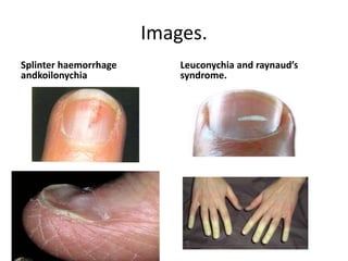
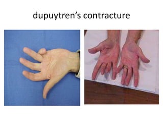
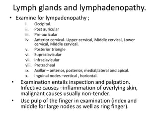
2. Introduction.
• Starts from greeting the patient and ends when the patient departs.
• There may be some physical findings that may prompt further
questioning
• When examining a patient its good to be open about your status –
medical student or junior doctor. Explain the reason that you might
take longer time in taking the patient history is ….due to less
experience.
• Examination should be done in a warm enviroment, private and
quiet. Day light is prefferd to artificial light-which may make the
subtle change in skin colour difficult. Could room increases anxiety
and shivering of muscles produces strange noises on auscultation of
chest.
• Adequately expose the patient to allow thorough examination.
Should undress completely or upto the thighs. Don’t forget to
exmine the coverd regions like the breast , genitalia perineum,
buttocks.
3. Cont,
• A chaperone is needed especially for intimate
examination like rectal and vaginal ,breasts. For
patient reassurance and protect the doctor on
subsequent accusations.
• Do examination from the right side of the
patientcouch. Don’t create more anxiousness to
the patient who already is anxious from critical
illness
• Left handed students may take longer to master
this.
• Try to expose the area of examination at a time
especially for very sick patients.
4. Cont,
• Always keep attention on patients comfort like addition of
pillows etc this improves patient doctor relationship.
• Make a quick assessment of severity of of patient illness -
examination could be postponed in very acute patients.
Posture & gait
• Observe the patient from the time heshe enters the room;
does he get easily out from the chair, walk freely, stiffly ,
limb, aided,
• In hospital note patient posture in bed, adjustment from
one point to another whether with ease or not.- ability is
lost in very ill patients.
• Patient with left heart failure –lying horizontally worsens
the situation.- orthopnoea
• peritonitis makes patient to lie supine, sometimes with
legs drawn up, still, quiet, with shallow breathing
movements.
5. cont
• Patients with renal colic- rolls around attempting to
find a comfortable position.
• With joint arthritis the affected limb. lies motionless .
Meningitis- neck may bend backwards and appear
burrow into the pillow.
Speech and interaction
• Does the patient smile…is it symmetrical.. facial
weakness
• Eye contact..Was the face expressionless(parkinsonism)
• Voice- hoarse could be due to laryngeal nerve palsy,
pressured- mania, thyrotoxicosis, monotonous –severe
depression, is it slurred from cerebellar disease or
previous stroke.
6. Physique and nutritional assessment
• The nutrition status of a patient may provide an important
indicator of a disease.
• HISTORY taking – diet history
• Note whether the patient is cachexic, slim, plump, obese.
• Check for anaemia
• Distribution of body fat – muscle bulk and oedema
• Dehydration –tongue, skin turgor,postural hypotension,
• Hypoalbuminaemia –occur due to the inflammatory
response, may lead to edema this makes overall body
weight unreliable marker for malnutrition.
• Blood pressure – standing / sitting and lying.( though BP is
invaluable in nutritional assessment)
• A drop in BP(systolic) while standing of >than 15mmhg is
postural hypotension. If accompanied by increase in pulse
rate –indicates blood vol. depletion.
7. CONT
• Wasting- temporalis, skeletal muscles of extremities, recent
weight loss may result to prominence of ribs.
• Dry cracked tongue and skin, loss of scalp hair, poor wound
healing, the limb muscles are thin and the distal reflexes
difficult to elicit, subcutaneous fat is atrophic.
• If patient is obese check whether centralized or generalized
distributed.
• Search for signs of mineral and vitamin def;
Eg, -night blindness- lack of vitamin A,
-pellagra – lack of vitamin B5 /niacin,
-scurvy – lack of vit,C,
-easy bleeding – lack of vit. K,
-smooth sore toungue without papillae (atrophic glossitis) –vit.B
defficiencies
-angular stomatitis (cheilosis) could be due to iron deficiency or B
vitamins
8. Cont,
• Should take the height and weight of the patient.
• Calculate the BMI, mid upper arm
circumference(MUAC), waist: hip ration, skin fold
thickness and malnutrition universal screening tool .
• BMI = wieght/height in metres.
Underwieght-<18.5
Healthy weight – 18.5 -24.5
Overweight – 25-29.9
Moderate obesity – 30 -34.9
Severely obese – 35-39.9
Morbidly obese - <40
9. cont
• Mid-upper circumference – patient should remove the
clothing, ms from acromion(tip of shoulder) to
olecranon process(tip of elbow) the mid point.
IF >25 the BMI is >20
>23.5 and <25cm BMI is <20
<23.5cm BMI is <18.5cm.
• MUAC can be used to estimate weight changeover
period of time. 10% its likely the weight and BMI have
changed by 10%.
• Skin fold thickness. – biceps, triceps, infrascapular, and
suprailiac,
• ==Waist hip ratio – men >94cm is increased, 102
Women >80cm is increased, 88cm very increased
10. The neck
• Inspect and palpate for swellings
Inspection– 1. swellings
• The thyroid- along the midline above the suprasternal
notch. Anterior part of the neck
How to examine
• Ask the patient to swallow
• Ask the patient to pull out the tongue . If the lump
moves up and down then it is of thyroid origin.
• If there is a swelling note whether it is involving the
two lobes by the shape it takes, change of skin colour.
• Note whether nodular
• Note lymph node swelling – site, whether unilateral or
bilateral, note the colour of underlying skin.
11. 2. JVP
• Check raised Jungular Venous Pressure.
• To examine incline the patient at 45 degrees. Assess
the waveform of the internal jungular vein which lies
adjacent to medial border of the sternocleidomastoid
muscle. Distented vein implies raised JVP. –could be
pathology of the thoracic cavity or heart pathology. If
patient lies flat it disappears in the cranium thoracic
cavity. (refer to cvs) exam.
3. Pulsations of other blood vessels
4. Use of accessory muscles of respiration–
sternocleidomastoid, trapeius.
5. Neck movement –note whether there is retraction or
neck stiffness. Ask the patient to touch the chest with
the chin. With neck stiffness or retraction this is
difficultresistance and painful.- stiffness could be due
to meningitis, subarachnoid haemorrhage, tetanus,
trauma, diseases
12. Palpation.
• Palpate for thyroid gland from behind ask the
patient to swallow ; feel the shape, size,
whether one lobe or both lobe involvement,
tender to touch, ms the diameter, Note the
surface whether smooth or rugged, nodular,
well defined, ill defined, regularity,
Concistency- hard soft.
• Palpate for lymphadenopathy, and the
trachea(refer to resp. exam)
13. Hands.
• Examine the hands carefully some diagnostic
information could evident.;
• The strength of the patient grip—could indicate
neurological musculoskeletal disorders.
• Tremors- fine associated with thyrotoxicosis,
rhythmical pill rolling –parkinsonism, coarse jerky
tremor –hepatic or uremic failure. (also referred to as
metabolic flap)
• Feel for dupuytren’s contracture in both hands,
thickening of tissue over the flexor tendon of the ring
finger at level of palmar crease.
• Finger clubbing- tissue thickening at the base of the
nail and the angle between the base of the nail and
adjacent skin is lost. The nail becomes convex both
transversally and longitudinally and in gross cases
(usually due to severe cyanotic heart disease,
bronchiectasis or empyema) the volume of finger pulp
increases). (schamroth’s window is lost)
14. How to examine for finger clubbing
• This is the drumstick appearance of the fingers.
1. Scamroth’s window test- Oppose the fingers to check
for the schamroth’s window.
2. Fluctuation test –if positive shows there is finger
clubbing.
3. The nail curvature –the horizontal and vertical
curvature.
Examine all fingers.
• In finger clubbing the angle between the nail and
adjacent tissue is lost.
What are the diseases associated with finger clubbing!
Chronic heart diseases-congenital cyanotic heart disease,
infective endocarditis
Lung diseases –cancer,bronciectases, lung absces,
empyma.
Liver cirrhosis, malabsorption syndrome
15. cont
• Lesser degrees of finger clubbing is seen in bronchial
carcinoma, inflammatory bowel disease, and infective
endocarditis.
• Osler’s nodes – transient tender swelling on the pulp of the
finger due to dermal infarcts from septic cardiac
vegetations. Examine on pulp of fingers
• Splinter hemorrhages and nail- folded infarctions -may be
signs of vasculitis or sign of trauma in normal individuals
hence they are non-specific. Examine on nail fold
• Tropic changes may be evident in the skin in certain
neurological diseases and in peripheral circulatory
disorders such as Reynaud's syndrome, in which vasospasm
of the of the digital arterioles causes the fingers to become
white and numb, then purplish, cyanosis and redness.
• Koilonynchia- nails are soft, thin, brittle, and normal
convexity replaced is replaced by spoon-shaped concavity
its due to long standing iron deficiency anaemia. On fingers
16. Cont,
• Leuconychia- opaque white nails. May occur in
chronic liver disease and other conditions
associated with hypo-albuminaemia.check on the
nails.
• Herberden’s nodes- at the distal interphalangeal
joints of all fingers
• Rheumatoid nodes –at the proximal
interphalangeal joints of all fingers.
• Hypertrophic osteoarthropathy – there is
clubbing and thickening of the radius, ulna, tibia,
and fibula.
22. Lymph glands and lymphadenopathy.
• Examine for lympadenopathy ;
i. Occipital.
ii. Post auricular
iii. Pre-auricular
iv. Anterior cervical- Upper cervical, Middle cervical, Lower
cervical, Middle cervical.
v. Posterior triangle
vi. Supraclavicular
vii. infraclavicular
viii. Pretracheal
ix. Axillar – anterior, posterior, medial,lateral and apical.
x. Inguinal nodes –vertical , horiontal.
• Examination entails inspection and palpation.
Infective causes –inflammation of overlying skin,
malignant causes usually non-tender.
• Use pulp of the finger in examination (index and
middle for large nodes as well as ring finger).
23. Cont,
• For the head and neck nodes, it is helpful to tilt the head
slightly towards the side of examination in order to relax
the muscles.
• Experience is required because examination could be
difficult.
• Small, mobile and discreet l.nodes are commonly found in
normal individuals.
• Determine the;
1. Site
2. size ,
3. position,
4. shape,
5. consistency, -whether the surface is smooth, rugged, nodular
regular.
6. Mobility fixed to the underlying structures or skin,
7. Tenderness –tender or non tender,
8. Matted or discreet.
9. whether isolated or whether several.
10. Edges –well defined or ill defined
11. rubbery
24. AXILLAE
• Examine for lymph nodes. Inspection could reveal an
absence of secondary sexual hair- could be due to
chronic liver disease.
• Support the weight of the patient’s arm by holding
his arm at the elbow with your non –examining hand,
so that the pectoral muscle are relaxed. With fingers
of your hand cupped together, probe the apex of the
left axillae, then slide them downwards against the
chest wall to feel for lymphadenopathy. Next sweep
your fingers along the inside of the anterior and
posterior axillary folds, feeling for enlargemant of the
pectoral and sub-scapular lymph nodes respectively.
Use your left hand in the same way to examine the
right axillae
25. Skin.
• Check for warmth with the back of your fingers.- there
could be generalised warmth in febrile illness or
thyrotoxicosis, or localised in regional inflammation.cold
skin- localised incase a limb is deprived blood or generalied
in circulatory failure==skin feels clammy and sweaty
• Check for skin;
– colour
– Texture
– Bleeding spots- echymosis, purpura
– Scars
– Lesions – macules, papules, vesicles, pustules, bullae, crusts
• Pallor, yellowness, pigmentation and cyanosis.
26. Leg and feet.
• It requires adequate exposure from from the groins and
buttocks to the toes. Note colour and texture of the skin.
• Peripheral vascular disease make the skin shiny, and hair
does not grow on ischemic legs. Pressure on the toes of
ischaemic feet causes blanching of purple colour with
subsequent slow return.
• Inspect for obvious oedema, and examine for pitting
oedema
• Press firmly but gently for 5 seconds behind the medial
malleolus, over the dorsum of the feet and on the sheen. If
oedema is present a depression is evident. Check whether
bilateral or unilateral
• Examine for varicose veins with patient standing. You can
asses the sufficiency of the saphenous vein by
trendelenburg’s test.( patient lies flat empty the vein by
raising the leg, occlude the vein at the upper end , if
incompetent valve there will be rapid refill from above,
when pr. Is released.
27.
28. Cont,
• Examine for DVT.
• Affected limb is swollen.
• Measure the circumference of the calf and
compared with the unaffected leg. Discrepancy of
more than 1cm is significant.
• To measure locate the tibia tuberosity mark 10cm
below it then take circumference of the limb. Ms
at the same distance for the other leg.
• Check for tenderness on the affected limb,
warmth,
• Perform Homan’s sign- forceful dorsiflexion of
foot may cause pain.
• It can also extend upto the thigh.
29. Odours.
• Certain oduors may provide diagnostic clues.
The odour of alcohol on the patient’s breath is
easily recognizable but do not assume that in
all patients it implies alcoholism.
• Diabetic ketoacidosis –acetone odour(pear
drops nail polish remover)
• Hepatic failuire– ammonia like mousy
• Halitosis –bad breath, common in supurative
lung disease and gingivitis.
30. Cont,
• For the head and neck nodes, it is helpful to
tilt the head slightly towards the side of
examination in order to relax the muscles.
• Experience is required co examination could
be difficult.
• Small, mobile and discreet l.nodes are
commonly found in normal individuals.
31. Breast.
• Should be done routinely. Especially on
performing chest exam.
• Requires tact and sensitivity and should be
conducted with a chaperone.
• Arms should be relaxed on the sides. By
inspection make note of the size, symmetry,
contour (dimpling), colour. Inflammatory breast
cancer with oedema of the overlying skin may
produce a characteristic look and texture termed
peau d’orange (orange peel skin). Note any
asymmetry or inversion of the nipples. Simple
long-standing inversion is often a normal
phenomen, but associated retraction of the
areola or recent nipple inversion are significant.
32. Cont.
• Ask the patient to raise her arms above her
head. This allows inspection of the
inframammatory fold and may expose subtle
contour abnormalities.
• To examine ask the patient to lie in supine
position ask the patient to rest one one arm
above her head. This helps spread the breast
tissue more evenly across the chest and makes
palpation of any nodules easier.
• The breast usually divided into four quadrants
with the upper outer quadrant extending into
an axillary tail.
33. Cont,
• Use the middle three fingers to palpate the breast, use
rotatory movements gently compress the tissue against
the chest wall. Proceed systematically to examine all
quadrants, the tail and areola. Sometimes it is useful to
support the breast with the other hand in order to aid
examination, especially when the breasts are large.
• Breast tissue is variable between patients, ranging from
smooth to granular, and may change in a given
individual with the menstrual cycle. For nodule;- note
size, shape, consistency, tenderness, mobility and the
presence of any tethering or skin ulceration
36. Summary.
General approach of routine exam.
• General appearance- wellunwell, neglected.
• Intelligence and education level.
• Metal state- consciousunconscious, confused
• Build and posture
• Nutrition, obesity, oedema
• Skin colour, cyanosis, anaemia, jaundice, pigmentation. Skin eruptions
• Dehydarion,
• Body hair
• Deformities, swellings
• Hair- texture and grooming
• Eyes – visual acuity compare one with the other
-Exophthalmos -ptosis
-oedema of the lids -conjuctiva- aneamia, inflammation
-pupils size, equality, -eye movement; nystagmus, strabismus
reaction to light, -ophthalmoscopic exam.
accommodation
37. Cont,
• Face- facies, jaw movements,
symmetryassymetry, rash
• Mouth and pharynx –breath odours, lips colour
and eruptions, tongue protusion and appearance,
teeth and gums-dentures, buccal mucous
mebrane- colour and pigmentatio
• Movement of thesoft palate, state of the tonsils
• Neck –movements, pain, and range, veins, lymph
nodes, thyroid, jungular venous pressure and
other pulsation.
• Upper limbs,
• Lower limbs
39. RESPIRATORY SYSTEM
• What are the symptoms..
– Cough
– Breathlessness
– Sputum
– Haemoptysis
– Wheezing
– Chest pain etc
• Smoking history- is important, note the pack
years.
• Family history- asthma, eczema…
• Occupational history…
45. General examination
• Look/watch at the patient as he/she enters the room ,
and during history taking, while undressing, climbing
the couch.
• If inpatient; Is there breathlessness, what is beside the
beds sputum mugs, pots, oxygen masks
• Physique, state of general nourishment
• Finger clubbing, cyanosis, check for central cyanosis on
the lips, tongue, for central cyanosis- indicates poor
oxygenationof blood by lungs.
• Peripheral cyanosis due to poor peripheral perfusion-
• Breathless patient- uses accessory muscles of
respiration(sternomastoid muscle)- like id COPD,
patient find easy to breath with pursed lips
• Listen to the voice-hoarse, cough is it normal explosive
or voice weak. Is there wheeze audible,loudest in
expiration , stridor?-high picthed inspiratory voice.
46. Chest exam
• Anatomy – bifurcation of trachea corresponds
the sternal angle anteriorly, posteriorly the 4th
and 5th disc thoracic vertebrae.
• Rt.lung 3 lobes lt. 2 lobes. A line from second
thoracic spine to the 6th rib in the mammary
line corresponds the upper border of the
lower lobe-major interlobular fissure.
48. INSPECTION - 1. appearance of the chest
Obvious scars from previous surgery, visible
lumps, lesions on the skin,
Therapeutic marks
Visible lumps/lesions
Bilaterally symetrical, elliptical in cross-section
49. 2. Movement of chest.
Is it symmetrical?
• Is it diminished on one side –could be abnormality
• Intercoastal recession- drawing of intercoastal spaces with
inspiration.- may indicate severe upper airway obstruction
like laryngeal disease, tumours of tracheal
• Sub costal recession in inspiration- like in COPD
Venous pulse;
• In the neck –if raised indicate rt. HF, or due to obstruction
of superior venacava may be due to malignancy in the
mediastinum.
3. Respiratory rate and rhythm
• Normal rate 14-16b/m
• Tachypnoea increased resp. rate, dyspnea - breathlessness
• Cheyne-stokes breathing- disturbance in respiratory
rythmn, there is cyclical deepening and quickening of resp.
followed by diminishing resp effort.(periodic breathing)
50. Cont,
• Eupnea- normal, good, unlabored breathing,
sometimes known as quiet breathing
• Bradypnea
• Biot’s -abnormal pattern of breathing
characterized by groups of quick, shallow
inspirations followed by regular or irregular
periods of apnea
• Cheynes-Stokes- disturbance in rhythm, where
ther is cyclical deepening and quickening of
respiration followed by diminishing respiratory
effort and rate.
• Kussmaul -is a deep and
labored breathing pattern often associated with
severe metabolic acidosis, particularly diabetic
ketoacidosis (DKA) but also kidney failure.
51. 4. Thoracic Contour
• Pectus Excavatum- abnormal development of the rib cage
where the breastbone (sternum) caves in, resulting in a
sunken chest wall deformity. Sometimes referred to as
"funnel chest,
• Pectus Carinatum- protrusion of the chest wall (the
opposite of pectus excavatum
• Kyphosis- forward bending),
• Scoliosis- lateral bending of the vertebral collumn
• Kyphoscoliosis - A combination of outward curvature
(kyphosis) and lateral curvature (scoliosis) of the spine
• Symmetry of chest movement
Deformities –kyphosis(forward bending),scoliosis(lateral
bending of the vertebral collumn),= this leads to assymetry
of the chest if severe may restrict lung movement
significantly.
Barrel chest-increased anteroposterior diameter
52. • This is seen in sever cardiac failure,neurological disorders.
PALPATION
1. LN in supraclavicular, cervical region,axillary region may
be secondary to malignancies in the chest.
2. Any obvious Swellings and tenderness, areas of pain feel
gently
3. Trachea and heart – position of cardiac impulse and
trachea.
= To examine trachea put the second and fourth fingers of
the examining hand on each edge of the sternal notch and
use the third finger to asses whether the trachea is
displaced/deviated to one side. Don’t use a lot of force.
In healthy pple you find slight deviation
• Displacement of the cardiac impulse without displacement
of trachea may due to scoliosis, congenital funnel
depression of the sternum or enlargement of the left
ventricle.
53. • In absence of these conditions, the signficance
displacement of either or both may suggest the
mediustinum has been altered by disease of
lungs or pleura. –
• pleural effusion, pneumothorax pushes
mediustinum away from affected side
• fibrosis of lungs, collaps pull to the affected side.
4. Chest expansion place the fingers on either side
of the chest of lower ribcage, so the tips of the
two thumbs meet in the midline in front but not
touching the chest. A deep breath by the patient
will increase the distant btn the thumbs and
indicate the degree of expansion. If one remains
remains closer to the midline- diminished
expansion on that side.
5. Tactile vocal fremitus- detected by palpation .
This is not aroutine examination technique.
54. Vocal fremitus
• Felt with hand on the chest its part of
palpation but heard after ausculation . Ask
patient to repeat words like ninety nine the
examining hand will percieve vibration. Some
pple use the ulna border of the hand but the
flat part of hand is more sensitive.
• It is more in consolidation, reduced in pleural
effusion.
55. PERCUSSION
1. Check for resonance
2. Dullness
3. Pain and tenderness
• The middle finger of the left is placed on the part to be
percussed and press firmly against it. The back of the
distal interphalengeal joint is then struck with tip of rt.
Hand middle finger. Movement should be at the wrist
not at the elbow.
• The percussing finger is flexed so that its terminal
phalanx os at right angles and it strikes the other finger
perpendicularly. The finer is raised as soon as the
finger is struck. Do tapping movements.
• Mistakes made; failure to place the finger firmly and
flat, striking from the elbow other than the elbow.
56. Cont,
• When air in the cavity of sufficient size and appropriate
shape is vibrating – resonant voice. This varies in
indivinduals. And different parts of the chest.
• More reasonant anteriorly below the clavicles and the
scapular posteriorly where muscles are thin and least one
the scapulae.
• At right side ther is loss of reasonance on inferiorly due to
the liver.
• On the left side the lower border overlaps the stomach so
there is a transition of lung reasonance to stomach
tympanicity.
Compare the percussion note on the two sides of the chest
systematically always, moving backward and forward from
one side to the other not all the way down one side then
the other.
• Percuss over the clavicles(traditionally done without finger
but it is uncomfortable to the pt. so do in usual way)
57. Cont,
• Percuss 3-4 areas in the anterior chest
wall.compare left with right
• Percuss the axillae, then 3-4 areas at the back
• If reasonance is reduced report as dullness –
occur when lung is more solid than
normal(consolidation), pleural cavity contains
fluid like p.effusion
• less common causes of dullness- thickened
pleura,
• Pleural effusion causes stony dullness. Elicit
whether unilateral or bilateral.
• Hyperresonance – pneumothorax,
58. AUSCULTATION
• Listen to the chest with diaphragm , not the
bell of the stethoscope which is more
sensitive.
• The chest should be fully exposed and the
stethoscope should not be sliding.
• Patient should be realxed not shivering. Learn
to disregard other sounds arising from the
heart when auscultating the resp.
• Ask the pt to take a deep breath, in and out of
the mouth.- demonstrate…
59. Cont,
• Check;
Breath sounds- intensity(loudness) and quality.
Intensity could be normal,reduced or increased.
1) Vesicular BS- Normal lung tissue makes the
sound quieter and filters higher frequencies. –
vesicular.
Intensity is reduced when there is localized
airway narrowing, if lung is extensively damaged
by eg emphysema , pleural thickening, pleural
fluid.
longer
Shorter and softerthan inspirational
Described as rustling sound.
60. Cont,
Breath sounds originate from turbulence of
airflow in the large airways.
2) broncial breathsounds -found in consolidation.
Tubular sounds produced by passage of air
through the trachea and partially through the
large bronchi. Both inspiration and expiration
BS are of the same duration and there is a
pause in between. Both are loud and clear. If
elicited over the trachea, its normal also called
trachea BS.
Inspiration. Expiration.
61. • If heard over the lung – denotes pathology like
cavitation
Carvenously BS – expiratory becomes louder
than inspiration
• Broncho vesicular breathsounds –doesn’t
denote any pathology occur at the tracheal
bifurcation.
• The vesicular BS could be altered and the
expiration becomes prolonged than the
inspiration especially in chronic obstructive
airway disease.-=asthma, empysema
62. Air entry- could be decreased, or diminished
rarely increased. This may be due to; pleural
effusion, pneumothorax, lung colapse, lung
fibrosis, pneumonia
Vocal reasonance- similar to vocal fremitus.
Sounds produced as air enters the lung. Ask the
patient to pronounce the words ninety nine as
you auscultate. They could be normal , increased
or decreased or absent. Compare both sides of
the chest.
If the patient pronounces the word and you can
clearly hear the word= increased vocal
reasonance= pectoloriloquy whispering
pectoloriloquy It is increased in PTB, pneumonia,
consolidation
It is decreased in pleural effusion, pneumothorax
63. • In consolidation or above level of pleural
effusion voice may be bleating this is known
as aegophony. High pitched nasal quality
sound. Sound produced by bleating of animal
like goat.
64. Added sounds
• Abnormal sounds- wheezes, crackles
• Wheezing – musical sounds associated with
air narrowing. high-pitched whistling sound
associated with Partial bronchial obstruction.
It is usually expirational. Polyphonic wheezes
are heard in COPD, diffuse air,flow obstruction
Are related to compression of bronchi, fixed
monophonic wheeze generated by localized
narrowing of a single bronchus. Eg in tumour
or FB, may be expiratory or inspiratory or
both.
65. • Crackles – short explosive sounds often
described as bubbling/clicking- when large
airway are full of sputum, crackle sing
beginning of inspiration is due to COPD,
localised loud and coarse- bronchiectasis,
• They are heard in – pulmonary oedema,are
fine in diffuse interstitial
• They could be fine or coarse.
Fine due to presence of exudate or fluid
material in the alveoli and terminal broncials.
Occurs in lobar pneumonia, CCF
66. Coarse – secretion within the bronchi eg
mucous. Usually bubbling of air through a
fluid medium. Mainly heard in children with
broncho pneumonia, patients with
immunosuppression., bronciectasis,
bronchitis,. Heard both in inspiration and
expiration.
Pleural rab – pleural inflammation, has
creaking /rubbing character as heard in
palpating hands. To confirm whether it is from
pleura ask the patient to stop breathing if the
sound doesn’t stop it means it is not pleural
rub….it could pericardial rub.
67. • Stridor –is a high-pitched, wheezing sound
caused by disrupted airflow. noisy produced
due to large partial airway obstruction. Could
be in larynx, trachea, main bronchi. Indicates
more serious condition hence require more
investigation heard in insp. But can be heard
on exp. Can be heard when mouth is open,
heard loudest on trachea
• Rhonchi – heard due to partial obstruction of
bronchial tree. Could be due to oedema of
bronchial wall, secretions, bronchiolitis,
bronchitis.
68. Investigations.
1).Sputums
Characteristics to check; mucoid, purulent, frothy,
bloodstained, rusty, quantity. Sputum should be
collected early in morning in a dry sterile bottle.
Collected 2 sputums spot and a morning sputum
• Mucoid sputum- charasteristic inpatients with
chronic bronchitis.
• Mucopurulent- bacterial infection is present in
patients with bronchitis, pneumonia,
bronchiectasis or lung abcess- foul smell sputum.
Asthmatic tinge yellow to the sputum due to
presence of many eosinophils.
69. • Foul smell sputum suspect presence of
anaerobic organism, bronchioctasis, lung
abcess.
• Pinkwhite frothy sputum – patients with
pulmonary oedema
• Rusty coloured sputum- lobar pneumonia
• Blood stained sputum- frank blood or blood
stained. Seen in bronchogenic carcinoma, PTB,
PTE, Pulmonary hypertension.
70. 2).Lung functions tests; Spirometer – will
measure how much air can be exhaled after
maximal inspiration. Ask the patient to breath
in as much as possible then blow the
spirometer untill no more air can come out.
• Terms to understand
a. VC(Vital capacity) - Amount of air that is
exhaled after maximal inspiration.
71. Total lung capacity – measured by amount of air
in the lungs at full inspirati
a. Residual volume – amount remaining in the
lungs after full expiration.
b. FVC (forced vital capacity)- measurement of
the amount of air exhaled , when a patient
blows a spirometer as hard and fast as
possible.
c. FEV- forced expiratory volume the volume
breathed out in the first second of the forced
expiration.
72. a. PEFR- peak expiratory flow rate.
Measures the size of lungs,how is easily air flows
and out of the airways, how lungs are efficient
in process of gaseous exchange
3). Arterial blood sampling –pao2, paco2, PH.
Paco2 is directly, related to the level of
alveolar ventilation, it raises when
al.ventilation is reduced.
73. 4). Imaging tests – xray,
How to view;
-position of the patient straight or rotated, if
straight the inner ends of clavicle will be
disposed symmetrically with reference to the
spinal collumn. any rotation alters this
appearance.
-outline of the heart and the mediustinum- size
shape and position
-position of trachea.
-diaphragm- assess whether it can be seen on
both sides, shape and position. The anterior end
of the sixth or seventh rib crosses the mid part of
diaphragm.
74. Cont,
-Lung fields- upper zone,mid zone, lower zone
-bony skeleton- symmetry of chest,scoliosis,ribs-
crowded or spaced,any ribs absent or eroded. AP and
Lateral view-
b) bronchoscopy- flexible bronchoscopy, passed through
the nose, pharynx and larynxtrachea and bronchial tree
then a bronchoscopy is used to obtain specimens.
c) CT scan
c) MRI
d)Ultra sound
Pleural biopsy and aspiration
Thoracoscopy- use of thoracoscope
Lung biopsy