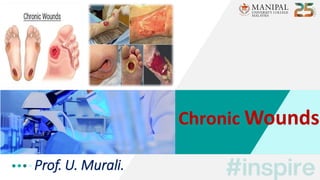
Chronic Wounds
•
0 likes•134 views

This topic is under the General Principles of Surgery for MBBS Students. It also deals with Scars & Contractures. The student should know to differentiate between Hypertrophic Scar & Keloid..

Recommended
More Related Content
What's hot
What's hot (20)
Similar to Chronic Wounds
Similar to Chronic Wounds (20)
More from Uthamalingam Murali
More from Uthamalingam Murali (20)
Recently uploaded
Recently uploaded (20)
Chronic Wounds
- 1. Prof. U. Murali. Chronic Wounds
- 2. Learning Objectives •Introduction •Leg ulcers / Pressure ulcers • Scars - Hypertrophic scar / Keloid • Contractures • Sinus & Fistula
- 3. • Chronic wounds fail to progress through the normal stages of wound healing in a timely manner. • They are often characterized by a prolonged inflammatory phase and persistent infections. • The management of chronic wounds therefore often involves debridement, control of infection and inflammation and appropriately selected dressings to correct moisture imbalances. • Chronic wounds can be categorized into – • Vascular ulcers (venous or arterial) • Diabetic ulcers & • Pressure ulcers. Introduction
- 5. • In developed countries, the most common chronic wounds are leg ulcers. • Defn: An ulcer is a break in the continuity of the covering epithelium, either skin or mucous membrane due to molecular death. • A prolonged inflammatory phase leads to overgrowth of granulation tissue and attempts to heal by scarring leave a fibrotic margin. Leg Ulcers
- 6. 6 Leg Ulcers
- 7. •CBP / Sr. Protein •Study of discharge – C/S • Edge biopsy • X-ray of the part • FNAC – Lymph node • Chest X-ray / Mantoux test • Arterial / Venous Doppler Leg Ulcers – Investigations
- 8. Leg Ulcers – Treatment • Cause should be found & treated. • Correction of anaemia, deficiencies like protein & vitamins. • Control of pain & infection. • Rest, immobilization, elevation & avoidance of repeated trauma. • Care of the ulcer by debridement, ulcer cleaning & dressing. • Once the ulcer granulates, defect is Closed with secondary suturing, skin graft (or) flaps.
- 10. • Pressure sore is tissue necrosis and ulceration due to prolonged pressure. It is more prominent between bony prominence and an external surface. • They can appear & extend rapidly in immobile patients and in those with debilitating illness. • It can also be called as – • Trophic ulcer. • Neuropathic / Neurogenic ulcer. • Bed sore & • Decubitus ulcers. Pressure Sore / Ulcer / Injury
- 11. SITES FACTORS Pressure Sore / Ulcer / Injury • Pressure • Sensory Loss • Anemia • Malnutrition • Moisture • Def. blood supply • Injury
- 12. Pressure Sore / Ulcer / Injury
- 13. Clinical Features • Occurs in 5% of in- patients. • Deep punched out edges. • Non-mobile ulcers – bone as its base. Type Investigations • Pus – C/S • Blood tests • Biopsy • X-ray - part Pressure Sore / Ulcer / Injury
- 14. • Postural changes: - Change in position – once in 2 hours - Lifting limb upwards – 10 s – once in10 mts • Use of – waterbed/air bed/air-fluidized bed. • Special pressure dispersion cushion / foams. • Absorbent porous clothing. • Regular use of talcum powder – skin dry. • Urinary & faecal care. • Good nutrition / Psychological counseling. • Treating the cause – Anemia/Diabetes…. • Antibiotics & Regular dressings - VAC. • Slough excision → skin grafting / flap cover to be done. P U - Treatment
- 16. • A scar is an area of fibrous tissue that replaces normal skin after an injury. Thus, scarring is a natural part of the healing process. • The remodelling and maturation phase of wound healing results in scar formation. • Scars result from the biological process of wound repair in the skin, as well as in other organs, and tissues of the body. • Exception of very minor lesions, every wound (e.g., after accident, disease, or surgery) results in some degree of scarring. Scar – Introduction
- 17. • Initially immature scar is formed during remodeling phase; this scar is raised, itchy, hard and pink in colour. • A mature scar is paler, acellular, softer, flat, without itching (diminishes). • An atrophic scar takes the form of a sunken recess in the skin. These are caused when underlying structures are lost. • A hypertrophic scar is excess scar but will not extend beyond the margin of the scar of the original wound. • Keloid is persistent excessive growth of the scar beyond its margin into the adjacent skin. Scar – Types
- 19. Scar – Treatment
- 20. • Hypertrophic scar is excessive formation of abnormal scar tissue which is raised, often vascular but confined within the margin of the original wound; usually its growth stops in 6 months and often regresses spontaneously. • They are more common in areas of increased tension, wounds crossing tension lines, deep dermal burns & wounds left to heal by secondary intention (longer than 3 weeks). Hypertrophic Scar / Keloid • Keloid scars extend beyond the boundaries of the original incision (or) wound, do not spontaneously regress and are difficult to treat. • The etiology is unknown but genetic predisposition is implicated. They often occur because of relatively minor trauma and mainly in those with darker skin pigmentation.
- 22. Mgt – Algorithm – Hypertrophic Scar
- 23. Mgt – Algorithm – Keloid
- 25. • Contracture is the result of a stiffness or constriction of muscles, joints, tendons, ligaments (or) skin that restricts normal movements. • Contracture develops when normally elastic connective tissues become replaced with inelastic fibrous tissues. Contracture • Contractures are either neurally (or) non- neurally mediated - • Neurally mediated – are due to spasticity and are a common sequalae of UMN lesions. • Non-neurally mediated – contractures are due to structural adaptations of soft tissues [Occur in response to prolonged immobilization of soft tissues].
- 26. • Inactivity & Scarring – from an injury / burn. • Muscular dystrophy. • Cerebral palsy. • Polio. • Rheumatoid arthritis. • L E – plantar flexion, hip flexion & knee flexion contractures are common. • UE – elbow flexion & supination / adduction & internal rotation contractures of shoulder. • Muscles that cross multiple joints – biceps, hamstrings, TFL & gastrocnemius are predisposed to contractures. Sites Types Contracture Causes
- 27. • Scar contractures can cause severe functional, psychological and aesthetic problems. • Contractures across joints may restrict the range of movement, leading to deformity, impairment and disability. • Surgical contracture release and reconstruction can be an effective treatment option. Release of contracture surgically and use of skin graft (or) "Z" plasty (or) different flaps. • Proper physiotherapy and rehabilitation is essential. Contracture
- 29. • It is a blind track lined by granulation tissue leading from an epithelial surface into the surrounding tissues. • Sinus means "hollow" or "a bay" (Latin). Sinus / Fistula • It is an abnormal communication between the lumen of one viscus to another or the body surface or between the vessels. • Fistula means "flute" or "a pipe or tube."
- 30. Sinus / Fistula
- 32. Sinus / Fistula
- 33. Sinus / Fistula
- 34. • Aetiology & treatment of Leg ulcers. • Basic Investigations & treatment aspects of Leg ulcers. • Sites & Factors causing Pressure ulcers. • Staging of Pressure injury | C/F & Treatment of Bedsore. • Scar – Types, Prevention & Treatment of scars. • Difference between Hypertrophic scar & Keloid. • Contracture – Causes, Sites & Types. • Defn – Sinus / Fistula | Causes for its persistent occurrence. To Summarize
- 35. References
- 36. • List 4 aetiological factors causing leg ulcers. • Mention the staging methods of decubitus ulcer. • Enumerate 3 sites & 3 factors causing pressure injuries. • Write the management algorithm for keloid. • Compare & Contrast hypertrophic scar from keloid. • Outline the various preventive methods of scar formation. • Write 5 causes for persistence of a sinus / fistula. • Mention the various sites & types of contractures. Question Time
- 37. The drug used for intralesional injection of keloid treatment is – ◼ a) Prednisolone. ◼ b) Triamcinolone. ◼ c) Androgen. ◼ d) Hydrocortisone. ◼
- 38. A patient with an injury that has full-thickness loss without exposure of underlying bone or muscle is at which of the following pressure ulcer stages? – ◼ a) Stage – 4. ◼ b) Stage – 3. ◼ c) Stage – 2. ◼ d) Stage – 1. ◼
- 39. Which one of the following is true regarding leg ulcers & their location? – ◼ a) Pressure ulcer – Tip of the toes. ◼ b) Arterial insufficiency – Medial side of leg. ◼ c) Venous insufficiency – Gaiter area. ◼ d) Diabetic ulcer – Above medial malleolus. ◼
- 40. A hirsute young male has a sinus just above the natal cleft. Which is the most likely diagnosis? – ◼ a) Tubercular sinus. ◼ b) Hidradenitis suppurativa. ◼ c) Fistula-in-ano. ◼ d) Pilonidal sinus. ◼
- 41. Which one of the following sites is more prone for contracture formation? – ◼ a) Hip flexion. ◼ b) Elbow pronation. ◼ c) External rotation of shoulder. ◼ d) Wrist flexion. ◼
- 46. THANK YOU