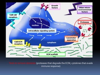
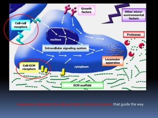
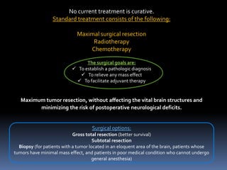
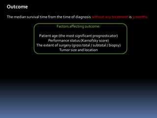
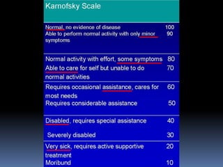
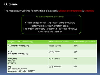
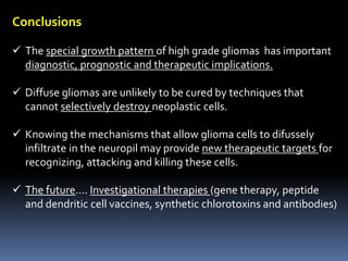
DIFFUSE GLIOMA GROWTH: a guerrilla war.
Dr JiménezArribas, Paloma
NSG 3st year resident. May, 2014.
Glioblastoma (GBM)
 The most common and most aggressive malignant primary brain tumor in
humans.
 Astrocitoma grade IVWHO.
 Incidence of 2–3 cases per 100,000 in
Europe and North America
Primary glioblastoma: the majority of GBMs.
- Arise without evidence of a less malignant precursor.
- Mean age 55 years.
- Short clinical history (<3 months)
Secondary glioblastoma:
- Develops by malignant degeneration of low grade (II, III) astrocitomas.
- Younger patients (mean age 40).
- Slower clinical course.
Histological findings:
 Nuclear atypia
 Mitosis
 Neovascularization with endothelial proliferation
 Areas of necrosis
 “Pseudopalisading cells”
• Overexpress hypoxia-inducible factor (HIF-1), and secrete
proangiogenic factors.
• Around areas of necrosis.
• Wave of tumor cells actively
migrating away from central hypoxia
Special growth pattern:
 In contrast to almost all other brain tumors, they infiltrate
extensively in the neuropil (network of neuronal and glial
cell processes)
 This growth pattern is:
 Almost unique in this kind of tumors.
 A major factor in therapeutic failure.
Special growth pattern (trough white matter):
Uncinate fasciculus (simultaneous
frontal and temporal lobe tumors)
Corpus callosum (butterfly glioma)
Radiological findings:
Ring-enhancing lesions
 Central necrotic área
 Enhancing rim (active tumoral cells)
 Severe perilesional edema
 Radiological visualization of the invasive front is difficult
 Tend to underestimate the extent of diffuse inflitrative
glioma growth.
Radiological findings:
 Multifocal gliomas: multiple lesions that come from an original
lesion. Usually located in the same brain hemisphere.
 Multicentric gliomas: multiple lesions not originated from the
same lesion.Widely separated.
To understand the growth pattern of these tumors….
Guerrilla war metaphor
Like guerrilla warriors….
 Tumors cells tend to invade individually or in small groups
in foreign territory and to abuse pre-existent supply lines.
 Visualization of the invasive front is problematic.
Like guerrilla warriors….
 Glioma cells have specific qualities that allow a diffuse infiltration
(Molecular background)
Internal system that coordinates inputs and outputs (membrane receptors).
Locomotor apparatus (dynamic remodeling of the cytoskeleton)
Trails to travel (myelinated fibers migration trough white matter tracts)
Tools to remove obstacles (proteases that degrade the ECM, cytokines that evade
immune response)
Interactions between the cells and their microenvironment that guide the way
Like guerrilla warriors….
 Conventional methods to fight glioma cells have limited effect or
cause too much collateral damage (they tend to blend with
normal brain), and a “search and destroy” tactic may be needed.
 Treatment involves surgery, chemotherapy and radiation.
 Surgery makes impossible a complete tumor removal in high
grade gliomas.
No current treatment is curative.
Standard treatment consists of the following:
Maximal surgical resection
Radiotherapy
Chemotherapy
The surgical goals are:
 To establish a pathologic diagnosis
 To relieve any mass effect
 To facilitate adjuvant therapy
Maximum tumor resection, without affecting the vital brain structures and
minimizing the risk of postoperative neurological deficits.
Surgical options:
Gross total resection (better survival)
Subtotal resection
Biopsy (for patients with a tumor located in an eloquent area of the brain, patients whose
tumors have minimal mass effect, and patients in poor medical condition who cannot undergo
general anesthesia)
Outcome
The median survival time from the time of diagnosis without any treatment is 3 months
Factors affecting outcome:
Patient age (the most significant prognosticator)
Performance status (Karnofsky score)
The extent of surgery (gross total / subtotal / biopsy)
Tumor size and location
Outcome
The median survival time from the time of diagnosis without any treatment is 3 months
Factors affecting outcome:
Patient age (the most significant prognosticator)
Performance status (Karnofsky score)
The extent of surgery (gross total / subtotal / biopsy)
Tumor size and location
Surgery + XRT + Chemoterapy Median survival (weeks) Estimated 2-year survival
< 40, frontal tumor (GTR) 132 (2,5 years) 65%
< 40,
Not frontal tumor (GTR)
71 (1,3 years) 35%
40<age<65
KPS > 70
GTR/STR
63 (1,2 years) 17%
>65
40< age<65 + KPS <80
40<age<65 + KPS >80 + BIOPSY
37 (5 months) 4%
Conclusions
 The special growth pattern of high grade gliomas has important
diagnostic, prognostic and therapeutic implications.
 Diffuse gliomas are unlikely to be cured by techniques that
cannot selectively destroy neoplastic cells.
 Knowing the mechanisms that allow glioma cells to difussely
infiltrate in the neuropil may provide new therapeutic targets for
recognizing, attacking and killing these cells.
 The future…. Investigational therapies (gene therapy, peptide
and dendritic cell vaccines, synthetic chlorotoxins and antibodies)
Glioblastoma - Diffuse guerilla war by Dr Paloma Jimenez Arribas

Glioblastoma - Diffuse guerilla war by Dr Paloma Jimenez Arribas

  • 1.
    DIFFUSE GLIOMA GROWTH:a guerrilla war. Dr JiménezArribas, Paloma NSG 3st year resident. May, 2014.
  • 3.
    Glioblastoma (GBM)  Themost common and most aggressive malignant primary brain tumor in humans.  Astrocitoma grade IVWHO.  Incidence of 2–3 cases per 100,000 in Europe and North America Primary glioblastoma: the majority of GBMs. - Arise without evidence of a less malignant precursor. - Mean age 55 years. - Short clinical history (<3 months) Secondary glioblastoma: - Develops by malignant degeneration of low grade (II, III) astrocitomas. - Younger patients (mean age 40). - Slower clinical course.
  • 4.
    Histological findings:  Nuclearatypia  Mitosis  Neovascularization with endothelial proliferation  Areas of necrosis  “Pseudopalisading cells” • Overexpress hypoxia-inducible factor (HIF-1), and secrete proangiogenic factors. • Around areas of necrosis. • Wave of tumor cells actively migrating away from central hypoxia
  • 5.
    Special growth pattern: In contrast to almost all other brain tumors, they infiltrate extensively in the neuropil (network of neuronal and glial cell processes)  This growth pattern is:  Almost unique in this kind of tumors.  A major factor in therapeutic failure.
  • 6.
    Special growth pattern(trough white matter): Uncinate fasciculus (simultaneous frontal and temporal lobe tumors) Corpus callosum (butterfly glioma)
  • 7.
    Radiological findings: Ring-enhancing lesions Central necrotic área  Enhancing rim (active tumoral cells)  Severe perilesional edema  Radiological visualization of the invasive front is difficult  Tend to underestimate the extent of diffuse inflitrative glioma growth.
  • 8.
    Radiological findings:  Multifocalgliomas: multiple lesions that come from an original lesion. Usually located in the same brain hemisphere.  Multicentric gliomas: multiple lesions not originated from the same lesion.Widely separated.
  • 9.
    To understand thegrowth pattern of these tumors…. Guerrilla war metaphor
  • 10.
    Like guerrilla warriors…. Tumors cells tend to invade individually or in small groups in foreign territory and to abuse pre-existent supply lines.  Visualization of the invasive front is problematic.
  • 11.
    Like guerrilla warriors…. Glioma cells have specific qualities that allow a diffuse infiltration (Molecular background)
  • 12.
    Internal system thatcoordinates inputs and outputs (membrane receptors).
  • 13.
    Locomotor apparatus (dynamicremodeling of the cytoskeleton) Trails to travel (myelinated fibers migration trough white matter tracts)
  • 14.
    Tools to removeobstacles (proteases that degrade the ECM, cytokines that evade immune response)
  • 15.
    Interactions between thecells and their microenvironment that guide the way
  • 16.
    Like guerrilla warriors…. Conventional methods to fight glioma cells have limited effect or cause too much collateral damage (they tend to blend with normal brain), and a “search and destroy” tactic may be needed.  Treatment involves surgery, chemotherapy and radiation.  Surgery makes impossible a complete tumor removal in high grade gliomas.
  • 19.
    No current treatmentis curative. Standard treatment consists of the following: Maximal surgical resection Radiotherapy Chemotherapy The surgical goals are:  To establish a pathologic diagnosis  To relieve any mass effect  To facilitate adjuvant therapy Maximum tumor resection, without affecting the vital brain structures and minimizing the risk of postoperative neurological deficits. Surgical options: Gross total resection (better survival) Subtotal resection Biopsy (for patients with a tumor located in an eloquent area of the brain, patients whose tumors have minimal mass effect, and patients in poor medical condition who cannot undergo general anesthesia)
  • 20.
    Outcome The median survivaltime from the time of diagnosis without any treatment is 3 months Factors affecting outcome: Patient age (the most significant prognosticator) Performance status (Karnofsky score) The extent of surgery (gross total / subtotal / biopsy) Tumor size and location
  • 22.
    Outcome The median survivaltime from the time of diagnosis without any treatment is 3 months Factors affecting outcome: Patient age (the most significant prognosticator) Performance status (Karnofsky score) The extent of surgery (gross total / subtotal / biopsy) Tumor size and location Surgery + XRT + Chemoterapy Median survival (weeks) Estimated 2-year survival < 40, frontal tumor (GTR) 132 (2,5 years) 65% < 40, Not frontal tumor (GTR) 71 (1,3 years) 35% 40<age<65 KPS > 70 GTR/STR 63 (1,2 years) 17% >65 40< age<65 + KPS <80 40<age<65 + KPS >80 + BIOPSY 37 (5 months) 4%
  • 23.
    Conclusions  The specialgrowth pattern of high grade gliomas has important diagnostic, prognostic and therapeutic implications.  Diffuse gliomas are unlikely to be cured by techniques that cannot selectively destroy neoplastic cells.  Knowing the mechanisms that allow glioma cells to difussely infiltrate in the neuropil may provide new therapeutic targets for recognizing, attacking and killing these cells.  The future…. Investigational therapies (gene therapy, peptide and dendritic cell vaccines, synthetic chlorotoxins and antibodies)