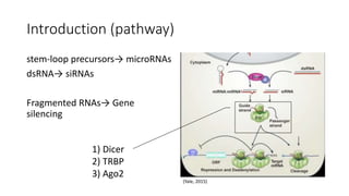
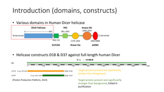
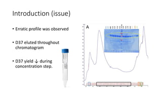
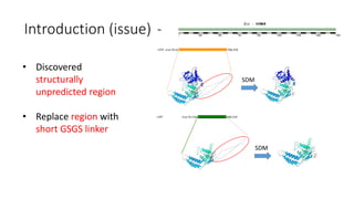
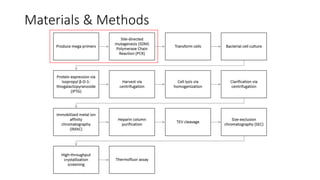
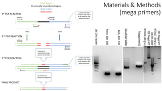
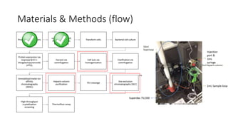
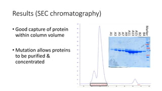
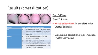
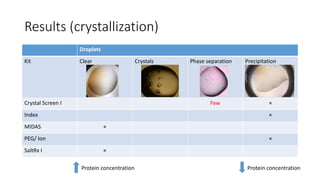
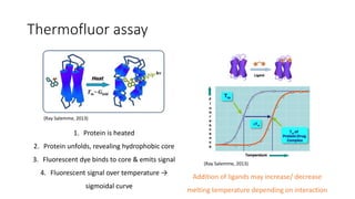
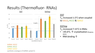
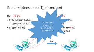
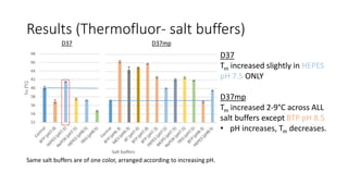
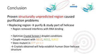
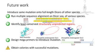
Expression, purification and
crystallization screening of human Dicer
helicase
Esther Tan Ding Qin (S10132125B)
Monisha Joy Gomez (S10122011E)
Supervisor: Asst. Prof. Dahai Luo (LKC-SOM)
Co-supervisor: Dr. Amy Ooi (NP) ss23
Contents
1. Introduction
• Pathway
• Domains, constructs
• Issue
2. Materials & Methods
• Flow
- Mega primer design
3. Results
• Heparin purification
• TEV cleavage
• Size-exclusion chromatography
3. Results
• Crystallization
• Thermoflour assay
• RNAs
• Buffers
4. Conclusion
5. Future work
Introduction (pathway)
stem-loop precursors→ microRNAs
dsRNA→ siRNAs
Fragmented RNAs→ Gene
silencing
(Yale, 2015)
1) Dicer
2) TRBP
3) Ago2
Introduction (domains, constructs)
• Various domains in Human Dicer helicase
• Helicase constructs D18 & D37 against full-length human Dicer
(Protein Production Platform, 2014)
Target protein present but significantly
weaker than background
Target protein present and significantly
stronger than background, Failed in
purification
Introduction (issue)
• Erratic profile was observed
• D37 eluted throughout
chromatogram
• D37 yield ↓ during
concentration step.
Introduction (issue)
• Discovered
structurally
unpredicted region
• Replace region with
short GSGS linker
SDM
SDM
Materials & Methods
Front Region
Structurally Unpredicted region
Back region
GSGS Linker
Materials & Methods
(mega primers)
Materials & Methods (flow)
Results (heparin purification)
Most proteins
captured in main
peak
Results (TEV cleavage)
15hrs TEV cleavage
- Some protein still contained
His6 tag.
22hrs TEV cleavage
- Most proteins were His6 tag
free.
Results (SEC chromatography)
• Good capture of protein
within column volume
• Mutation allows proteins
to be purified &
concentrated
Results (crystallization)
Apo D37mp
After 28 days,
• Phase separation in droplets with
Crystal Screen I
• Optimizing conditions may increase
crystal formation
Fraction Drop Condition (salt, buffer, precipitant)
SEC A 0.2M Ammonium acetate, 0.1M Sodium citrate
tribasic dehydrate pH 5.6 30% w/v Polyethylene
glycol 4,000
Heparin B 0.1 M TRIS hydrochloride pH 8.5, 8% w/v
Polyethylene glycol 8,000
C 0.05 M Cesium chloride, 0.1 M MES
monohydrate pH 6.5, 30% v/v Jeffamine M-600
D 0.2 M Magnesium chloride hexahydrate, 0.1 M
TRIS pH 8.5, 3.4 M 1,6-Hexanediol
Results (crystallization)
Droplets
Kit Clear Crystals Phase separation Precipitation
Crystal Screen I Few Ă—
Index Ă—
MIDAS Ă—
PEG/ Ion Ă—
SaltRx I Ă—
Protein concentration Protein concentration
Thermofluor assay
1. Protein is heated
2. Protein unfolds, revealing hydrophobic core
3. Fluorescent dye binds to core & emits signal
4. Fluorescent signal over temperature →
sigmoidal curve
Addition of ligands may increase/ decrease
melting temperature depending on interaction
(Ray Salemme, 2013)
(Ray Salemme, 2013)
Results (Thermofluor- RNAs)
D37
Tm increased 1-2°C when coupled
to GG12, AG12 & SLA12
D37mp
Tm increased 7-15°C in RNAs
• >45.0°C, ↑ crystallization (Dupeux,
2011)
• RNA binding ↑
dsRNA: GG12
dsRNA: AG12
ssRNA: SLA12
synthetic analogue of dsRNA: poly(I:C)
D37 D37mp
Results (decreased Tm of mutant)
D37- 40.1°C
• 422mM NaCl buffer
- Q-column fraction
• Bigger (348aa)
D37mp- 37.2°C
• 300mM NaCl buffer
- SEC fraction
• Smaller (348- 48+ 4aa)
• ↓ heat to unfold
SDM
>1 variable,
reason for
decreased Tm
inconclusive
Results (Thermofluor- salt buffers)
D37
Tm increased slightly in HEPES
pH 7.5 ONLY
D37mp
Tm increased 2-9°C across ALL
salt buffers except BTP pH 8.5
• pH increases, Tm decreases.
Same salt buffers are of one color, arranged according to increasing pH.
D37 D37mp
Conclusion
Proven structurally unpredicted region caused
purification problems
• Replacing region → purify & study part of helicase
• Region removed interferes with RNA binding
• Optimize Crystal Screen I droplet conditions
• Couple mutant with GG12, AG12, SLA12
• Store mutant in BTP pH 6.3
→ Crystals obtained will help establish human Dicer helicase
structure
Future work
Introduce same mutation onto full-length Dicers of other species
1. Run multiple sequence alignment on Dicer seq. of various species.
2. Identify least conserved structurally unpredicted regions.
3. Design mega primers to introduce mutation.
4. Obtain colonies with successful mutations.
Mus musculus Drosophila melanogaster Dcr1 Drosophila melanogaster Dcr2
Acknowledgements
Asst. Prof. Dahai Luo
Lee Kong Chian School of Medicine
Email: LUODAHAI@NTU.EDU.SG
Phone: (+65)6586 9705
Office: Proteos #07-03B, Biopolis
Dr. Ming Wei Chen
Lee Kong Chian School of Medicine
Dr. Amy Ooi
Ngee Ann Polytechnic
We also deeply appreciate assistance from Dr. Chong Wai Liew, Hui Yee
Yong, Kouhan Li, Wint Wint Phoo, Yee Hwa Wong & Yongqian Zhao, which
has helped us along the way.
References
• Salemme, R. (2013). B1. Label Free Drug Screening. [online] Available at: http://www.beta-
sheet.org/page11/page17/page18/index.html [Accessed 2 Feb. 2015].
• Salemme, R. (2013). E. Thermofluor Updates. [online] Available at: http://www.beta-
sheet.org/page11/page45/index.html [Accessed 2 Feb. 2015].
• Dupeux, F., M. Rower, G. Seroul, D. Blot and J.A Marquez. (2011) A thermal stability assay can help
to estimate the crystallization likelihood of biological samples. CrossMark, D67, 915-919
• n.a., (2015). Hampton Research. [online] Available at:
http://hamptonresearch.com/tip_detail.aspx?id=150 [Accessed 16 Jan. 2015].
• Soding, J., A. Biegert and A.N. Lupas. (2005) The HHpred interactive server for protein homology
detection and structure prediction. Nucleic Acids Res, 33, W244-248.
• Wostenberg, C., J.W. Lary, D. Sahu, R. Acevedo, K.A. Quarles, J.L. Cole and S.A. Showalter. (2012)
The Role of Human Dicer-dsRBD in Processing Small Regulatory RNAs. PLOS ONE, 7 (12), 1-12.
• Yale.edu, (2015). [online] Available at:
http://www.yale.edu/giraldezlab/miRNA_files/shapeimage_4.png [Accessed 3 Feb. 2015].

FYP Internal Presentation (COMPILED)

  • 1.
    Expression, purification and crystallizationscreening of human Dicer helicase Esther Tan Ding Qin (S10132125B) Monisha Joy Gomez (S10122011E) Supervisor: Asst. Prof. Dahai Luo (LKC-SOM) Co-supervisor: Dr. Amy Ooi (NP) ss23
  • 2.
    Contents 1. Introduction • Pathway •Domains, constructs • Issue 2. Materials & Methods • Flow - Mega primer design 3. Results • Heparin purification • TEV cleavage • Size-exclusion chromatography 3. Results • Crystallization • Thermoflour assay • RNAs • Buffers 4. Conclusion 5. Future work
  • 3.
    Introduction (pathway) stem-loop precursors→microRNAs dsRNA→ siRNAs Fragmented RNAs→ Gene silencing (Yale, 2015) 1) Dicer 2) TRBP 3) Ago2
  • 4.
    Introduction (domains, constructs) •Various domains in Human Dicer helicase • Helicase constructs D18 & D37 against full-length human Dicer (Protein Production Platform, 2014) Target protein present but significantly weaker than background Target protein present and significantly stronger than background, Failed in purification
  • 5.
    Introduction (issue) • Erraticprofile was observed • D37 eluted throughout chromatogram • D37 yield ↓ during concentration step.
  • 6.
    Introduction (issue) • Discovered structurally unpredictedregion • Replace region with short GSGS linker SDM SDM
  • 7.
  • 8.
    Front Region Structurally Unpredictedregion Back region GSGS Linker Materials & Methods (mega primers)
  • 9.
  • 10.
    Results (heparin purification) Mostproteins captured in main peak
  • 11.
    Results (TEV cleavage) 15hrsTEV cleavage - Some protein still contained His6 tag. 22hrs TEV cleavage - Most proteins were His6 tag free.
  • 12.
    Results (SEC chromatography) •Good capture of protein within column volume • Mutation allows proteins to be purified & concentrated
  • 13.
    Results (crystallization) Apo D37mp After28 days, • Phase separation in droplets with Crystal Screen I • Optimizing conditions may increase crystal formation Fraction Drop Condition (salt, buffer, precipitant) SEC A 0.2M Ammonium acetate, 0.1M Sodium citrate tribasic dehydrate pH 5.6 30% w/v Polyethylene glycol 4,000 Heparin B 0.1 M TRIS hydrochloride pH 8.5, 8% w/v Polyethylene glycol 8,000 C 0.05 M Cesium chloride, 0.1 M MES monohydrate pH 6.5, 30% v/v Jeffamine M-600 D 0.2 M Magnesium chloride hexahydrate, 0.1 M TRIS pH 8.5, 3.4 M 1,6-Hexanediol
  • 14.
    Results (crystallization) Droplets Kit ClearCrystals Phase separation Precipitation Crystal Screen I Few Ă— Index Ă— MIDAS Ă— PEG/ Ion Ă— SaltRx I Ă— Protein concentration Protein concentration
  • 15.
    Thermofluor assay 1. Proteinis heated 2. Protein unfolds, revealing hydrophobic core 3. Fluorescent dye binds to core & emits signal 4. Fluorescent signal over temperature → sigmoidal curve Addition of ligands may increase/ decrease melting temperature depending on interaction (Ray Salemme, 2013) (Ray Salemme, 2013)
  • 16.
    Results (Thermofluor- RNAs) D37 Tmincreased 1-2°C when coupled to GG12, AG12 & SLA12 D37mp Tm increased 7-15°C in RNAs • >45.0°C, ↑ crystallization (Dupeux, 2011) • RNA binding ↑ dsRNA: GG12 dsRNA: AG12 ssRNA: SLA12 synthetic analogue of dsRNA: poly(I:C) D37 D37mp
  • 17.
    Results (decreased Tmof mutant) D37- 40.1°C • 422mM NaCl buffer - Q-column fraction • Bigger (348aa) D37mp- 37.2°C • 300mM NaCl buffer - SEC fraction • Smaller (348- 48+ 4aa) • ↓ heat to unfold SDM >1 variable, reason for decreased Tm inconclusive
  • 18.
    Results (Thermofluor- saltbuffers) D37 Tm increased slightly in HEPES pH 7.5 ONLY D37mp Tm increased 2-9°C across ALL salt buffers except BTP pH 8.5 • pH increases, Tm decreases. Same salt buffers are of one color, arranged according to increasing pH. D37 D37mp
  • 19.
    Conclusion Proven structurally unpredictedregion caused purification problems • Replacing region → purify & study part of helicase • Region removed interferes with RNA binding • Optimize Crystal Screen I droplet conditions • Couple mutant with GG12, AG12, SLA12 • Store mutant in BTP pH 6.3 → Crystals obtained will help establish human Dicer helicase structure
  • 20.
    Future work Introduce samemutation onto full-length Dicers of other species 1. Run multiple sequence alignment on Dicer seq. of various species. 2. Identify least conserved structurally unpredicted regions. 3. Design mega primers to introduce mutation. 4. Obtain colonies with successful mutations. Mus musculus Drosophila melanogaster Dcr1 Drosophila melanogaster Dcr2
  • 21.
    Acknowledgements Asst. Prof. DahaiLuo Lee Kong Chian School of Medicine Email: LUODAHAI@NTU.EDU.SG Phone: (+65)6586 9705 Office: Proteos #07-03B, Biopolis Dr. Ming Wei Chen Lee Kong Chian School of Medicine Dr. Amy Ooi Ngee Ann Polytechnic We also deeply appreciate assistance from Dr. Chong Wai Liew, Hui Yee Yong, Kouhan Li, Wint Wint Phoo, Yee Hwa Wong & Yongqian Zhao, which has helped us along the way.
  • 22.
    References • Salemme, R.(2013). B1. Label Free Drug Screening. [online] Available at: http://www.beta- sheet.org/page11/page17/page18/index.html [Accessed 2 Feb. 2015]. • Salemme, R. (2013). E. Thermofluor Updates. [online] Available at: http://www.beta- sheet.org/page11/page45/index.html [Accessed 2 Feb. 2015]. • Dupeux, F., M. Rower, G. Seroul, D. Blot and J.A Marquez. (2011) A thermal stability assay can help to estimate the crystallization likelihood of biological samples. CrossMark, D67, 915-919 • n.a., (2015). Hampton Research. [online] Available at: http://hamptonresearch.com/tip_detail.aspx?id=150 [Accessed 16 Jan. 2015]. • Soding, J., A. Biegert and A.N. Lupas. (2005) The HHpred interactive server for protein homology detection and structure prediction. Nucleic Acids Res, 33, W244-248. • Wostenberg, C., J.W. Lary, D. Sahu, R. Acevedo, K.A. Quarles, J.L. Cole and S.A. Showalter. (2012) The Role of Human Dicer-dsRBD in Processing Small Regulatory RNAs. PLOS ONE, 7 (12), 1-12. • Yale.edu, (2015). [online] Available at: http://www.yale.edu/giraldezlab/miRNA_files/shapeimage_4.png [Accessed 3 Feb. 2015].

Editor's Notes

  • #2 Good afternoon Prof. Sanjay Swarup, I am Esther and this is my partner, Monisha. Today we will be presenting to you our FYP project on the expression, purification and crystallization screening of the human Dicer helicase.
  • #3 In this presentation, we will be covering the introduction, materials and methods used, results obtained, the conclusion and the future work.
  • #4 So first and foremost, what is a Dicer? Dicer is a multi-domain ribonuclease *point to picture* which plays an important role in RNA interference pathway by cleaving stem loop precursors and dsRNAs *click* into microRNAs and siRNAs respectively. These fragmented RNAs, together with RNA-induced silencing complex (RISC), plays an important role in gene silencing *click*. *click* RISC is made of Dicer, TRBP (trans-activation response RNA binding protein, also known as dsRNA binding protein) and Argonaute family of proteins (also known as Ago2). TRBP will recruit Ago2 (the core of RISC) where siRNA and mircoRNa will be loaded onto and used as guide for gene silencing via base pair recognition.
  • #5 Earlier studies have focused on Dicers from lower eukaryotic species as they have lesser domains which are thus, less complex compared to the human Dicer, consisting of 5 more domains; the N-terminal helicase, the TRBP, the domain of unknown function (DUF283), the Argonaute family of proteins and the carboxyl-terminal (C-terminal) *point to respective image*. After establishing the functions from lower eukaryotes, research on various domains of the human Dicer was made possible to understand their roles in the dicing activity. Our project focuses on the study of its helicase domain as its role and structure has yet to be established. *click* We have worked on 2 protein constructs encompassing different parts of the human Dicer helicase domain, D18 and D37, which is a shorter version *point to respective image*. In order to carry out biochemical analysis, we tried to obtain soluble proteins from both constructs. *click* We were able to obtain D37 proteins but not D18. Even so, we face issues purifying D37.
  • #6 As mention from the previous slide, we were unable to purify the D37 proteins. During Size Exclusion Chromatography, an erratic profile was observed. Proteins were supposed to be eluted at column volume of 60-70mL based on molecular weight, but instead, *click* eluted throughout the chromatogram as confirmed from the SDS-PAGE of the eluted fractions as well. *click* Furthermore, when we tried to concentrate the proteins via a concentrator, we were unable to recover much of the proteins as it seemed to have adhered to the membrane of concentrator. Thus, D37 is not amendable to purification: its apparent hydrophobicity may render it unstable.
  • #7 By sequence analysis and structure modelling by Prof. Dahai, we discovered a structurally unpredicted region which may have been causing the purification problems. We proceeded to replace that region (E415-F462aa) with a GSGS linker via Site-directed Mutagenesis (SDM) using mega primers.
  • #8 *click* Now I will talk about materials and methods used starting with *click* producing the mega primers and the subsequent Site Directed Mutagenesis.
  • #9 In total, 2 pairs of primers were used to create our mega primer from a D18 or D37 template. We named them mp1, 2 ,3 and 4. mp1 and 2 amplifies front region while mp3 and 4 amplifies the back regions surrounding the structurally unpredicted region *point to areas* Amplicons obtained *click* were mixed together to serve as a template for second 2nd PCR reaction, and this time round, primers mp1 and 4 were used to produce our mega primer which is seen in this band here *click*. *click* Lastly, the site-directed mutagenesis was performed on D18 & D37 templates with the mega primer produced. The SDM product was sent for sequencing to check for the correct and successful mutation. We were able to successfully replace the regions in both D18 and D37 with GSGS and created D18mp and D37mp respectively.
  • #10 Upon successful mutation, we introduced the mutant plasmids into Rossetta 2 competent cells and then plated it onto LB/Kanamycin agar plates for selection. We then grew 2L cultures in Terrific Broth, induced protein expression with .5mM of IPTG, harvest the cells after 19 hours, *click* lysed the cells via homogenizer and removed cell debris via centrifugation. We conducted Immobilized Metal Affinity Chromatography to remove unspecific cellular proteins. We were only able to obtain soluble proteins for D37mp but not for D18mp. We then proceeded with protein purification of D37mp via *click* heparin column. Next, we performed TEV cleavage followed by *click* Size Exclusion Chromatography, the final polishing step for purification which removes aggregated proteins and separate possible oligomers. The D37mp proteins obtained were then used for crystal screening and biochemical analysis. Now we will be looking at the results obtained.
  • #11 For the heparin purification, a sharp, distinct peak was observed in the chromatogram, indicating a good separation of proteins. *click* SDS-PAGE conducted for the alternating elution fractions shows that there is a good capture of D37mp proteins in the main peak and thus, we carried on with TEV cleavage.
  • #12 After 15hrs of TEV cleavage we realized that some of the proteins still contained the His tags, and hence *click* we took another TEV cleavage sample after 22hrs, and realized that the proteins were almost His-tag free. However, we had already proceeded onto SEC.
  • #13 We were now able to elute proteins within the expected column volume in distinct peaks in contrast with the D37 construct where an erratic chromatogram was obtained. *click* SDS-PAGE results of alternating fractions shows that there is a good capture of D37mp proteins in the main peak. Proteins can also be concentrated without sticking to concentrator membrane. *click* Thus, this may indicate that replacement of the structurally unpredicted region may have aided the purification process. Now I will hand over the time to Monisha to discuss the experiments conducted on the D37mp proteins we obtained.
  • #14 Crystal screening was conducted with the apo mutant proteins, which is D37mp alone. Crystals obtained can be used to determine the structure of the mutant protein. However, phase separation was observed in a few droplets screened in Crystal Screen 1, a crystal screening kit with varying salts, buffers and precipitants. Phase separation is a metastable area between obtaining precipitated and clear droplets. Increasing the temperature, varying the pH and reagent concentrations may increase the likelihood of crystal formation.
  • #15 Also, droplets with a lot of precipitations were observed mainly in Crystal Screen I, Index, & PEG/Ion screens. This shows that protein concentration may have been too high, and need to be reduced *click*. Droplets in MIDAS & SaltRx I screens on the other hand were mostly clear, suggesting that protein concentration may be too low and thus, need to be increased *click*.
  • #16 The biochemical properties of the proteins were then assessed using the Thermofluor assay. It measures the melting temperature of a protein using a fluorescent dye. As the protein is heated, its structure unfolds and reveals the hydrophobic core. This enables the florescent dye to bind to it and emit a light signal which is then used to plot the fluorescent signal picked up over temperature. *click* The resulting sigmoidal curve is used to determine the protein’s melting temperature. This Tm may increase with the addition of ligands. The higher the melting temperature obtained, the higher the protein’s stability and ultimately, the higher the possibility of obtaining crystals through crystal screening.
  • #17 The melting temperature of the protein before and after mutation was measured with dsRNAs [GG12, AG12 & poly(I:C)] & ssRNA SLA12. *click* The melting temperature of the wild-type protein increased slightly by about 1-2°C when coupled to GG12, AG12 & SLA12. *click* The melting temperature of the mutant protein on the other hand increased significantly by about 7-15°C when coupled to ALL the different ligands. *click* Thus, by coupling the mutant to GG12, AG12 or SLA12, the high increment in the melting temperature resulting in above 45.0°C will give a higher chance of crystallization. We have also observed that there is an increased affinity towards the short RNAs as compared to before the mutation. It can be inferred that the 48 amino acid may have interfered in RNA binding. Lastly, we found out that the melting temperature of the wild-type to the mutant protein alone decreased slightly.
  • #18 This may be because of various factors besides the removal of the 48 amino acid region. Firstly, the proteins were stored in different sodium chloride concentrations. Furthermore, the mutant is smaller after mutation compared to the wild-type and thus, may require less heat to unfold. *click* Hence, due to the presence of more than one variable, the decreased melting temperature of the protein after mutation is inconclusive. It has to be coupled to the short RNAs to improve its melting temperature significantly.
  • #19 The melting temperature of the protein before and after mutation was also measured in different salt buffers of varying pH. *click* The melting temperature of the wild-type protein increased slightly in HEPES pH 7.5 only. *click* The melting temperature of the mutant protein on the other hand increased in all salts of varying pH except in Bis-TRIS Propane, BTP, pH 8.5. A trend was also observed, as the pH increases, the melting temperature decreases. *click* With that, the mutant could greatly increase in stability if its buffer were to be exchanged to BTP pH 6.3. The buffer storing the mutant during crystallization was instead HEPES pH 7.5, which may explain why we failed to obtain crystals due to the lower melting temperature and hence, lower stability.
  • #20 In conclusion, we have proven that IT WAS the structurally unpredicted region causing the purification problems of the human dicer helicase construct. By replacing that region, we were able to successfully purify and study a part of it. *click* We have learnt that the region removed interferes in RNA binding and have gathered information that by optimizing Crystal Screen I droplet conditions, coupling the mutant with either GG12, AG12 or SLA12 and storing the mutant in BTP pH 6.3 instead of HEPES pH 7.5, we may be able to increase our chances of getting crystals. *click* These crystals then can be used to help establish the structure of the human dicer helicase.
  • #21 Besides that, since the mutation helped purify a region of the human Dicer helicase, we are trying to introduce the same mutation onto full-length Dicers of other species. *click* We have ran multiple sequence alignment on Dicer sequences of various species and have also *click* identified the least conserved structurally unpredicted regions on the Mus musculus Dicer and Drosophila melanogaster Dicer-1 & 2. *click* Although we have designed the mega primers to replace those regions with GSGS, *click* we have yet to obtain colonies with the successful mutations.
  • #22 With that, we’ve come to the end of our presentation. We are grateful to Asst. Prof. Dahai Luo, our supervisor, Dr. Amy Ooi, our co-supervisor and Dr. Ming Wei Chen, our post-doctoral research fellow, for their constant support and guidance throughout our final year project. We also deeply appreciate the assistance from our fellow researchers which has helped us along the way. We would also like to acknowledge the Protein Production Platform over at Biopolis for providing the constructs, Lee Kong Chian School of Medicine for hosting us and Ngee Ann Polytechnic for providing the opportunity to embark on this journey. Thank you.
  • #23 Ray Salemme