Downloaded 89 times

![J.P. Nolan et al. / Resuscitation 81 (2010) 1219–1276 1223
The International Consensus on Cardiopulmonary with the science in the consensus document, but will also con-
Science sider geographic, economic and system differences in practice,
and the availability of medical devices and drugs. These 2010
The International Liaison Committee on Resuscitation (ILCOR) ERC Resuscitation Guidelines are derived from the 2010 CoSTR
includes representatives from the American Heart Association document but represent consensus among members of the ERC
(AHA), the European Resuscitation Council (ERC), the Heart and Executive Committee. The ERC Executive Committee considers
Stroke Foundation of Canada (HSFC), the Australian and New these new recommendations to be the most effective and easily
Zealand Committee on Resuscitation (ANZCOR), Resuscitation learned interventions that can be supported by current knowl-
Council of Southern Africa (RCSA), the Inter-American Heart Foun- edge, research and experience. Inevitably, even within Europe,
dation (IAHF), and the Resuscitation Council of Asia (RCA). Since differences in the availability of drugs, equipment, and person-
2000, researchers from the ILCOR member councils have evalu- nel will necessitate local, regional and national adaptation of these
ated resuscitation science in 5-yearly cycles. The conclusions and guidelines. Many of the recommendations made in the ERC Guide-
recommendations of the 2005 International Consensus Conference lines 2005 remain unchanged in 2010, either because no new
on Cardiopulmonary Resuscitation and Emergency Cardiovascular studies have been published or because new evidence since 2005
Care With Treatment Recommendations were published at the end has merely strengthened the evidence that was already avail-
of 2005.37,38 The most recent International Consensus Conference able.
was held in Dallas in February 2010 and the published conclusions
and recommendations from this process form the basis of these
2010 ERC Guidelines.2 Conflict of interest policy for the 2010 ERC Guidelines
Each of the six ILCOR task forces [basic life support (BLS);
advanced life support (ALS); acute coronary syndromes (ACS); All authors of these 2010 ERC Resuscitation Guidelines have
paediatric life support (PLS); neonatal life support (NLS); and edu- signed COI declarations (Appendix B).
cation, implementation and teams (EIT)] identified topics requiring
evidence evaluation and invited international experts to review
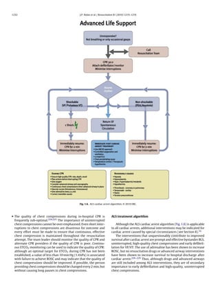
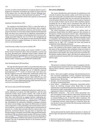
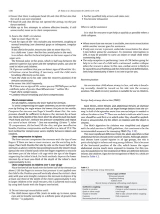
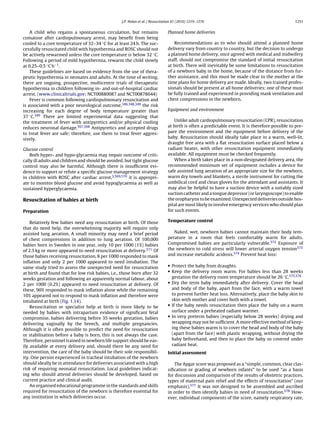
them. The literature reviews followed a standardised ‘worksheet’ The Chain of Survival
template including a specifically designed grading system to define
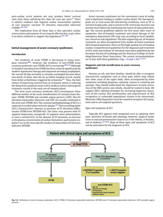
the level of evidence of each study.39 When possible, two expert The actions linking the victim of sudden cardiac arrest with sur-
reviewers were invited to undertake independent evaluations for vival are called the Chain of Survival (Fig. 1.1). The first link of this
each topic. The 2010 International Consensus Conference involved chain indicates the importance of recognising those at risk of car-
313 experts from 30 countries. During the 3 years leading up diac arrest and calling for help in the hope that early treatment
to this conference, 356 worksheet authors reviewed thousands can prevent arrest. The central links depict the integration of CPR
of relevant, peer-reviewed publications to address 277 specific and defibrillation as the fundamental components of early resus-
resuscitation questions, each in standard PICO (Population, Inter- citation in an attempt to restore life. Immediate CPR can double or
vention, Comparison Outcome) format.2 Each science statement triple survival from VF OHCA.42–45 Performing chest-compression-
summarised the experts’ interpretation of all relevant data on a spe- only CPR is better than giving no CPR at all.46,47 Following VF OHCA,
cific topic and consensus draft treatment recommendations were cardiopulmonary resuscitation plus defibrillation within 3–5 min
added by the relevant ILCOR task force. Final wording of science of collapse can produce survival rates as high as 49–75%.48–55 Each
statements and treatment recommendations was completed after minute of delay before defibrillation reduces the probability of sur-
further review by ILCOR member organisations and the editorial vival to discharge by 10–12%.42,56 The final link in the Chain of
board.2 Survival, effective post-resuscitation care, is targeted at preserving
The comprehensive conflict of interest (COI) policy that was function, particularly of the brain and heart. In hospital, the impor-
created for the 2005 International Consensus Conference40 was tance of early recognition of the critically ill patient and activation
revised for 2010.41 Representatives of manufacturers and industry of a medical emergency or rapid response team, with treatment
did not participate in either of the 2005 and the 2010 conferences. aimed at preventing cardiac arrest, is now well accepted.6 Over the
last few years, the importance of the post-cardiac arrest phase of
From science to guidelines treatment, depicted in the fourth ring of the Chain of Survival, has
been increasingly recognised.3 Differences in post-cardiac arrest
As in 2005, the resuscitation organisations forming ILCOR will treatment may account for some of the inter-hospital variability in
publish individual resuscitation guidelines that are consistent outcome after cardiac arrest.57–63
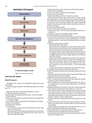
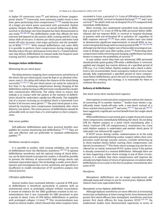
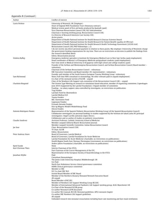
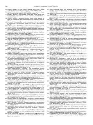
Fig. 1.1. Chain of Survival.](https://image.slidesharecdn.com/fullerc2010guidelines-120109122937-phpapp02/85/Full-erc-2010_guidelines-5-320.jpg)
![1240 J.P. Nolan et al. / Resuscitation 81 (2010) 1219–1276
the use of induced hypothermia in comatose survivors of out-of- Biochemical markers
hospital cardiac arrest caused by VF. One randomised trial355 and a Evidence does not support the use of serum (e.g., neuronal spe-
pseudo-randomised trial356 demonstrated improved neurological cific enolase, S100 protein) or CSF biomarkers alone as predictors
outcome at hospital discharge or at 6 months in comatose patients of poor outcomes in comatose patients after cardiac arrest with or
after out-of-hospital VF cardiac arrest. Cooling was initiated within without treatment with therapeutic hypothermia (TH). Limitations
minutes to hours after ROSC and a temperature range of 32–34 ◦ C included small numbers of patients studied and/or inconsistency in
was maintained for 12–24 h. Extrapolation of these data to other cut-off values for predicting poor outcome.
cardiac arrests (e.g., other initial rhythms, in-hospital arrests, pae-
diatric patients) seems reasonable but is supported by only lower Electrophysiological studies
level data.317,357–363 No electrophysiological study reliably predicts outcome of a
The practical application of therapeutic hypothermia is divided comatose patient within the first 24 h after cardiac arrest. If
into three phases: induction, maintenance, and rewarming.364 Ani- somatosensory evoked potentials (SSEP) are measured after 24 h
mal data indicate that earlier cooling after ROSC produces better in comatose cardiac arrest survivors not treated with therapeu-
outcomes.365 External and/or internal cooling techniques can be tic hypothermia, bilateral absence of the N20 cortical response to
used to initiate cooling. An infusion of 30 ml kg−1 of 4 ◦ C saline or median nerve stimulation predicts poor outcome (death or CPC 3
Hartmann’s solution decreases core temperature by approximately or 4) with a FPR of 0.7% (95% CI: 0.1–3.7%).376
1.5 ◦ C. Other methods of inducing and/or maintaining hypothermia
include: simple ice packs and/or wet towels; cooling blankets or Imaging studies
pads; water or air circulating blankets; water circulating gel-coated Many imaging modalities (magnetic resonance imaging [MRI],
pads; intravascular heat exchanger; and cardiopulmonary bypass. computed tomography [CT], single photon emission com-
In the maintenance phase, a cooling method with effective puted tomography [SPECT], cerebral angiography, transcranial
temperature monitoring that avoids temperature fluctuations is Doppler, nuclear medicine, near infra-red spectroscopy [NIRS])
preferred. This is best achieved with external or internal cooling have been studied to determine their utility for prediction
devices that include continuous temperature feedback to achieve a of outcome in adult cardiac arrest survivors.15 There are
set target temperature. Plasma electrolyte concentrations, effective no high-level studies that support the use of any imaging
intravascular volume and metabolic rate can change rapidly dur- modality to predict outcome of comatose cardiac arrest sur-
ing rewarming, as they do during cooling. Thus, rewarming must be vivors.
achieved slowly: the optimal rate is not known, but the consensus
is currently about 0.25–0.5 ◦ C of warming per hour.362 Impact of therapeutic hypothermia on prognostication
The well-recognised physiological effects of hypothermia need There is inadequate evidence to recommend a specific approach
to be managed carefully.364 to prognosticating poor outcome in post-cardiac arrest patients
treated with therapeutic hypothermia. There are no clinical neuro-
logical signs, electrophysiological studies, biomarkers, or imaging
Prognostication
modalities that can reliably predict neurological outcome in the
first 24 h after cardiac arrest. Based on limited available evidence,
Two-thirds of those dying after admission to ICU following out-
potentially reliable prognosticators of poor outcome in patients
of-hospital cardiac arrest die from neurological injury; this has been
treated with therapeutic hypothermia after cardiac arrest include
shown both with227 and without305 therapeutic hypothermia. A
bilateral absence of N20 peak on SSEP ≥24 h after cardiac arrest
quarter of those dying after admission to ICU following in-hospital
(FPR 0%, 95% CI 0–69%) and the absence of both corneal and pupil-
cardiac arrest die from neurological injury. A means of predicting
lary reflexes 3 or more days after cardiac arrest (FPR 0%, 95% CI
neurological outcome that can be applied to individual patients
0–48%).368,377 Limited available evidence also suggests that a Glas-
immediately after ROSC is required. Many studies have focused on
gow Motor Score of 2 or less at 3 days post-ROSC (FPR 14% [95% CI
prediction of poor long-term outcome (vegetative state or death),
3–44%])368 and the presence of status epilepticus (FPR of 7% [95%
based on clinical or test findings that indicate irreversible brain
CI 1–25%] to 11.5% [95% CI 3–31%])378,379 are potentially unreliable
injury, to enable clinicians to limit care or withdraw organ sup-
prognosticators of poor outcome in post-cardiac arrest patients
port. The implications of these prognostic tests are such that they
treated with therapeutic hypothermia. Given the limited available
should have 100% specificity or zero false positive rate (FPR), i.e.,
evidence, decisions to limit care should not be made based on the
proportion of individuals who eventually have a ‘good’ long-term
results of a single prognostication tool.
outcome despite the prediction of a poor outcome.
Organ donation
Clinical examination
There are no clinical neurological signs that predict poor out- Solid organs have been successfully transplanted after cardiac
come (Cerebral Performance Category [CPC] 3 or 4, or death) death.380 This group of patients offers an untapped opportunity
reliably less than 24 h after cardiac arrest. In adult patients who to increase the organ donor pool. Organ retrieval from non-heart
are comatose after cardiac arrest, and who have not been treated beating donors is classified as controlled or uncontrolled.381 Con-
with hypothermia and who do not have confounding factors (such trolled donation occurs after planned withdrawal of treatment
as hypotension, sedatives or muscle relaxants), the absence of both following non-survivable injuries/illnesses. Uncontrolled dona-
pupillary light and corneal reflex at ≥72 h reliably predicts poor tion describes donation after a patient is brought in dead or
outcome (FPR 0%; 95% CI 0–9%).330 Absence of vestibulo-ocular with on-going CPR that fails to restore a spontaneous circula-
reflexes at ≥24 h (FPR 0%; 95% CI 0–14%)366,367 and a GCS motor tion.
score of 2 or less at ≥72 h (FPR 5%; 95% CI 2–9%)330 are less reliable.
Other clinical signs, including myoclonus, are not recommended Cardiac arrest centres
for predicting poor outcome. The presence of myoclonus status in
adults is strongly associated with poor outcome,329,330,368–370 but There is wide variability in survival among hospitals caring
rare cases of good neurological recovery have been described and for patients after resuscitation from cardiac arrest.57–63 There
accurate diagnosis is problematic.371–375 is some low-level evidence that ICUs admitting more than 50](https://image.slidesharecdn.com/fullerc2010guidelines-120109122937-phpapp02/85/Full-erc-2010_guidelines-22-320.jpg)
![J.P. Nolan et al. / Resuscitation 81 (2010) 1219–1276 1243
Treatment of acute coronary syndromes—symptoms as soon as possible is recommended for patients presenting with
STEMI and planned PCI. Prasugrel or ticagrelor can be used instead
Glyceryl trinitrate is an effective treatment for ischaemic chest of clopidogrel before planned PCI. Patients with STEMI treated with
pain and has beneficial haemodynamic effects, such as dilation of fibrinolysis should be treated with clopidogrel (300 mg loading
the venous capacitance vessels, dilation of the coronary arteries dose up to an age of 75 years and 75 mg without loading dose if
and, to a minor extent, the peripheral arteries. Glyceryl trinitrate >75 years of age) in addition to ASA and an antithrombin.
may be considered if the systolic blood pressure is above 90 mm Hg
and the patient has ongoing ischaemic chest pain. Glyceryl trini- Glycoprotein (Gp) IIB/IIIA inhibitors
trate can also be useful in the treatment of acute pulmonary Gp IIB/IIIA receptor is the common final link of platelet aggre-
congestion. Nitrates should not be used in patients with hypoten- gation. Eptifibatide and tirofiban lead to reversible inhibition,
sion (systolic blood pressure ≤90 mm Hg), particularly if combined while abciximab leads to irreversible inhibition of the Gp IIB/IIIA
with bradycardia, and in patients with inferior infarction and sus- receptor. There are insufficient data to support routine pre-
pected right ventricular involvement. Use of nitrates under these treatment with Gp IIB/IIIA inhibitors in patients with STEMI or
circumstances can decrease the blood pressure and cardiac output. non-STEMI-ACS.
Morphine is the analgesic of choice for nitrate-refractory pain
and also has calming effects on the patient making sedatives
Antithrombins
unnecessary in most cases. Since morphine is a dilator of venous
capacitance vessels, it may have additional benefit in patients with
Unfractionated heparin (UFH) is an indirect inhibitor of throm-
pulmonary congestion. Give morphine in initial doses of 3–5 mg
bin, which in combination with ASA is used as an adjunct with
intravenously and repeat every few minutes until the patient is
fibrinolytic therapy or primary PCI (PPCI) and is an important part
pain-free. Non-steroidal anti-inflammatory drugs (NSAIDs) should
of treatment of unstable angina and STEMI. There are now several
be avoided for analgesia because of their pro-thrombotic effects.423
alternative antithrombins for the treatment of patients with ACS.
Monitoring of the arterial oxygen saturation (SaO2 ) with pulse
In comparison with UFH, these alternatives have a more specific
oximetry will help to determine the need for supplemental oxy-
factor Xa activity (low molecular weight heparins [LMWH], fonda-
gen. These patients do not need supplemental oxygen unless
parinux) or are direct thrombin inhibitors (bivalirudin). With these
they are hypoxaemic. Limited data suggest that high-flow oxy-
newer antithrombins, in general, there is no need to monitor the
gen may be harmful in patients with uncomplicated myocardial
coagulation system and there is a reduced risk of thrombocytope-
infarction.424–426 Aim to achieve an oxygen saturation of 94–98%, or
nia.
88–92% if the patient is at risk of hypercapnic respiratory failure.427
In comparison with UFH, enoxaparin reduces the combined end-
point of mortality, myocardial infarction and the need for urgent
Treatment of acute coronary syndromes—cause
revascularisation, if given within the first 24–36 h of onset of symp-
toms of non-STEMI-ACS.432,433 For patients with a planned initial
Inhibitors of platelet aggregation
conservative approach, fondaparinux and enoxaparin are reason-
able alternatives to UFH. For patients with an increased bleeding
Inhibition of platelet aggregation is of primary importance for
risk consider giving fondaparinux or bivalirudin, which cause less
initial treatment of coronary syndromes as well as for secondary
bleeding than UFH.434–436 For patients with a planned invasive
prevention, since platelet activation and aggregation is the key
approach, enoxaparin or bivalirudin are reasonable alternatives to
process initiating an ACS.
UFH.
Several randomised studies of patients with STEMI under-
Acetylsalicylic acid (ASA)
going fibrinolysis have shown that additional treatment with
Large randomised controlled trials indicate decreased mortal-
enoxaparin instead of UFH produced better clinical outcomes
ity when ASA (75–325 mg) is given to hospitalised patients with
(irrespective of the fibrinolytic used) but a slightly increased
ACS. A few studies have suggested reduced mortality if ASA is given
bleeding rate in elderly (≥75 years) and low weight patients
earlier.428,429 Therefore, give ASA as soon as possible to all patients
(BW < 60 kg).437–439
with suspected ACS unless the patient has a known true allergy to
Enoxaparin is a safe and effective alternative to UFH for con-
ASA. ASA may be given by the first healthcare provider, bystander
temporary PPCI (i.e., broad use of thienopyridines and/or Gp IIB/IIIA
or by dispatcher assistance according to local protocols. The initial
receptor blockers).440,441 There are insufficient data to recommend
dose of chewable ASA is 160–325 mg. Other forms of ASA (soluble,
any LMWH other than enoxaparin for PPCI in STEMI. Bivalirudin is
IV) may be as effective as chewed tablets.
also a safe alternative to UFH for STEMI and planned PCI.
ADP receptor inhibitors
Thienopyridines (clopidogrel, prasugrel) and the cyclo-pentyl- Strategies and systems of care
triazolo-pyrimidine, ticagrelor, inhibit the ADP receptor irre-
versibly, which further reduces platelet aggregation in addition to Several systematic strategies to improve quality of out-of-
that produced by ASA. hospital care for patients with ACS have been investigated. These
If given in addition to heparin and ASA in high-risk non-STEMI- strategies are principally intended to promptly identify patients
ACS patients, clopidogrel improves outcome.430,431 Clopidogrel with STEMI in order to shorten the delay to reperfusion treatment.
should be given as early as possible in addition to ASA and an Also triage criteria have been developed to select high-risk patients
antithrombin to all patients presenting with non-STEMI-ACS. If a with non-STEMI-ACS for transport to tertiary care centres offering
conservative approach is selected, give a loading dose of 300 mg; 24/7 PCI services. In this context, several specific decisions have to
with a planned PCI strategy, an initial dose of 600 mg may be pre- be made during initial care beyond the basic diagnostic steps nec-
ferred. Prasugrel or ticagrelor can be given instead of clopidogrel. essary for clinical evaluation of the patient and interpretation of a
Although there is no large study on the use of clopidogrel for 12-lead ECG. These decisions relate to:
pre-treatment of patients presenting with STEMI and planned PCI,
it is likely that this strategy is beneficial. Since platelet inhibition (1) Reperfusion strategy in patients with STEMI i.e., PPCI vs. pre-
is more profound with a higher dose, a 600 mg loading dose given hospital fibrinolysis.](https://image.slidesharecdn.com/fullerc2010guidelines-120109122937-phpapp02/85/Full-erc-2010_guidelines-25-320.jpg)
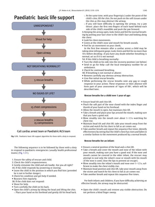
![1248 J.P. Nolan et al. / Resuscitation 81 (2010) 1219–1276
• detection of end-tidal CO2 if the child has a perfusing rhythm Circulation
(this may also be seen with effective CPR, but it is not completely • Establish cardiac monitoring [first line—pulse oximetry (SpO2 ),
reliable); ECG and non-invasive blood pressure (NIBP)].
• observation of symmetrical chest wall movement during positive • Secure vascular access. This may be by peripheral IV or IO cannu-
pressure ventilation; lation. If already in situ, a central intravenous catheter should be
• observation of mist in the tube during the expiratory phase of used.
ventilation; • Give a fluid bolus (20 ml kg−1 ) and/or drugs (e.g., inotropes, vaso-
• absence of gastric distension; pressors, anti-arrhythmics) as required.
• equal air entry heard on bilateral auscultation in the axillae and • Isotonic crystalloids are recommended as initial resuscitation
apices of the chest; fluid in infants and children with any type of shock, including
• absence of air entry into the stomach on auscultation; septic shock.515–518
• improvement or stabilisation of SpO2 in the expected range • Assess and re-assess the child continuously, commencing each
(delayed sign!); time with the airway before proceeding to breathing and then
• heart rate moving closer to the age-expected value (or remaining the circulation.
within the normal range) (delayed sign!). • During treatment, capnography, invasive monitoring of arterial
blood pressure, blood gas analysis, cardiac output monitoring,
echocardiography and central venous oxygen saturation (ScvO2 )
If the child is in cardiopulmonary arrest and exhaled CO2 is not may be useful to guide the management of respiratory and/or
detected despite adequate chest compressions, or if there is any circulatory failure.
doubt, confirm tracheal tube position by direct laryngoscopy.
Vascular access. Venous access can be difficult to establish dur-
ing resuscitation of an infant or child: if attempts at establishing
IV access are unsuccessful after one minute, insert an IO needle
Breathing. Give oxygen at the highest concentration (i.e., 100%) instead.519,520 Intraosseous or IV access is much preferred to the
during initial resuscitation. Once circulation is restored, give suffi- tracheal route for giving drugs.521
cient oxygen to maintain an arterial oxygen saturation (SaO2 ) in
the range of 94–98%.498,499
Healthcare providers commonly provide excessive ventila- Adrenaline. The recommended IV/IO dose of adrenaline in chil-
tion during CPR and this may be harmful. Hyperventilation dren for the first and for subsequent doses is 10 g kg−1 . The
causes increased intra-thoracic pressure, decreased cerebral and maximum single dose is 1 mg. If needed, give further doses of
coronary perfusion, and poorer survival rates in animals and adrenaline every 3–5 min. Intratracheal adrenaline is no longer
adults.224,225,286,500–503 Although normoventilation is the objec- recommended,522–525 but if this route is ever used, the dose is ten
tive during resuscitation, it is difficult to know the precise minute times this (100 g kg−1 ).
volume that is being delivered. A simple guide to deliver an accept-
able tidal volume is to achieve modest chest wall rise. Once the Advanced management of cardiopulmonary arrest
airway is protected by tracheal intubation, continue positive pres-
sure ventilation at 10–12 breaths min−1 without interrupting chest 1. When a child becomes unresponsive, without signs of life (no
compressions. When circulation is restored, or if the child still has breathing, cough or any detectable movement), start CPR imme-
a perfusing rhythm, ventilate at 12–20 breaths min−1 to achieve a diately.
normal arterial carbon dioxide tension (PaCO2 ). 2. Provide BMV with 100% oxygen.
Monitoring end-tidal CO2 (ETCO2 ) with a colorimetric detec- 3. Commence monitoring. Send for a manual defibrillator or an AED
tor or capnometer confirms tracheal tube placement in the child to identify and treat shockable rhythms as quickly as possible
weighing more than 2 kg, and may be used in pre- and in-hospital (Fig. 1.13).
settings, as well as during any transportation of the child.504–507
A colour change or the presence of a capnographic waveform for ABC
more than four ventilated breaths indicates that the tube is in the
tracheobronchial tree both in the presence of a perfusing rhythm
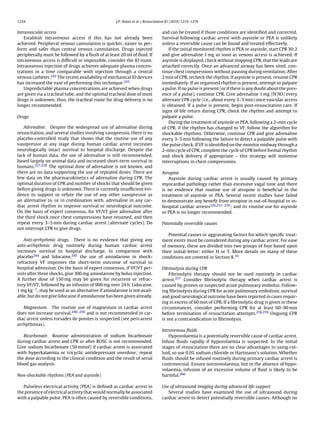
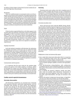
Commence and continue with basic life support
and during cardiopulmonary arrest. Capnography does not rule
Oxygenate and ventilate with BMV
out intubation of a bronchus. The absence of exhaled CO2 dur-
Provide positive pressure ventilation with a high inspired oxy-
ing cardiopulmonary arrest does not guarantee tube misplacement
gen concentration
since a low or absent end tidal CO2 may reflect low or absent
Give five rescue breaths followed by external chest compression
pulmonary blood flow.235,508–510 Capnography may also provide
and positive pressure ventilation in the ratio of 15:2
information on the efficiency of chest compressions and can give an
Avoid rescuer fatigue by frequently changing the rescuer per-
early indication of ROSC.511,512 Efforts should be made to improve
forming chest compressions
chest compression quality if the ETCO2 remains below 15 mm Hg
Establish cardiac monitoring
(2 kPa). Current evidence does not support the use of a threshold
Assess cardiac rhythm and signs of life
ETCO2 value as an indicator for the discontinuation of resuscitation
(±Check for a central pulse for no more than 10 s)
efforts.
The self-inflating bulb or aspirating syringe (oesophageal detec-
Non-shockable—asystole, PEA
tor device, ODD) may be used for the secondary confirmation of
tracheal tube placement in children with a perfusing rhythm.513,514
There are no studies on the use of the ODD in children who are in • Give adrenaline IV or IO (10 g kg−1 ) and repeat every 3–5 min.
cardiopulmonary arrest. • Identify and treat any reversible causes (4Hs & 4Ts).
Clinical evaluation of the oxygen saturation of arterial blood
(SaO2 ) is unreliable; therefore, monitor the child’s peripheral oxy- Shockable—VF/pulseless VT
gen saturation continuously by pulse oximetry (SpO2 ). Attempt defibrillation immediately (4 J kg−1 ):](https://image.slidesharecdn.com/fullerc2010guidelines-120109122937-phpapp02/85/Full-erc-2010_guidelines-30-320.jpg)
![1266 J.P. Nolan et al. / Resuscitation 81 (2010) 1219–1276
97. White RD, Bunch TJ, Hankins DG. Evolution of a community-wide early defib- 126. Strohmenger HU, Lindner KH, Brown CG. Analysis of the ventricular fibrillation
rillation programme experience over 13 years using police/fire personnel and ECG signal amplitude and frequency parameters as predictors of countershock
paramedics as responders. Resuscitation 2005;65:279–83. success in humans. Chest 1997;111:584–9.
98. Mosesso Jr VN, Davis EA, Auble TE, Paris PM, Yealy DM. Use of automated 127. Strohmenger HU, Eftestol T, Sunde K, et al. The predictive value of ventricu-
external defibrillators by police officers for treatment of out-of-hospital cardiac lar fibrillation electrocardiogram signal frequency and amplitude variables in
arrest. Ann Emerg Med 1998;32:200–7. patients with out-of-hospital cardiac arrest. Anesth Analg 2001;93:1428–33.
99. The Public Access Defibrillation Trial Investigators. Public-access defib- 128. Podbregar M, Kovacic M, Podbregar-Mars A, Brezocnik M. Predicting defibrilla-
rillation and survival after out-of-hospital cardiac arrest. N Engl J Med tion success by ‘genetic’ programming in patients with out-of-hospital cardiac
2004;351:637–46. arrest. Resuscitation 2003;57:153–9.
100. Kitamura T, Iwami T, Kawamura T, Nagao K, Tanaka H, Hiraide A. Nationwide 129. Menegazzi JJ, Callaway CW, Sherman LD, et al. Ventricular fibrillation scaling
public-access defibrillation in Japan. N Engl J Med 2010;362:994–1004. exponent can guide timing of defibrillation and other therapies. Circulation
101. Bardy GH, Lee KL, Mark DB, et al. Home use of automated external defibrillators 2004;109:926–31.
for sudden cardiac arrest. N Engl J Med 2008;358:1793–804. 130. Povoas HP, Weil MH, Tang W, Bisera J, Klouche K, Barbatsis A. Predicting
102. Zafari AM, Zarter SK, Heggen V, et al. A program encouraging early defibril- the success of defibrillation by electrocardiographic analysis. Resuscitation
lation results in improved in-hospital resuscitation efficacy. J Am Coll Cardiol 2002;53:77–82.
2004;44:846–52. 131. Noc M, Weil MH, Tang W, Sun S, Pernat A, Bisera J. Electrocardiographic pre-
103. Destro A, Marzaloni M, Sermasi S, Rossi F. Automatic external defibrillators in diction of the success of cardiac resuscitation. Crit Care Med 1999;27:708–14.
the hospital as well? Resuscitation 1996;31:39–43. 132. Strohmenger HU, Lindner KH, Keller A, Lindner IM, Pfenninger EG. Spectral
104. Spearpoint KG, Gruber PC, Brett SJ. Impact of the Immediate Life Support course analysis of ventricular fibrillation and closed-chest cardiopulmonary resusci-
on the incidence and outcome of in-hospital cardiac arrest calls: an observa- tation. Resuscitation 1996;33:155–61.
tional study over 6 years. Resuscitation 2009;80:638–43. 133. Noc M, Weil MH, Gazmuri RJ, Sun S, Biscera J, Tang W. Ventricular fibrillation
105. Cummins RO, Eisenberg MS, Litwin PE, Graves JR, Hearne TR, Hallstrom AP. voltage as a monitor of the effectiveness of cardiopulmonary resuscitation. J
Automatic external defibrillators used by emergency medical technicians: a Lab Clin Med 1994;124:421–6.
controlled clinical trial. JAMA 1987;257:1605–10. 134. Lightfoot CB, Nremt P, Callaway CW, et al. Dynamic nature of electrocar-
106. Stults KR, Brown DD, Kerber RE. Efficacy of an automated external defibrillator diographic waveform predicts rescue shock outcome in porcine ventricular
in the management of out-of-hospital cardiac arrest: validation of the diagnos- fibrillation. Ann Emerg Med 2003;42:230–41.
tic algorithm and initial clinical experience in a rural environment. Circulation 135. Marn-Pernat A, Weil MH, Tang W, Pernat A, Bisera J. Optimizing timing of
1986;73:701–9. ventricular defibrillation. Crit Care Med 2001;29:2360–5.
107. Kramer-Johansen J, Edelson DP, Abella BS, Becker LB, Wik L, Steen PA. Pauses 136. Hamprecht FA, Achleitner U, Krismer AC, et al. Fibrillation power, an alternative
in chest compression and inappropriate shocks: a comparison of manual and method of ECG spectral analysis for prediction of countershock success in a
semi-automatic defibrillation attempts. Resuscitation 2007;73:212–20. porcine model of ventricular fibrillation. Resuscitation 2001;50:287–96.
108. Pytte M, Pedersen TE, Ottem J, Rokvam AS, Sunde K. Comparison of hands-off 137. Amann A, Achleitner U, Antretter H, et al. Analysing ventricular fibrilla-
time during CPR with manual and semi-automatic defibrillation in a manikin tion ECG-signals and predicting defibrillation success during cardiopulmonary
model. Resuscitation 2007;73:131–6. resuscitation employing N(alpha)-histograms. Resuscitation 2001;50:77–85.
109. Forcina MS, Farhat AY, O’Neil WW, Haines DE. Cardiac arrest survival after 138. Brown CG, Griffith RF, Van Ligten P, et al. Median frequency—a new parameter
implementation of automated external defibrillator technology in the in- for predicting defibrillation success rate. Ann Emerg Med 1991;20:787–9.
hospital setting. Crit Care Med 2009;37:1229–36. 139. Amann A, Rheinberger K, Achleitner U, et al. The prediction of defibrillation
110. Edelson DP, Abella BS, Kramer-Johansen J, et al. Effects of compression depth outcome using a new combination of mean frequency and amplitude in porcine
and pre-shock pauses predict defibrillation failure during cardiac arrest. Resus- models of cardiac arrest. Anesth Analg 2002;95:716–22 [table of contents].
citation 2006;71:137–45. 140. Deakin CD, Nolan JP. European Resuscitation Council guidelines for
111. Yu T, Weil MH, Tang W, et al. Adverse outcomes of interrupted precordial resuscitation 2005. Section 3. Electrical therapies: automated external defib-
compression during automated defibrillation. Circulation 2002;106:368–72. rillators, defibrillation, cardioversion and pacing. Resuscitation 2005;67(Suppl.
112. Gundersen K, Kvaloy JT, Kramer-Johansen J, Steen PA, Eftestol T. Development 1):S25–37.
of the probability of return of spontaneous circulation in intervals without 141. Cobb LA, Fahrenbruch CE, Walsh TR, et al. Influence of cardiopulmonary resus-
chest compressions during out-of-hospital cardiac arrest: an observational citation prior to defibrillation in patients with out-of-hospital ventricular
study. BMC Med 2009;7:6. fibrillation. JAMA 1999;281:1182–8.
113. Lloyd MS, Heeke B, Walter PF, Langberg JJ. Hands-on defibrillation: an analy- 142. Wik L, Hansen TB, Fylling F, et al. Delaying defibrillation to give basic cardiopul-
sis of electrical current flow through rescuers in direct contact with patients monary resuscitation to patients with out-of-hospital ventricular fibrillation:
during biphasic external defibrillation. Circulation 2008;117:2510–4. a randomized trial. JAMA 2003;289:1389–95.
114. Bojar RM, Payne DD, Rastegar H, Diehl JT, Cleveland RJ. Use of self-adhesive 143. Baker PW, Conway J, Cotton C, et al. Defibrillation or cardiopulmonary
external defibrillator pads for complex cardiac surgical procedures. Ann Thorac resuscitation first for patients with out-of-hospital cardiac arrests found by
Surg 1988;46:587–8. paramedics to be in ventricular fibrillation? A randomised control trial. Resus-
115. Bradbury N, Hyde D, Nolan J. Reliability of ECG monitoring with a gel citation 2008;79:424–31.
pad/paddle combination after defibrillation. Resuscitation 2000;44:203–6. 144. Jacobs IG, Finn JC, Oxer HF, Jelinek GA. CPR before defibrillation in
116. Brown J, Rogers J, Soar J. Cardiac arrest during surgery and ventilation out-of-hospital cardiac arrest: a randomized trial. Emerg Med Australas
in the prone position: a case report and systematic review. Resuscitation 2005;17:39–45.
2001;50:233–8. 145. Hayakawa M, Gando S, Okamoto H, Asai Y, Uegaki S, Makise H. Shortening
117. Perkins GD, Davies RP, Soar J, Thickett DR. The impact of manual defibrillation of cardiopulmonary resuscitation time before the defibrillation worsens the
technique on no-flow time during simulated cardiopulmonary resuscitation. outcome in out-of-hospital VF patients. Am J Emerg Med 2009;27:470–4.
Resuscitation 2007;73:109–14. 146. Bradley SM, Gabriel EE, Aufderheide TP, et al. Survival Increases with CPR by
118. Wilson RF, Sirna S, White CW, Kerber RE. Defibrillation of high-risk patients Emergency Medical Services before defibrillation of out-of-hospital ventricu-
during coronary angiography using self-adhesive, preapplied electrode pads. lar fibrillation or ventricular tachycardia: observations from the resuscitation
Am J Cardiol 1987;60:380–2. outcomes consortium. Resuscitation 2010;81:155–62.
119. Stults KR, Brown DD, Cooley F, Kerber RE. Self-adhesive monitor/defibrillation 147. Christenson J, Andrusiek D, Everson-Stewart S, et al. Chest compression fraction
pads improve prehospital defibrillation success. Ann Emerg Med determines survival in patients with out-of-hospital ventricular fibrillation.
1987;16:872–7. Circulation 2009;120:1241–7.
120. Callaway CW, Sherman LD, Mosesso Jr VN, Dietrich TJ, Holt E, Clarkson MC. 148. Olasveengen TM, Vik E, Kuzovlev A, Sunde K. Effect of implementation of new
Scaling exponent predicts defibrillation success for out-of-hospital ventricular resuscitation guidelines on quality of cardiopulmonary resuscitation and sur-
fibrillation cardiac arrest. Circulation 2001;103:1656–61. vival. Resuscitation 2009;80:407–11.
121. Eftestol T, Sunde K, Aase SO, Husoy JH, Steen PA. Predicting outcome of 149. Bobrow BJ, Clark LL, Ewy GA, et al. Minimally interrupted cardiac resuscita-
defibrillation by spectral characterization and nonparametric classification of tion by emergency medical services for out-of-hospital cardiac arrest. JAMA
ventricular fibrillation in patients with out-of-hospital cardiac arrest. Circula- 2008;299:1158–65.
tion 2000;102:1523–9. 150. Rea TD, Helbock M, Perry S, et al. Increasing use of cardiopulmonary resuscita-
122. Eftestol T, Wik L, Sunde K, Steen PA. Effects of cardiopulmonary resuscitation tion during out-of-hospital ventricular fibrillation arrest: survival implications
on predictors of ventricular fibrillation defibrillation success during out-of- of guideline changes. Circulation 2006;114:2760–5.
hospital cardiac arrest. Circulation 2004;110:10–5. 151. Steinmetz J, Barnung S, Nielsen SL, Risom M, Rasmussen LS. Improved survival
123. Weaver WD, Cobb LA, Dennis D, Ray R, Hallstrom AP, Copass MK. Amplitude of after an out-of-hospital cardiac arrest using new guidelines. Acta Anaesthesiol
ventricular fibrillation waveform and outcome after cardiac arrest. Ann Intern Scand 2008;52:908–13.
Med 1985;102:53–5. 152. Jost D, Degrange H, Verret C, et al. DEFI 2005: a randomized controlled trial
124. Brown CG, Dzwonczyk R. Signal analysis of the human electrocardiogram dur- of the effect of automated external defibrillator cardiopulmonary resusci-
ing ventricular fibrillation: frequency and amplitude parameters as predictors tation protocol on outcome from out-of-hospital cardiac arrest. Circulation
of successful countershock. Ann Emerg Med 1996;27:184–8. 2010;121:1614–22.
125. Callaham M, Braun O, Valentine W, Clark DM, Zegans C. Prehospital car- 153. van Alem AP, Chapman FW, Lank P, Hart AA, Koster RW. A prospective,
diac arrest treated by urban first-responders: profile of patient response and randomised and blinded comparison of first shock success of monopha-
prediction of outcome by ventricular fibrillation waveform. Ann Emerg Med sic and biphasic waveforms in out-of-hospital cardiac arrest. Resuscitation
1993;22:1664–77. 2003;58:17–24.](https://image.slidesharecdn.com/fullerc2010guidelines-120109122937-phpapp02/85/Full-erc-2010_guidelines-48-320.jpg)
![1270 J.P. Nolan et al. / Resuscitation 81 (2010) 1219–1276
neurological recovery in human cardiac arrest survivors. J Cereb Blood Flow 368. Al Thenayan E, Savard M, Sharpe M, Norton L, Young B. Predictors of poor
Metab 1997;17:430–6. neurologic outcome after induced mild hypothermia following cardiac arrest.
336. Calle PA, Buylaert WA, Vanhaute OA. Glycemia in the post-resuscitation Neurology 2008;71:1535–7.
period. The Cerebral Resuscitation Study Group. Resuscitation 369. Wijdicks EF, Parisi JE, Sharbrough FW. Prognostic value of myoclonus status in
1989;17(Suppl.):S181–8 [discussion S99–S206]. comatose survivors of cardiac arrest. Ann Neurol 1994;35:239–43.
337. Longstreth Jr WT, Diehr P, Inui TS. Prediction of awakening after out-of-hospital 370. Thomke F, Marx JJ, Sauer O, et al. Observations on comatose survivors
cardiac arrest. N Engl J Med 1983;308:1378–82. of cardiopulmonary resuscitation with generalized myoclonus. BMC Neurol
338. Longstreth Jr WT, Inui TS. High blood glucose level on hospital admission and 2005;5:14.
poor neurological recovery after cardiac arrest. Ann Neurol 1984;15:59–63. 371. Arnoldus EP, Lammers GJ. Postanoxic coma: good recovery despite myoclonus
339. Finfer S, Chittock DR, Su SY, et al. Intensive versus conventional glucose control status. Ann Neurol 1995;38:697–8.
in critically ill patients. N Engl J Med 2009;360:1283–97. 372. Celesia GG, Grigg MM, Ross E. Generalized status myoclonicus in acute anoxic
340. Preiser JC, Devos P, Ruiz-Santana S, et al. A prospective randomised multi- and toxic-metabolic encephalopathies. Arch Neurol 1988;45:781–4.
centre controlled trial on tight glucose control by intensive insulin therapy 373. Morris HR, Howard RS, Brown P. Early myoclonic status and outcome
in adult intensive care units: the Glucontrol study. Intensive Care Med after cardiorespiratory arrest. J Neurol Neurosurg Psychiatry 1998;64:
2009;35:1738–48. 267–8.
341. Griesdale DE, de Souza RJ, van Dam RM, et al. Intensive insulin therapy and 374. Datta S, Hart GK, Opdam H, Gutteridge G, Archer J. Post-hypoxic myoclonic
mortality among critically ill patients: a meta-analysis including NICE-SUGAR status: the prognosis is not always hopeless. Crit Care Resusc 2009;11:39–41.
study data. CMAJ 2009;180:821–7. 375. English WA, Giffin NJ, Nolan JP. Myoclonus after cardiac arrest: pitfalls in diag-
342. Wiener RS, Wiener DC, Larson RJ. Benefits and risks of tight glucose control in nosis and prognosis. Anaesthesia 2009;64:908–11.
critically ill adults: a meta-analysis. JAMA 2008;300:933–44. 376. Wijdicks EF, Hijdra A, Young GB, Bassetti CL, Wiebe S. Practice parameter: pre-
343. Krinsley JS, Grover A. Severe hypoglycemia in critically ill patients: risk factors diction of outcome in comatose survivors after cardiopulmonary resuscitation
and outcomes. Crit Care Med 2007;35:2262–7. (an evidence-based review): report of the Quality Standards Subcommittee of
344. Meyfroidt G, Keenan DM, Wang X, Wouters PJ, Veldhuis JD, Van den Berghe the American Academy of Neurology. Neurology 2006;67:203–10.
G. Dynamic characteristics of blood glucose time series during the course of 377. Tiainen M, Kovala TT, Takkunen OS, Roine RO. Somatosensory and brainstem
critical illness: effects of intensive insulin therapy and relative association with auditory evoked potentials in cardiac arrest patients treated with hypothermia.
mortality. Crit Care Med 2010;38:1021–9. Crit Care Med 2005;33:1736–40.
345. Padkin A. Glucose control after cardiac arrest. Resuscitation 2009;80:611–2. 378. Rossetti AO, Oddo M, Liaudet L, Kaplan PW. Predictors of awakening
346. Takino M, Okada Y. Hyperthermia following cardiopulmonary resuscitation. from postanoxic status epilepticus after therapeutic hypothermia. Neurology
Intensive Care Med 1991;17:419–20. 2009;72:744–9.
347. Hickey RW, Kochanek PM, Ferimer H, Alexander HL, Garman RH, Graham 379. Rossetti AO, Logroscino G, Liaudet L, et al. Status epilepticus: an independent
SH. Induced hyperthermia exacerbates neurologic neuronal histologic damage outcome predictor after cerebral anoxia. Neurology 2007;69:255–60.
after asphyxial cardiac arrest in rats. Crit Care Med 2003;31:531–5. 380. Fieux F, Losser MR, Bourgeois E, et al. Kidney retrieval after sudden out of
348. Takasu A, Saitoh D, Kaneko N, Sakamoto T, Okada Y. Hyperthermia: is it an hospital refractory cardiac arrest: a cohort of uncontrolled non heart beating
ominous sign after cardiac arrest? Resuscitation 2001;49:273–7. donors. Crit Care 2009;13:R141.
349. Zeiner A, Holzer M, Sterz F, et al. Hyperthermia after cardiac arrest is associated 381. Kootstra G. Statement on non-heart-beating donor programs. Transplant Proc
with an unfavorable neurologic outcome. Arch Intern Med 2001;161:2007–12. 1995;27:2965.
350. Hickey RW, Kochanek PM, Ferimer H, Graham SH, Safar P. Hypothermia and 382. Vermeer F, Oude Ophuis AJ, vd Berg EJ, et al. Prospective randomised compar-
hyperthermia in children after resuscitation from cardiac arrest. Pediatrics ison between thrombolysis, rescue PTCA, and primary PTCA in patients with
2000;106:118–22. extensive myocardial infarction admitted to a hospital without PTCA facilities:
351. Diringer MN, Reaven NL, Funk SE, Uman GC. Elevated body temperature inde- a safety and feasibility study. Heart 1999;82:426–31.
pendently contributes to increased length of stay in neurologic intensive care 383. Widimsky P, Groch L, Zelizko M, Aschermann M, Bednar F, Suryapranata H.
unit patients. Crit Care Med 2004;32:1489–95. Multicentre randomized trial comparing transport to primary angioplasty vs
352. Gunn AJ, Thoresen M. Hypothermic neuroprotection. NeuroRx 2006;3:154–69. immediate thrombolysis vs combined strategy for patients with acute myocar-
353. Froehler MT, Geocadin RG. Hypothermia for neuroprotection after car- dial infarction presenting to a community hospital without a catheterization
diac arrest: mechanisms, clinical trials and patient care. J Neurol Sci laboratory. The PRAGUE study. Eur Heart J 2000;21:823–31.
2007;261:118–26. 384. Widimsky P, Budesinsky T, Vorac D, et al. Long distance transport for primary
354. McCullough JN, Zhang N, Reich DL, et al. Cerebral metabolic suppression during angioplasty vs immediate thrombolysis in acute myocardial infarction. Final
hypothermic circulatory arrest in humans. Ann Thorac Surg 1999;67:1895–9 results of the randomized national multicentre trial—PRAGUE-2. Eur Heart J
[discussion 919–21]. 2003;24:94–104.
355. Mild therapeutic hypothermia to improve the neurologic outcome after cardiac 385. Le May MR, So DY, Dionne R, et al. A citywide protocol for primary PCI in
arrest. N Engl J Med 2002;346:549–56. ST-segment elevation myocardial infarction. N Engl J Med 2008;358:231–40.
356. Bernard SA, Gray TW, Buist MD, et al. Treatment of comatose survivors 386. Abernathy 3rd JH, McGwin Jr G, Acker 3rd JE, Rue 3rd LW. Impact of a voluntary
of out-of-hospital cardiac arrest with induced hypothermia. N Engl J Med trauma system on mortality, length of stay, and cost at a level I trauma center.
2002;346:557–63. Am Surg 2002;68:182–92.
357. Bernard SA, Jones BM, Horne MK. Clinical trial of induced hypothermia 387. Clemmer TP, Orme Jr JF, Thomas FO, Brooks KA. Outcome of critically injured
in comatose survivors of out-of-hospital cardiac arrest. Ann Emerg Med patients treated at Level I trauma centers versus full-service community hos-
1997;30:146–53. pitals. Crit Care Med 1985;13:861–3.
358. Oddo M, Schaller MD, Feihl F, Ribordy V, Liaudet L. From evidence to clini- 388. Culica D, Aday LA, Rohrer JE. Regionalized trauma care system in Texas: impli-
cal practice: effective implementation of therapeutic hypothermia to improve cations for redesigning trauma systems. Med Sci Monit 2007;13:SR9–18.
patient outcome after cardiac arrest. Crit Care Med 2006;34:1865–73. 389. Hannan EL, Farrell LS, Cooper A, Henry M, Simon B, Simon R. Physiologic trauma
359. Busch M, Soreide E, Lossius HM, Lexow K, Dickstein K. Rapid implementation of triage criteria in adult trauma patients: are they effective in saving lives by
therapeutic hypothermia in comatose out-of-hospital cardiac arrest survivors. transporting patients to trauma centers? J Am Coll Surg 2005;200:584–92.
Acta Anaesthesiol Scand 2006;50:1277–83. 390. Harrington DT, Connolly M, Biffl WL, Majercik SD, Cioffi WG. Transfer times to
360. Storm C, Steffen I, Schefold JC, et al. Mild therapeutic hypothermia shortens definitive care facilities are too long: a consequence of an immature trauma
intensive care unit stay of survivors after out-of-hospital cardiac arrest com- system. Ann Surg 2005;241:961–6 [discussion 6–8].
pared to historical controls. Crit Care 2008;12:R78. 391. Liberman M, Mulder DS, Lavoie A, Sampalis JS. Implementation of a trauma
361. Don CW, Longstreth Jr WT, Maynard C, et al. Active surface cooling protocol care system: evolution through evaluation. J Trauma 2004;56:1330–5.
to induce mild therapeutic hypothermia after out-of-hospital cardiac arrest: a 392. MacKenzie EJ, Rivara FP, Jurkovich GJ, et al. A national evaluation of the effect
retrospective before-and-after comparison in a single hospital. Crit Care Med of trauma-center care on mortality. N Engl J Med 2006;354:366–78.
2009;37:3062–9. 393. Mann NC, Cahn RM, Mullins RJ, Brand DM, Jurkovich GJ. Survival among injured
362. Arrich J. Clinical application of mild therapeutic hypothermia after cardiac geriatric patients during construction of a statewide trauma system. J Trauma
arrest. Crit Care Med 2007;35:1041–7. 2001;50:1111–6.
363. Holzer M, Mullner M, Sterz F, et al. Efficacy and safety of endovascular 394. Mullins RJ, Veum-Stone J, Hedges JR, et al. Influence of a statewide trauma
cooling after cardiac arrest: cohort study and Bayesian approach. Stroke system on location of hospitalization and outcome of injured patients. J Trauma
2006;37:1792–7. 1996;40:536–45 [discussion 45–46].
364. Polderman KH, Herold I. Therapeutic hypothermia and controlled normother- 395. Mullins RJ, Mann NC, Hedges JR, Worrall W, Jurkovich GJ. Preferential benefit
mia in the intensive care unit: practical considerations, side effects, and cooling of implementation of a statewide trauma system in one of two adjacent states.
methods. Crit Care Med 2009;37:1101–20. J Trauma 1998;44:609–16 [discussion 17].
365. Kuboyama K, Safar P, Radovsky A, et al. Delay in cooling negates the beneficial 396. Mullins RJ, Veum-Stone J, Helfand M, et al. Outcome of hospitalized injured
effect of mild resuscitative cerebral hypothermia after cardia arrest in dogs: a patients after institution of a trauma system in an urban area. JAMA
prospective, randomized study. Crit Care Med 1993;21:1348–58. 1994;271:1919–24.
366. Edgren E, Hedstrand U, Nordin M, Rydin E, Ronquist G. Prediction of outcome 397. Mullner R, Goldberg J. An evaluation of the Illinois trauma system. Med Care
after cardiac arrest. Crit Care Med 1987;15:820–5. 1978;16:140–51.
367. Young GB, Doig G, Ragazzoni A. Anoxic-ischemic encephalopathy: clin- 398. Mullner R, Goldberg J. Toward an outcome-oriented medical geography: an
ical and electrophysiological associations with outcome. Neurocrit Care evaluation of the Illinois trauma/emergency medical services system. Soc Sci
2005;2:159–64. Med 1978;12:103–10.](https://image.slidesharecdn.com/fullerc2010guidelines-120109122937-phpapp02/85/Full-erc-2010_guidelines-52-320.jpg)
![J.P. Nolan et al. / Resuscitation 81 (2010) 1219–1276 1271
399. Nathens AB, Jurkovich GJ, Rivara FP, Maier RV. Effectiveness of state trauma 427. O’Driscoll BR, Howard LS, Davison AG. BTS guideline for emergency oxygen use
systems in reducing injury-related mortality: a national evaluation. J Trauma in adult patients. Thorax 2008;63(Suppl. 6):vi1–68.
2000;48:25–30 [discussion 1]. 428. Freimark D, Matetzky S, Leor J, et al. Timing of aspirin administration as a
400. Nathens AB, Maier RV, Brundage SI, Jurkovich GJ, Grossman DC. The effect determinant of survival of patients with acute myocardial infarction treated
of interfacility transfer on outcome in an urban trauma system. J Trauma with thrombolysis. Am J Cardiol 2002;89:381–5.
2003;55:444–9. 429. Barbash IM, Freimark D, Gottlieb S, et al. Outcome of myocardial infarction
401. Nicholl J, Turner J. Effectiveness of a regional trauma system in reducing mor- in patients treated with aspirin is enhanced by pre-hospital administration.
tality from major trauma: before and after study. BMJ 1997;315:1349–54. Cardiology 2002;98:141–7.
402. Potoka DA, Schall LC, Gardner MJ, Stafford PW, Peitzman AB, Ford HR. Impact 430. Yusuf S, Zhao F, Mehta SR, Chrolavicius S, Tognoni G, Fox KK. Effects of clopido-
of pediatric trauma centers on mortality in a statewide system. J Trauma grel in addition to aspirin in patients with acute coronary syndromes without
2000;49:237–45. ST-segment elevation. N Engl J Med 2001;345:494–502.
403. Sampalis JS, Lavoie A, Boukas S, et al. Trauma center designation: initial impact 431. Mehta SR, Yusuf S, Peters RJ, et al. Effects of pretreatment with clopi-
on trauma-related mortality. J Trauma 1995;39:232–7 [discussion 7–9]. dogrel and aspirin followed by long-term therapy in patients undergoing
404. Sampalis JS, Denis R, Frechette P, Brown R, Fleiszer D, Mulder D. Direct transport percutaneous coronary intervention: the PCI-CURE study. Lancet 2001;358:
to tertiary trauma centers versus transfer from lower level facilities: impact 527–33.
on mortality and morbidity among patients with major trauma. J Trauma 432. TIMI-11B Investigators, Antman EM, McCabe CH, et al. Enoxaparin prevents
1997;43:288–95 [discussion 95–96]. death and cardiac ischemic events in unstable angina/non-Q-wave myocardial
405. Nichol G, Aufderheide TP, Eigel B, et al. Regional systems of care for out- infarction. Results of the thrombolysis in myocardial infarction (TIMI) 11B trial.
of-hospital cardiac arrest: a policy statement from the American Heart Circulation 1999;100:1593–601.
Association. Circulation 2010;121:709–29. 433. Cohen M, Demers C, Gurfinkel EP, et al. A comparison of low-molecular-weight
406. Nichol G, Soar J. Regional cardiac resuscitation systems of care. Curr Opin Crit heparin with unfractionated heparin for unstable coronary artery disease. Effi-
Care 2010;16:223–30. cacy and safety of subcutaneous enoxaparin in non-Q-wave coronary events
407. Soar J, Packham S. Cardiac arrest centres make sense. Resuscitation study group. N Engl J Med 1997;337:447–52.
2010;81:507–8. 434. Yusuf S, Mehta SR, Chrolavicius S, et al. Comparison of fondaparinux and enoxa-
408. Tunstall-Pedoe H, Vanuzzo D, Hobbs M, et al. Estimation of contribution of parin in acute coronary syndromes. N Engl J Med 2006;354:1464–76.
changes in coronary care to improving survival, event rates, and coronary 435. Mehta SR, Boden WE, Eikelboom JW, et al. Antithrombotic therapy with fon-
heart disease mortality across the WHO MONICA Project populations. Lancet daparinux in relation to interventional management strategy in patients with
2000;355:688–700. ST- and non-ST-segment elevation acute coronary syndromes: an individual
409. Fox KA, Cokkinos DV, Deckers J, Keil U, Maggioni A, Steg G. The ENACT study: patient-level combined analysis of the Fifth and Sixth Organization to Assess
a pan-European survey of acute coronary syndromes. European Network for Strategies in Ischemic Syndromes (OASIS 5 and 6) randomized trials. Circula-
Acute Coronary Treatment. Eur Heart J 2000;21:1440–9. tion 2008;118:2038–46.
410. Goodman SG, Huang W, Yan AT, et al. The expanded global registry of 436. Lincoff AM, Bittl JA, Harrington RA, et al. Bivalirudin and provisional glycopro-
acute coronary events: baseline characteristics, management practices, and tein IIb/IIIa blockade compared with heparin and planned glycoprotein IIb/IIIa
hospital outcomes of patients with acute coronary syndromes. Am Heart J blockade during percutaneous coronary intervention: REPLACE-2 randomized
2009;158:193–201, e1–e5. trial. JAMA 2003;289:853–63.
411. Lowel H, Meisinger C, Heier M, et al. Sex specific trends of sudden car- 437. Efficacy and safety of tenecteplase in combination with enoxaparin, abciximab,
diac death and acute myocardial infarction: results of the population-based or unfractionated heparin: the ASSENT-3 randomised trial in acute myocardial
KORA/MONICA-Augsburg register 1985 to 1998. Dtsch Med Wochenschr infarction. Lancet 2001;358:605–13.
2002;127:2311–6. 438. Eikelboom JW, Quinlan DJ, Mehta SR, Turpie AG, Menown IB, Yusuf S. Unfrac-
412. Thygesen K, Alpert JS, White HD. Universal definition of myocardial infarction. tionated and low-molecular-weight heparin as adjuncts to thrombolysis in
Eur Heart J 2007;28:2525–38. aspirin-treated patients with ST-elevation acute myocardial infarction: a meta-
413. Van de Werf F, Bax J, Betriu A, et al. Management of acute myocardial infarction analysis of the randomized trials. Circulation 2005;112:3855–67.
in patients presenting with persistent ST-segment elevation: the Task Force on 439. Wallentin L, Goldstein P, Armstrong PW, et al. Efficacy and safety of
the Management of ST-Segment Elevation Acute Myocardial Infarction of the tenecteplase in combination with the low-molecular-weight heparin enoxa-
European Society of Cardiology. Eur Heart J 2008;29:2909–45. parin or unfractionated heparin in the prehospital setting: the Assessment
414. Antman EM, Anbe DT, Armstrong PW, et al. ACC/AHA guidelines for the of the Safety and Efficacy of a New Thrombolytic Regimen (ASSENT)-3
management of patients with ST-elevation myocardial infarction—executive PLUS randomized trial in acute myocardial infarction. Circulation 2003;108:
summary: a report of the American College of Cardiology/American Heart 135–42.
Association Task Force on Practice Guidelines (Writing Committee to Revise 440. Zeymer U, Gitt A, Junger C, et al. Efficacy and safety of enoxaparin in unselected
the 1999 Guidelines for the Management of Patients With Acute Myocardial patients with ST-segment elevation myocardial infarction. Thromb Haemost
Infarction). Circulation 2004;110:588–636. 2008;99:150–4.
415. Douglas PS, Ginsburg GS. The evaluation of chest pain in women. N Engl J Med 441. Zeymer U, Gitt A, Zahn R, et al. Efficacy and safety of enoxaparin in combination
1996;334:1311–5. with and without GP IIb/IIIa inhibitors in unselected patients with ST segment
416. Solomon CG, Lee TH, Cook EF, et al. Comparison of clinical presentation of elevation myocardial infarction treated with primary percutaneous coronary
acute myocardial infarction in patients older than 65 years of age to younger intervention. EuroIntervention 2009;4:524–8.
patients: the Multicenter Chest Pain Study experience. Am J Cardiol 1989;63: 442. Bassand JP, Hamm CW, Ardissino D, et al. Guidelines for the diagnosis and
772–6. treatment of non-ST-segment elevation acute coronary syndromes. Eur Heart
417. Ioannidis JP, Salem D, Chew PW, Lau J. Accuracy and clinical effect of out- J 2007;28:1598–660.
of-hospital electrocardiography in the diagnosis of acute cardiac ischemia: a 443. Anderson JL, Adams CD, Antman EM, et al. ACC/AHA 2007 guidelines for
meta-analysis. Ann Emerg Med 2001;37:461–70. the management of patients with unstable angina/non ST-elevation myocar-
418. Kudenchuk PJ, Ho MT, Weaver WD, et al. Accuracy of computer-interpreted dial infarction: a report of the American College of Cardiology/American
electrocardiography in selecting patients for thrombolytic therapy. MITI Heart Association Task Force on Practice Guidelines (Writing Committee to
Project Investigators. J Am Coll Cardiol 1991;17:1486–91. Revise the 2002 Guidelines for the Management of Patients With Unstable
419. Dhruva VN, Abdelhadi SI, Anis A, et al. ST-Segment Analysis Using Wireless Angina/Non ST-Elevation Myocardial Infarction): developed in collaboration
Technology in Acute Myocardial Infarction (STAT-MI) trial. J Am Coll Cardiol with the American College of Emergency Physicians, the Society for Cardiovas-
2007;50:509–13. cular Angiography and Interventions, and the Society of Thoracic Surgeons:
420. Antman EM, Tanasijevic MJ, Thompson B, et al. Cardiac-specific troponin I levels endorsed by the American Association of Cardiovascular and Pulmonary Reha-
to predict the risk of mortality in patients with acute coronary syndromes. N bilitation and the Society for Academic Emergency Medicine. Circulation
Engl J Med 1996;335:1342–9. 2007;116:e148–304.
421. Hess EP, Thiruganasambandamoorthy V, Wells GA, et al. Diagnostic accuracy of 444. Kushner FG, Hand M, Smith SCJJr, et al. 2009 Focused Updates: ACC/AHA
clinical prediction rules to exclude acute coronary syndrome in the emergency guidelines for the management of patients with ST-elevation myocardial
department setting: a systematic review. CJEM 2008;10:373–82. infarction (updating the 2004 Guideline and 2007 Focused Update) and
422. Ramakrishna G, Milavetz JJ, Zinsmeister AR, et al. Effect of exercise treadmill ACC/AHA/SCAI Guidelines on Percutaneous Coronary Intervention (updating
testing and stress imaging on the triage of patients with chest pain: CHEER the 2005 Guideline and 2007 Focused Update): a report of the American
substudy. Mayo Clin Proc 2005;80:322–9. College of Cardiology Foundation/American Heart Association Task Force on
423. Kearney PM, Baigent C, Godwin J, Halls H, Emberson JR, Patrono C. Do selective Practice Guidelines. Circulation 2009;120:2271–306. Erratum in: Circulation.
cyclo-oxygenase-2 inhibitors and traditional non-steroidal anti-inflammatory 010 March 30;121(12):e257. Dosage error in article text.
drugs increase the risk of atherothrombosis? Meta-analysis of randomised 445. Boersma E, Maas AC, Deckers JW, Simoons ML. Early thrombolytic treat-
trials. BMJ 2006;332:1302–8. ment in acute myocardial infarction: reappraisal of the golden hour. Lancet
424. Rawles JM, Kenmure AC. Controlled trial of oxygen in uncomplicated myocar- 1996;348:771–5.
dial infarction. BMJ 1976;1:1121–3. 446. Prehospital thrombolytic therapy in patients with suspected acute myocardial
425. Wijesinghe M, Perrin K, Ranchord A, Simmonds M, Weatherall M, Beasley R. infarction. The European Myocardial Infarction Project Group. N Engl J Med
Routine use of oxygen in the treatment of myocardial infarction: systematic 1993;329:383–9.
review. Heart 2009;95:198–202. 447. Weaver WD, Cerqueira M, Hallstrom AP, et al. Prehospital-initiated vs
426. Cabello JB, Burls A, Emparanza JI, Bayliss S, Quinn T. Oxygen therapy for acute hospital-initiated thrombolytic therapy. The Myocardial Infarction Triage and
myocardial infarction. Cochrane Database Syst Rev 2010;6:CD007160. Intervention Trial. JAMA 1993;270:1211–6.](https://image.slidesharecdn.com/fullerc2010guidelines-120109122937-phpapp02/85/Full-erc-2010_guidelines-53-320.jpg)
![1272 J.P. Nolan et al. / Resuscitation 81 (2010) 1219–1276
448. Feasibility, safety, and efficacy of domiciliary thrombolysis by general 477. Brilli RJ, Gibson R, Luria JW, et al. Implementation of a medical emergency
practitioners: Grampian region early anistreplase trial. GREAT Group. BMJ team in a large pediatric teaching hospital prevents respiratory and car-
1992;305:548–53. diopulmonary arrests outside the intensive care unit. Pediatr Crit Care Med
449. Welsh RC, Travers A, Senaratne M, Williams R, Armstrong PW. Feasibility 2007;8:236–46 [quiz 47].
and applicability of paramedic-based prehospital fibrinolysis in a large North 478. Tibballs J, Kinney S, Duke T, Oakley E, Hennessy M. Reduction of paediatric in-
American center. Am Heart J 2006;152:1007–14. patient cardiac arrest and death with a medical emergency team: preliminary
450. Pedley DK, Bissett K, Connolly EM, et al. Prospective observational cohort study results. Arch Dis Child 2005;90:1148–52.
of time saved by prehospital thrombolysis for ST elevation myocardial infarc- 479. Sagarin MJ, Chiang V, Sakles JC, et al. Rapid sequence intubation for pediatric
tion delivered by paramedics. BMJ 2003;327:22–6. emergency airway management. Pediatr Emerg Care 2002;18:417–23.
451. Grijseels EW, Bouten MJ, Lenderink T, et al. Pre-hospital thrombolytic ther- 480. Moynihan RJ, Brock-Utne JG, Archer JH, Feld LH, Kreitzman TR. The effect of
apy with either alteplase or streptokinase. Practical applications, complications cricoid pressure on preventing gastric insufflation in infants and children.
and long-term results in 529 patients. Eur Heart J 1995;16:1833–8. Anesthesiology 1993;78:652–6.
452. Morrison LJ, Verbeek PR, McDonald AC, Sawadsky BV, Cook DJ. Mortality and 481. Salem MR, Joseph NJ, Heyman HJ, Belani B, Paulissian R, Ferrara TP. Cricoid
prehospital thrombolysis for acute myocardial infarction: a meta-analysis. compression is effective in obliterating the esophageal lumen in the presence
JAMA 2000;283:2686–92. of a nasogastric tube. Anesthesiology 1985;63:443–6.
453. Keeley EC, Boura JA, Grines CL. Primary angioplasty versus intravenous throm- 482. Walker RW, Ravi R, Haylett K. Effect of cricoid force on airway calibre in chil-
bolytic therapy for acute myocardial infarction: a quantitative review of 23 dren: a bronchoscopic assessment. Br J Anaesth 2010;104:71–4.
randomised trials. Lancet 2003;361:13–20. 483. Khine HH, Corddry DH, Kettrick RG, et al. Comparison of cuffed and uncuffed
454. Dalby M, Bouzamondo A, Lechat P, Montalescot G. Transfer for primary endotracheal tubes in young children during general anesthesia. Anesthesiol-
angioplasty versus immediate thrombolysis in acute myocardial infarction: ogy 1997;86:627–31 [discussion 27A].
a meta-analysis. Circulation 2003;108:1809–14. 484. Weiss M, Dullenkopf A, Fischer JE, Keller C, Gerber AC. Prospective randomized
455. Steg PG, Bonnefoy E, Chabaud S, et al. Impact of time to treatment on mortality controlled multi-centre trial of cuffed or uncuffed endotracheal tubes in small
after prehospital fibrinolysis or primary angioplasty: data from the CAPTIM children. Br J Anaesth 2009;103:867–73.
randomized clinical trial. Circulation 2003;108:2851–6. 485. Duracher C, Schmautz E, Martinon C, Faivre J, Carli P, Orliaguet G. Evaluation of
456. Bonnefoy E, Steg PG, Boutitie F, et al. Comparison of primary angioplasty and cuffed tracheal tube size predicted using the Khine formula in children. Paediatr
pre-hospital fibrinolysis in acute myocardial infarction (CAPTIM) trial: a 5-year Anaesth 2008;18:113–8.
follow-up. Eur Heart J 2009;30:1598–606. 486. Dullenkopf A, Kretschmar O, Knirsch W, et al. Comparison of tracheal tube cuff
457. Kalla K, Christ G, Karnik R, et al. Implementation of guidelines improves diameters with internal transverse diameters of the trachea in children. Acta
the standard of care: the Viennese registry on reperfusion strategies Anaesthesiol Scand 2006;50:201–5.
in ST-elevation myocardial infarction (Vienna STEMI registry). Circulation 487. Dullenkopf A, Gerber AC, Weiss M. Fit and seal characteristics of a new pae-
2006;113:2398–405. diatric tracheal tube with high volume-low pressure polyurethane cuff. Acta
458. Primary versus tenecteplase-facilitated percutaneous coronary intervention Anaesthesiol Scand 2005;49:232–7.
in patients with ST-segment elevation acute myocardial infarction (ASSENT-4 488. Salgo B, Schmitz A, Henze G, et al. Evaluation of a new recommendation for
PCI): randomised trial. Lancet 2006;367:569–78. improved cuffed tracheal tube size selection in infants and small children. Acta
459. Pinto DS, Kirtane AJ, Nallamothu BK, et al. Hospital delays in reperfusion for Anaesthesiol Scand 2006;50:557–61.
ST-elevation myocardial infarction: implications when selecting a reperfusion 489. Luten RC, Wears RL, Broselow J, et al. Length-based endotracheal tube and
strategy. Circulation 2006;114:2019–25. emergency equipment in pediatrics. Ann Emerg Med 1992;21:900–4.
460. Keeley EC, Boura JA, Grines CL. Comparison of primary and facilitated per- 490. Deakers TW, Reynolds G, Stretton M, Newth CJ. Cuffed endotracheal tubes in
cutaneous coronary interventions for ST-elevation myocardial infarction: pediatric intensive care. J Pediatr 1994;125:57–62.
quantitative review of randomised trials. Lancet 2006;367:579–88. 491. Newth CJ, Rachman B, Patel N, Hammer J. The use of cuffed versus
461. Herrmann HC, Lu J, Brodie BR, et al. Benefit of facilitated percutaneous coronary uncuffed endotracheal tubes in pediatric intensive care. J Pediatr 2004;144:
intervention in high-risk ST-segment elevation myocardial infarction patients 333–7.
presenting to nonpercutaneous coronary intervention hospitals. JACC Cardio- 492. Dorsey DP, Bowman SM, Klein MB, Archer D, Sharar SR. Perioperative use of
vasc Interv 2009;2:917–24. cuffed endotracheal tubes is advantageous in young pediatric burn patients.
462. Gershlick AH, Stephens-Lloyd A, Hughes S, et al. Rescue angioplasty after Burns 2010.
failed thrombolytic therapy for acute myocardial infarction. N Engl J Med 493. Mhanna MJ, Zamel YB, Tichy CM, Super DM. The “air leak” test around the
2005;353:2758–68. endotracheal tube, as a predictor of postextubation stridor, is age dependent
463. Danchin N, Coste P, Ferrieres J, et al. Comparison of thrombolysis followed by in children. Crit Care Med 2002;30:2639–43.
broad use of percutaneous coronary intervention with primary percutaneous 494. Gausche M, Lewis RJ, Stratton SJ, et al. Effect of out-of-hospital pediatric endo-
coronary intervention for ST-segment-elevation acute myocardial infarction: tracheal intubation on survival and neurological outcome: a controlled clinical
data from the french registry on acute ST-elevation myocardial infarction trial. JAMA 2000;283:783–90.
(FAST-MI). Circulation 2008;118:268–76. 495. Kelly JJ, Eynon CA, Kaplan JL, de Garavilla L, Dalsey WC. Use of tube con-
464. Cantor WJ, Fitchett D, Borgundvaag B, et al. Routine early angioplasty after densation as an indicator of endotracheal tube placement. Ann Emerg Med
fibrinolysis for acute myocardial infarction. N Engl J Med 2009;360:2705–18. 1998;31:575–8.
465. Redding JS. The choking controversy: critique of evidence on the Heimlich 496. Andersen KH, Hald A. Assessing the position of the tracheal tube: the reliability
maneuver. Crit Care Med 1979;7:475–9. of different methods. Anaesthesia 1989;44:984–5.
466. Kuisma M, Suominen P, Korpela R. Paediatric out-of-hospital cardiac 497. Andersen KH, Schultz-Lebahn T. Oesophageal intubation can be undetected by
arrests—epidemiology and outcome. Resuscitation 1995;30:141–50. auscultation of the chest. Acta Anaesthesiol Scand 1994;38:580–2.
467. Sirbaugh PE, Pepe PE, Shook JE, et al. A prospective, population-based study 498. Van de Louw A, Cracco C, Cerf C, et al. Accuracy of pulse oximetry in the inten-
of the demographics, epidemiology, management, and outcome of out-of- sive care unit. Intensive Care Med 2001;27:1606–13.
hospital pediatric cardiopulmonary arrest. Ann Emerg Med 1999;33:174–84. 499. Seguin P, Le Rouzo A, Tanguy M, Guillou YM, Feuillu A, Malledant Y. Evidence
468. Hickey RW, Cohen DM, Strausbaugh S, Dietrich AM. Pediatric patients requiring for the need of bedside accuracy of pulse oximetry in an intensive care unit.
CPR in the prehospital setting. Ann Emerg Med 1995;25:495–501. Crit Care Med 2000;28:703–6.
469. Young KD, Seidel JS. Pediatric cardiopulmonary resuscitation: a collective 500. Aufderheide TP, Sigurdsson G, Pirrallo RG, et al. Hyperventilation-
review. Ann Emerg Med 1999;33:195–205. induced hypotension during cardiopulmonary resuscitation. Circulation
470. Reis AG, Nadkarni V, Perondi MB, Grisi S, Berg RA. A prospective investigation 2004;109:1960–5.
into the epidemiology of in-hospital pediatric cardiopulmonary resuscitation 501. Aufderheide TP, Lurie KG. Death by hyperventilation: a common and life-
using the international Utstein reporting style. Pediatrics 2002;109:200–9. threatening problem during cardiopulmonary resuscitation. Crit Care Med
471. Young KD, Gausche-Hill M, McClung CD, Lewis RJ. A prospective, population- 2004;32:S345–51.
based study of the epidemiology and outcome of out-of-hospital pediatric 502. Borke WB, Munkeby BH, Morkrid L, Thaulow E, Saugstad OD. Resuscitation
cardiopulmonary arrest. Pediatrics 2004;114:157–64. with 100% O(2) does not protect the myocardium in hypoxic newborn piglets.
472. Richman PB, Nashed AH. The etiology of cardiac arrest in children and Arch Dis Child Fetal Neonatal Ed 2004;89:F156–60.
young adults: special considerations for ED management. Am J Emerg Med 503. O’Neill JF, Deakin CD. Do we hyperventilate cardiac arrest patients? Resuscita-
1999;17:264–70. tion 2007;73:82–5.
473. Engdahl J, Bang A, Karlson BW, Lindqvist J, Herlitz J. Characteristics and 504. Bhende MS, Thompson AE, Orr RA. Utility of an end-tidal carbon dioxide
outcome among patients suffering from out of hospital cardiac arrest of non- detector during stabilization and transport of critically ill children. Pediatrics
cardiac aetiology. Resuscitation 2003;57:33–41. 1992;89:1042–4.
474. Tibballs J, Kinney S. Reduction of hospital mortality and of preventable car- 505. Bhende MS, LaCovey DC. End-tidal carbon dioxide monitoring in the prehos-
diac arrest and death on introduction of a pediatric medical emergency team. pital setting. Prehosp Emerg Care 2001;5:208–13.
Pediatr Crit Care Med 2009;10:306–12. 506. Ornato JP, Shipley JB, Racht EM, et al. Multicenter study of a portable, hand-
475. Hunt EA, Zimmer KP, Rinke ML, et al. Transition from a traditional code team size, colorimetric end-tidal carbon dioxide detection device. Ann Emerg Med
to a medical emergency team and categorization of cardiopulmonary arrests 1992;21:518–23.
in a children’s center. Arch Pediatr Adolesc Med 2008;162:117–22. 507. Gonzalez del Rey JA, Poirier MP, Digiulio GA. Evaluation of an ambu-
476. Sharek PJ, Parast LM, Leong K, et al. Effect of a rapid response team on hospital- bag valve with a self-contained, colorimetric end-tidal CO2 system in the
wide mortality and code rates outside the ICU in a Children’s Hospital. JAMA detection of airway mishaps: an animal trial. Pediatr Emerg Care 2000;16:
2007;298:2267–74. 121–3.](https://image.slidesharecdn.com/fullerc2010guidelines-120109122937-phpapp02/85/Full-erc-2010_guidelines-54-320.jpg)
![J.P. Nolan et al. / Resuscitation 81 (2010) 1219–1276 1273
508. Bhende MS, Karasic DG, Karasic RB. End-tidal carbon dioxide changes during unexplained death: a molecular autopsy of 49 medical examiner/coroner’s
cardiopulmonary resuscitation after experimental asphyxial cardiac arrest. Am cases. Mayo Clin Proc 2004;79:1380–4.
J Emerg Med 1996;14:349–50. 541. Graham EM, Forbus GA, Bradley SM, Shirali GS, Atz AM. Incidence and outcome
509. DeBehnke DJ, Hilander SJ, Dobler DW, Wickman LL, Swart GL. The hemody- of cardiopulmonary resuscitation in patients with shunted single ventricle:
namic and arterial blood gas response to asphyxiation: a canine model of advantage of right ventricle to pulmonary artery shunt. J Thorac Cardiovasc
pulseless electrical activity. Resuscitation 1995;30:169–75. Surg 2006;131:e7–8.
510. Ornato JP, Garnett AR, Glauser FL. Relationship between cardiac output and the 542. Charpie JR, Dekeon MK, Goldberg CS, Mosca RS, Bove EL, Kulik TJ. Postoperative
end-tidal carbon dioxide tension. Ann Emerg Med 1990;19:1104–6. hemodynamics after Norwood palliation for hypoplastic left heart syndrome.
511. Mauer D, Schneider T, Elich D, Dick W. Carbon dioxide levels during Am J Cardiol 2001;87:198–202.
pre-hospital active compression–decompression versus standard cardiopul- 543. Hoffman GM, Mussatto KA, Brosig CL, et al. Systemic venous oxygen saturation
monary resuscitation. Resuscitation 1998;39:67–74. after the Norwood procedure and childhood neurodevelopmental outcome. J
512. Kolar M, Krizmaric M, Klemen P, Grmec S. Partial pressure of end-tidal car- Thorac Cardiovasc Surg 2005;130:1094–100.
bon dioxide successful predicts cardiopulmonary resuscitation in the field: a 544. Johnson BA, Hoffman GM, Tweddell JS, et al. Near-infrared spectroscopy in
prospective observational study. Crit Care 2008;12:R115. neonates before palliation of hypoplastic left heart syndrome. Ann Thorac Surg
513. Sharieff GQ, Rodarte A, Wilton N, Bleyle D. The self-inflating bulb as an airway 2009;87:571–7 [discussion 7–9].
adjunct: is it reliable in children weighing less than 20 kilograms? Acad Emerg 545. Hoffman GM, Tweddell JS, Ghanayem NS, et al. Alteration of the critical arteri-
Med 2003;10:303–8. ovenous oxygen saturation relationship by sustained afterload reduction after
514. Sharieff GQ, Rodarte A, Wilton N, Silva PD, Bleyle D. The self-inflating bulb as an the Norwood procedure. J Thorac Cardiovasc Surg 2004;127:738–45.
esophageal detector device in children weighing more than twenty kilograms: 546. De Oliveira NC, Van Arsdell GS. Practical use of alpha blockade strategy in the
a comparison of two techniques. Ann Emerg Med 2003;41:623–9. management of hypoplastic left heart syndrome following stage one palliation
515. Dung NM, Day NPJ, Tam DTH, et al. Fluid replacement in dengue shock syn- with a Blalock-Taussig shunt. Semin Thorac Cardiovasc Surg Pediatr Card Surg
drome: a randomized, double-blind comparison of four intravenous-fluid Annu 2004;7:11–5.
regimens. Clin Infect Dis 1999;29:787–94. 547. Tweddell JS, Hoffman GM, Mussatto KA, et al. Improved survival of patients
516. Ngo NT, Cao XT, Kneen R, et al. Acute management of dengue shock syndrome: undergoing palliation of hypoplastic left heart syndrome: lessons learned from
a randomized double-blind comparison of 4 intravenous fluid regimens in the 115 consecutive patients. Circulation 2002;106:I82–9.
first hour. Clin Infect Dis 2001;32:204–13. 548. Shekerdemian LS, Shore DF, Lincoln C, Bush A, Redington AN. Negative-
517. Wills BA, Nguyen MD, Ha TL, et al. Comparison of three fluid solutions for pressure ventilation improves cardiac output after right heart surgery.
resuscitation in dengue shock syndrome. N Engl J Med 2005;353:877–89. Circulation 1996;94:II49–55.
518. Upadhyay M, Singhi S, Murlidharan J, Kaur N, Majumdar S. Randomized eval- 549. Shekerdemian LS, Bush A, Shore DF, Lincoln C, Redington AN. Cardiopulmonary
uation of fluid resuscitation with crystalloid (saline) and colloid (polymer interactions after Fontan operations: augmentation of cardiac output using
from degraded gelatin in saline) in pediatric septic shock. Indian Pediatr negative pressure ventilation. Circulation 1997;96:3934–42.
2005;42:223–31. 550. Booth KL, Roth SJ, Thiagarajan RR, Almodovar MC, del Nido PJ, Laussen PC.
519. Lillis KA, Jaffe DM. Prehospital intravenous access in children. Ann Emerg Med Extracorporeal membrane oxygenation support of the Fontan and bidirectional
1992;21:1430–4. Glenn circulations. Ann Thorac Surg 2004;77:1341–8.
520. Kanter RK, Zimmerman JJ, Strauss RH, Stoeckel KA. Pediatric emergency intra- 551. Polderman FN, Cohen J, Blom NA, et al. Sudden unexpected death in chil-
venous access. Evaluation of a protocol. Am J Dis Child 1986;140:132–4. dren with a previously diagnosed cardiovascular disorder. Int J Cardiol
521. Kleinman ME, Oh W, Stonestreet BS. Comparison of intravenous and endotra- 2004;95:171–6.
cheal epinephrine during cardiopulmonary resuscitation in newborn piglets. 552. Sanatani S, Wilson G, Smith CR, Hamilton RM, Williams WG, Adatia I. Sud-
Crit Care Med 1999;27:2748–54. den unexpected death in children with heart disease. Congenit Heart Dis
522. Roberts JR, Greenburg MI, Knaub M, Baskin SI. Comparison of the pharmacolog- 2006;1:89–97.
ical effects of epinephrine administered by the intravenous and endotracheal 553. Morris K, Beghetti M, Petros A, Adatia I, Bohn D. Comparison of hyperventilation
routes. JACEP 1978;7:260–4. and inhaled nitric oxide for pulmonary hypertension after repair of congenital
523. Zaritsky A. Pediatric resuscitation pharmacology. Members of the medications heart disease. Crit Care Med 2000;28:2974–8.
in pediatric resuscitation panel. Ann Emerg Med 1993;22:445–55. 554. Hoeper MM, Galie N, Murali S, et al. Outcome after cardiopulmonary resusci-
524. Manisterski Y, Vaknin Z, Ben-Abraham R, et al. Endotracheal epinephrine: a tation in patients with pulmonary arterial hypertension. Am J Respir Crit Care
call for larger doses. Anesth Analg 2002;95:1037–41 [table of contents]. Med 2002;165:341–4.
525. Efrati O, Ben-Abraham R, Barak A, et al. Endobronchial adrenaline: should it be 555. Rimensberger PC, Spahr-Schopfer I, Berner M, et al. Inhaled nitric oxide ver-
reconsidered? Dose response and haemodynamic effect in dogs. Resuscitation sus aerosolized iloprost in secondary pulmonary hypertension in children with
2003;59:117–22. congenital heart disease: vasodilator capacity and cellular mechanisms. Circu-
526. Saul JP, Scott WA, Brown S, et al. Intravenous amiodarone for incessant tach- lation 2001;103:544–8.
yarrhythmias in children: a randomized, double-blind, antiarrhythmic drug 556. Liu KS, Tsai FC, Huang YK, et al. Extracorporeal life support: a simple and
trial. Circulation 2005;112:3470–7. effective weapon for postcardiotomy right ventricular failure. Artif Organs
527. Tsung JW, Blaivas M. Feasibility of correlating the pulse check with focused 2009;33:504–8.
point-of-care echocardiography during pediatric cardiac arrest: a case series. 557. Dhillon R, Pearson GA, Firmin RK, Chan KC, Leanage R. Extracorporeal mem-
Resuscitation 2008;77:264–9. brane oxygenation and the treatment of critical pulmonary hypertension in
528. Cummins RO, Graves JR, Larsen MP, et al. Out-of-hospital transcutaneous pac- congenital heart disease. Eur J Cardiothorac Surg 1995;9:553–6.
ing by emergency medical technicians in patients with asystolic cardiac arrest. 558. Arpesella G, Loforte A, Mikus E, Mikus PM. Extracorporeal membrane oxygena-
N Engl J Med 1993;328:1377–82. tion for primary allograft failure. Transplant Proc 2008;40:3596–7.
529. Sreeram N, Wren C. Supraventricular tachycardia in infants: response to initial 559. Strueber M, Hoeper MM, Fischer S, et al. Bridge to thoracic organ transplanta-
treatment. Arch Dis Child 1990;65:127–9. tion in patients with pulmonary arterial hypertension using a pumpless lung
530. Benson Jr D, Smith W, Dunnigan A, Sterba R, Gallagher J. Mechanisms of regular assist device. Am J Transplant 2009;9:853–7.
wide QRS tachycardia in infants and children. Am J Cardiol 1982;49:1778–88. 560. Gluckman PD, Wyatt JS, Azzopardi D, et al. Selective head cooling with mild sys-
531. Ackerman MJ, Siu BL, Sturner WQ, et al. Postmortem molecular analysis of temic hypothermia after neonatal encephalopathy: multicentre randomised
SCN5A defects in sudden infant death syndrome. JAMA 2001;286:2264–9. trial. Lancet 2005;365:663–70.
532. Arnestad M, Crotti L, Rognum TO, et al. Prevalence of long-QT syndrome gene 561. Battin MR, Penrice J, Gunn TR, Gunn AJ. Treatment of term infants with head
variants in sudden infant death syndrome. Circulation 2007;115:361–7. cooling and mild systemic hypothermia (35.0 degrees C and 34.5 degrees C)
533. Cronk LB, Ye B, Kaku T, et al. Novel mechanism for sudden infant death syn- after perinatal asphyxia. Pediatrics 2003;111:244–51.
drome: persistent late sodium current secondary to mutations in caveolin-3. 562. Compagnoni G, Pogliani L, Lista G, Castoldi F, Fontana P, Mosca F. Hypothermia
Heart Rhythm 2007;4:161–6. reduces neurological damage in asphyxiated newborn infants. Biol Neonate
534. Millat G, Kugener B, Chevalier P, et al. Contribution of long-QT syn- 2002;82:222–7.
drome genetic variants in sudden infant death syndrome. Pediatr Cardiol 563. Gunn AJ, Gunn TR, Gunning MI, Williams CE, Gluckman PD. Neuroprotec-
2009;30:502–9. tion with prolonged head cooling started before postischemic seizures in fetal
535. Otagiri T, Kijima K, Osawa M, et al. Cardiac ion channel gene mutations in sheep. Pediatrics 1998;102:1098–106.
sudden infant death syndrome. Pediatr Res 2008;64:482–7. 564. Debillon T, Daoud P, Durand P, et al. Whole-body cooling after perinatal
536. Plant LD, Bowers PN, Liu Q, et al. A common cardiac sodium channel variant asphyxia: a pilot study in term neonates. Dev Med Child Neurol 2003;45:
associated with sudden infant death in African Americans. SCN5A S1103Y. J 17–23.
Clin Invest 2006;116:430–5. 565. Shankaran S, Laptook AR, Ehrenkranz RA, et al. Whole-body hypother-
537. Tester DJ, Dura M, Carturan E, et al. A mechanism for sudden infant death mia for neonates with hypoxic-ischemic encephalopathy. N Engl J Med
syndrome (SIDS): stress-induced leak via ryanodine receptors. Heart Rhythm 2005;353:1574–84.
2007;4:733–9. 566. Doherty DR, Parshuram CS, Gaboury I, et al. Hypothermia therapy after pedi-
538. Albert CM, Nam EG, Rimm EB, et al. Cardiac sodium channel gene variants and atric cardiac arrest. Circulation 2009;119:1492–500.
sudden cardiac death in women. Circulation 2008;117:16–23. 567. Coimbra C, Boris-Moller F, Drake M, Wieloch T. Diminished neuronal damage
539. Chugh SS, Senashova O, Watts A, et al. Postmortem molecular screening in in the rat brain by late treatment with the antipyretic drug dipyrone or cooling
unexplained sudden death. J Am Coll Cardiol 2004;43:1625–9. following cerebral ischemia. Acta Neuropathol 1996;92:447–53.
540. Tester DJ, Spoon DB, Valdivia HH, Makielski JC, Ackerman MJ. Targeted muta- 568. Coimbra C, Drake M, Boris-Moller F, Wieloch T. Long-lasting neuro-
tional analysis of the RyR2-encoded cardiac ryanodine receptor in sudden protective effect of postischemic hypothermia and treatment with an](https://image.slidesharecdn.com/fullerc2010guidelines-120109122937-phpapp02/85/Full-erc-2010_guidelines-55-320.jpg)
![1274 J.P. Nolan et al. / Resuscitation 81 (2010) 1219–1276
anti-inflammatory/antipyretic drug. Evidence for chronic encephalopathic 605. Bouchama A, Dehbi M, Chaves-Carballo E. Cooling and hemodynamic manage-
processes following ischemia. Stroke 1996;27:1578–85. ment in heatstroke: practical recommendations. Crit Care 2007;11:R54.
569. Losert H, Sterz F, Roine RO, et al. Strict normoglycaemic blood glucose levels in 606. Eshel G, Safar P, Sassano J, Stezoski W. Hyperthermia-induced cardiac arrest in
the therapeutic management of patients within 12 h after cardiac arrest might dogs and monkeys. Resuscitation 1990;20:129–43.
not be necessary. Resuscitation 2008;76:214–20. 607. Eshel G, Safar P, Radovsky A, Stezoski SW. Hyperthermia-induced cardiac
570. Oksanen T, Skrifvars MB, Varpula T, et al. Strict versus moderate glucose arrest in monkeys: limited efficacy of standard CPR. Aviat Space Environ Med
control after resuscitation from ventricular fibrillation. Intensive Care Med 1997;68:415–20.
2007;33:2093–100. 608. Masoli M, Fabian D, Holt S, Beasley R. The global burden of asthma:
571. Palme-Kilander C. Methods of resuscitation in low-Apgar-score newborn executive summary of the GINA Dissemination Committee report. Allergy
infants—a national survey. Acta Paediatr 1992;81:739–44. 2004;59:469–78.
572. Dahm LS, James LS. Newborn temperature and calculated heat loss in the deliv- 609. Global Strategy for Asthma Management and Prevention 2009 [accessed
ery room. Pediatrics 1972;49:504–13. 24.06.10].
573. Stephenson J, Du J, Oliver TK. The effect of cooling on blood gas tensions in 610. Williams TJ, Tuxen DV, Scheinkestel CD, Czarny D, Bowes G. Risk factors for
newborn infants. J Pediatr 1970;76:848–52. morbidity in mechanically ventilated patients with acute severe asthma. Am
574. Gandy GM, Adamsons Jr K, Cunningham N, Silverman WA, James LS. Thermal Rev Respir Dis 1992;146:607–15.
environment and acid–base homeostasis in human infants during the first few 611. Bowman FP, Menegazzi JJ, Check BD, Duckett TM. Lower esophageal sphincter
hours of life. J Clin Invest 1964;43:751–8. pressure during prolonged cardiac arrest and resuscitation. Ann Emerg Med
575. Kent AL, Williams J. Increasing ambient operating theatre temperature and 1995;26:216–9.
wrapping in polyethylene improves admission temperature in premature 612. Leatherman JW, McArthur C, Shapiro RS. Effect of prolongation of expira-
infants. J Paediatr Child Health 2008;44:325–31. tory time on dynamic hyperinflation in mechanically ventilated patients with
576. Knobel RB, Wimmer Jr JE, Holbert D. Heat loss prevention for preterm infants severe asthma. Crit Care Med 2004;32:1542–5.
in the delivery room. J Perinatol 2005;25:304–8. 613. Lapinsky SE, Leung RS. Auto-PEEP and electromechanical dissociation. N Engl
577. Apgar V. A proposal for a new method of evaluation of the newborn infant. Curr J Med 1996;335:674.
Res Anesth Analg 1953:32. 614. Rogers PL, Schlichtig R, Miro A, Pinsky M. Auto-PEEP during CPR. An “occult”
578. Chamberlain G, Banks J. Assessment of the Apgar score. Lancet 1974;2:1225–8. cause of electromechanical dissociation? Chest 1991;99:492–3.
579. Owen CJ, Wyllie JP. Determination of heart rate in the baby at birth. Resuscita- 615. Rosengarten PL, Tuxen DV, Dziukas L, Scheinkestel C, Merrett K, Bowes G. Circu-
tion 2004;60:213–7. latory arrest induced by intermittent positive pressure ventilation in a patient
580. Kamlin CO, Dawson JA, O’Donnell CP, et al. Accuracy of pulse oximetry mea- with severe asthma. Anaesth Intensive Care 1991;19:118–21.
surement of heart rate of newborn infants in the delivery room. J Pediatr 616. Sprung J, Hunter K, Barnas GM, Bourke DL. Abdominal distention is not always a
2008;152:756–60. sign of esophageal intubation: cardiac arrest due to “auto-PEEP”. Anesth Analg
581. O’Donnell CP, Kamlin CO, Davis PG, Carlin JB, Morley CJ. Clinical assessment of 1994;78:801–4.
infant colour at delivery. Arch Dis Child Fetal Neonatal Ed 2007;92:F465–7. 617. Harrison R. Chest compression first aid for respiratory arrest due to acute
582. Cordero Jr L, Hon EH. Neonatal bradycardia following nasopharyngeal stimu- asphyxic asthma. Emerg Med J 2010;27:59–61.
lation. J Pediatr 1971;78:441–7. 618. Deakin CD, McLaren RM, Petley GW, Clewlow F, Dalrymple-Hay MJ. Effects
583. Houri PK, Frank LR, Menegazzi JJ, Taylor R. A randomized, controlled trial of of positive end-expiratory pressure on transthoracic impedance—implications
two-thumb vs two-finger chest compression in a swine infant model of cardiac for defibrillation. Resuscitation 1998;37:9–12.
arrest [see comment]. Prehosp Emerg Care 1997;1:65–7. 619. Galbois A, Ait-Oufella H, Baudel JL, et al. Pleural ultrasound compared to chest
584. David R. Closed chest cardiac massage in the newborn infant. Pediatrics radiographic detection of pneumothorax resolution after drainage. Chest 2010.
1988;81:552–4. 620. Mabuchi N, Takasu H, Ito S, et al. Successful extracorporeal lung assist (ECLA)
585. Menegazzi JJ, Auble TE, Nicklas KA, Hosack GM, Rack L, Goode JS. Two-thumb for a patient with severe asthma and cardiac arrest. Clin Intensive Care
versus two-finger chest compression during CRP in a swine infant model of 1991;2:292–4.
cardiac arrest. Ann Emerg Med 1993;22:240–3. 621. Martin GB, Rivers EP, Paradis NA, Goetting MG, Morris DC, Nowak RM. Emer-
586. Thaler MM, Stobie GH. An improved technique of external caridac compression gency department cardiopulmonary bypass in the treatment of human cardiac
in infants and young children. N Engl J Med 1963;269:606–10. arrest. Chest 1998;113:743–51.
587. Meyer A, Nadkarni V, Pollock A, et al. Evaluation of the Neonatal Resuscita- 622. Soar J, Pumphrey R, Cant A, et al. Emergency treatment of anaphylac-
tion Program’s recommended chest compression depth using computerized tic reactions—guidelines for healthcare providers. Resuscitation 2008;77:
tomography imaging. Resuscitation 2010;81:544–8. 157–69.
588. Wyckoff MH, Perlman JM, Laptook AR. Use of volume expansion during delivery 623. Soar J. Emergency treatment of anaphylaxis in adults: concise guidance. Clin
room resuscitation in near-term and term infants. Pediatrics 2005;115:950–5. Med 2009;9:181–5.
589. Soar J, Deakin CD, Nolan JP, et al. European Resuscitation Council guidelines for 624. Charalambous CP, Zipitis CS, Keenan DJ. Chest reexploration in the intensive
resuscitation 2005. Section 7. Cardiac arrest in special circumstances. Resusci- care unit after cardiac surgery: a safe alternative to returning to the operating
tation 2005;67(Suppl. 1):S135–70. theater. Ann Thorac Surg 2006;81:191–4.
590. Bronstein AC, Spyker DA, Cantilena Jr LR, Green JL, Rumack BH, Giffin SL. 625. McKowen RL, Magovern GJ, Liebler GA, Park SB, Burkholder JA, Maher TD. Infec-
2008 Annual Report of the American Association of Poison Control Centers’ tious complications and cost-effectiveness of open resuscitation in the surgical
National Poison Data System (NPDS): 26th Annual Report. Clin Toxicol (Phila) intensive care unit after cardiac surgery. Ann Thorac Surg 1985;40:388–92.
2009;47:911–1084. 626. Pottle A, Bullock I, Thomas J, Scott L. Survival to discharge following Open Chest
591. Yanagawa Y, Sakamoto T, Okada Y. Recovery from a psychotropic drug over- Cardiac Compression (OCCC). A 4-year retrospective audit in a cardiothoracic
dose tends to depend on the time from ingestion to arrival, the Glasgow specialist centre – Royal Brompton and Harefield NHS Trust, United Kingdom.
Coma Scale, and a sign of circulatory insufficiency on arrival. Am J Emerg Med Resuscitation 2002;52:269–72.
2007;25:757–61. 627. Mackay JH, Powell SJ, Osgathorp J, Rozario CJ. Six-year prospective audit of
592. Zimmerman JL. Poisonings and overdoses in the intensive care unit: general chest reopening after cardiac arrest. Eur J Cardiothorac Surg 2002;22:421–5.
and specific management issues. Crit Care Med 2003;31:2794–801. 628. Birdi I, Chaudhuri N, Lenthall K, Reddy S, Nashef SA. Emergency reinstitution of
593. Warner DS, Bierens JJ, Beerman SB, Katz LM. Drowning: a cry for help. Anes- cardiopulmonary bypass following cardiac surgery: outcome justifies the cost.
thesiology 2009;110:1211–3. Eur J Cardiothorac Surg 2000;17:743–6.
594. Danzl D. Accidental hypothermia. In: Auerbach P, editor. Wilderness medicine. 629. el-Banayosy A, Brehm C, Kizner L, et al. Cardiopulmonary resuscitation after
St. Louis: Mosby; 2007. p. 125–60. cardiac surgery: a two-year study. J Cardiothorac Vasc Anesth 1998;12:390–2.
595. Paal P, Beikircher W, Brugger H. Avalanche emergencies. Review of the current 630. Anthi A, Tzelepis GE, Alivizatos P, Michalis A, Palatianos GM, Geroulanos
situation. Anaesthesist 2006;55:314–24. S. Unexpected cardiac arrest after cardiac surgery: incidence, predisposing
596. Mattu A, Brady WJ, Perron AD. Electrocardiographic manifestations of causes, and outcome of open chest cardiopulmonary resuscitation. Chest
hypothermia. Am J Emerg Med 2002;20:314–26. 1998;113:15–9.
597. Ujhelyi MR, Sims JJ, Dubin SA, Vender J, Miller AW. Defibrillation energy 631. Wahba A, Gotz W, Birnbaum DE. Outcome of cardiopulmonary resuscitation
requirements and electrical heterogeneity during total body hypothermia. Crit following open heart surgery. Scand Cardiovasc J 1997;31:147–9.
Care Med 2001;29:1006–11. 632. Kaiser GC, Naunheim KS, Fiore AC, et al. Reoperation in the intensive care unit.
598. Walpoth BH, Walpoth-Aslan BN, Mattle HP, et al. Outcome of survivors of acci- Ann Thorac Surg 1990;49:903–7 [discussion 8].
dental deep hypothermia and circulatory arrest treated with extracorporeal 633. Rhodes JF, Blaufox AD, Seiden HS, et al. Cardiac arrest in infants after congenital
blood warming. N Engl J Med 1997;337:1500–5. heart surgery. Circulation 1999;100:II194–9.
599. Bouchama A, Knochel JP. Heat stroke. N Engl J Med 2002;346:1978–88. 634. Kempen PM, Allgood R. Right ventricular rupture during closed-chest car-
600. Bouchama A. The 2003 European heat wave. Intensive Care Med 2004;30:1–3. diopulmonary resuscitation after pneumonectomy with pericardiotomy: a
601. Coris EE, Ramirez AM, Van Durme DJ. Heat illness in athletes: the dangerous case report. Crit Care Med 1999;27:1378–9.
combination of heat, humidity and exercise. Sports Med 2004;34:9–16. 635. Bohrer H, Gust R, Bottiger BW. Cardiopulmonary resuscitation after cardiac
602. Grogan H, Hopkins PM. Heat stroke: implications for critical care and anaes- surgery. J Cardiothorac Vasc Anesth 1995;9:352.
thesia. Br J Anaesth 2002;88:700–7. 636. Klintschar M, Darok M, Radner H. Massive injury to the heart after attempted
603. Hadad E, Weinbroum AA, Ben-Abraham R. Drug-induced hyperthermia and active compression–decompression cardiopulmonary resuscitation. Int J Legal
muscle rigidity: a practical approach. Eur J Emerg Med 2003;10:149–54. Med 1998;111:93–6.
604. Halloran LL, Bernard DW. Management of drug-induced hyperthermia. Curr 637. Fosse E, Lindberg H. Left ventricular rupture following external chest compres-
Opin Pediatr 2004;16:211–5. sion. Acta Anaesthesiol Scand 1996;40:502–4.](https://image.slidesharecdn.com/fullerc2010guidelines-120109122937-phpapp02/85/Full-erc-2010_guidelines-56-320.jpg)
![J.P. Nolan et al. / Resuscitation 81 (2010) 1219–1276 1275
638. Li Y, Wang H, Cho JH, et al. Defibrillation delivered during the upstroke 671. Cardosi RJ, Porter KB. Cesarean delivery of twins during maternal cardiopul-
phase of manual chest compression improves shock success. Crit Care Med monary arrest. Obstet Gynecol 1998;92:695–7.
2010;38:910–5. 672. Johnson MD, Luppi CJ, Over DC. Cardiopulmonary resuscitation. In: Gam-
639. Li Y, Yu T, Ristagno G, et al. The optimal phasic relationship between bling DR, Douglas MJ, editors. Obstetric anesthesia and uncommon disorders.
synchronized shock and mechanical chest compressions. Resuscitation Philadelphia: W.B. Saunders; 1998. p. 51–74.
2010;81:724–9. 673. Nanson J, Elcock D, Williams M, Deakin CD. Do physiological changes in preg-
640. Rosemurgy AS, Norris PA, Olson SM, Hurst JM, Albrink MH. Prehospital trau- nancy change defibrillation energy requirements? Br J Anaesth 2001;87:237–9.
matic cardiac arrest: the cost of futility. J Trauma 1993;35:468–73. 674. Katz VL, Dotters DJ, Droegemueller W. Perimortem cesarean delivery. Obstet
641. Shimazu S, Shatney CH. Outcomes of trauma patients with no vital signs on Gynecol 1986;68:571–6.
hospital admission. J Trauma 1983;23:213–6. 675. American Heart Association in collaboration with International Liaison Com-
642. Battistella FD, Nugent W, Owings JT, Anderson JT. Field triage of the pulseless mittee on Resuscitation. Guidelines 2000 for Cardiopulmonary Resuscitation
trauma patient. Arch Surg 1999;134:742–5. and Emergency Cardiovascular Care. Circulation 2000;102(Suppl.):I1–384.
643. Stockinger ZT, McSwain Jr NE. Additional evidence in support of withholding 676. Chapter 4; Part 6: cardiac arrest associated with pregnancy.Cummins R, Hazin-
or terminating cardiopulmonary resuscitation for trauma patients in the field. ski M, Field J, editors. ACLS—the reference textbook. Dallas: American Heart
J Am Coll Surg 2004;198:227–31. Association; 2003. p. 143–58.
644. Fulton RL, Voigt WJ, Hilakos AS. Confusion surrounding the treatment of trau- 677. Budnick LD. Bathtub-related electrocutions in the United States, 1979 to 1982.
matic cardiac arrest. J Am Coll Surg 1995;181:209–14. JAMA 1984;252:918–20.
645. Pasquale MD, Rhodes M, Cipolle MD, Hanley T, Wasser T. Defining “dead on 678. Lightning-associated deaths—United States, 1980–1995. MMWR Morb Mortal
arrival”: impact on a level I trauma center. J Trauma 1996;41:726–30. Wkly Rep 1998;47:391–4.
646. Stratton SJ, Brickett K, Crammer T. Prehospital pulseless, unconscious pene- 679. Geddes LA, Bourland JD, Ford G. The mechanism underlying sudden death from
trating trauma victims: field assessments associated with survival. J Trauma electric shock. Med Instrum 1986;20:303–15.
1998;45:96–100. 680. Cooper MA. Lightning injuries: prognostic signs for death. Ann Emerg Med
647. Maron BJ, Estes 3rd NA. Commotio cordis. N Engl J Med 2010;362:917–27. 1980;9:134–8.
648. Maron BJ, Gohman TE, Kyle SB, Estes 3rd NA, Link MS. Clinical profile and 681. Kleinschmidt-DeMasters BK. Neuropathology of lightning-strike injuries.
spectrum of commotio cordis. JAMA 2002;287:1142–6. Semin Neurol 1995;15:323–8.
649. Maron BJ, Estes 3rd NA, Link MS. Task Force 11: commotio cordis. J Am Coll 682. Stewart CE. When lightning strikes. Emerg Med Serv 2000;29:57–67 [quiz 103].
Cardiol 2005;45:1371–3. 683. Cooper MA. Emergent care of lightning and electrical injuries. Semin Neurol
650. Nesbitt AD, Cooper PJ, Kohl P. Rediscovering commotio cordis. Lancet 1995;15:268–78.
2001;357:1195–7. 684. Duclos PJ, Sanderson LM. An epidemiological description of lightning-related
651. Link MS, Estes M, Maron BJ. Sudden death caused by chest wall trauma (com- deaths in the United States. Int J Epidemiol 1990;19:673–9.
motio cordis). In: Kohl P, Sachs F, Franz MR, editors. Cardiac mechano-electric 685. Epperly TD, Stewart JR. The physical effects of lightning injury. J Fam Pract
feedback and arrhythmias: from pipette to patient. Philadelphia: Elsevier Saun- 1989;29:267–72.
ders; 2005. p. 270–6. 686. Whitcomb D, Martinez JA, Daberkow D. Lightning injuries. South Med J
652. Cera SM, Mostafa G, Sing RF, Sarafin JL, Matthews BD, Heniford BT. Physiologic 2002;95:1331–4.
predictors of survival in post-traumatic arrest. Am Surg 2003;69:140–4. 687. Chamberlain DA, Hazinski MF. Education in resuscitation. Resuscitation
653. Esposito TJ, Jurkovich GJ, Rice CL, Maier RV, Copass MK, Ashbaugh DG. Reap- 2003;59:11–43.
praisal of emergency room thoracotomy in a changing environment. J Trauma 688. Yeung J, Perkins GD. Timing of drug administration during CPR and the role of
1991;31:881–5 [discussion 5–7]. simulation. Resuscitation 2010;81:265–6.
654. Martin SK, Shatney CH, Sherck JP, et al. Blunt trauma patients with pre- 689. Berdowski J, Schmohl A, Tijssen JG, Koster RW. Time needed for a regional
hospital pulseless electrical activity (PEA): poor ending assured. J Trauma emergency medical system to implement resuscitation Guidelines 2005—The
2002;53:876–80 [discussion 80–81]. Netherlands experience. Resuscitation 2009;80:1336–41.
655. Domeier RM, McSwain Jr NE, Hopson LR, et al. Guidelines for withholding or 690. Bigham BL, Koprowicz K, Aufderheide TP, et al. Delayed Prehospital Implemen-
termination of resuscitation in prehospital traumatic cardiopulmonary arrest. tation of the 2005 American Heart Association Guidelines for Cardiopulmonary
J Am Coll Surg 2003;196:475–81. Resuscitation and Emergency Cardiac Care. Prehosp Emerg Care 2010.
656. Gervin AS, Fischer RP. The importance of prompt transport of salvage of patients 691. Andersen PO, Jensen MK, Lippert A, Ostergaard D. Identifying non-technical
with penetrating heart wounds. J Trauma 1982;22:443–8. skills and barriers for improvement of teamwork in cardiac arrest teams. Resus-
657. Branney SW, Moore EE, Feldhaus KM, Wolfe RE. Critical analysis of two citation 2010;81:695–702.
decades of experience with postinjury emergency department thoracotomy 692. Flin R, Patey R, Glavin R, Maran N. Anaesthetists’ non-technical skills. Br J
in a regional trauma center. J Trauma 1998;45:87–94 [discussion 5]. Anaesth 2010.
658. Durham III LA, Richardson RJ, Wall Jr MJ, Pepe PE, Mattox KL. Emergency center 693. Axelsson A, Thoren A, Holmberg S, Herlitz J. Attitudes of trained Swedish lay
thoracotomy: impact of prehospital resuscitation. J Trauma 1992;32:775–9. rescuers toward CPR performance in an emergency: a survey of 1012 recently
659. Frezza EE, Mezghebe H. Is 30 minutes the golden period to perform emergency trained CPR rescuers. Resuscitation 2000;44:27–36.
room thoratomy (ERT) in penetrating chest injuries? J Cardiovasc Surg (Torino) 694. Hubble MW, Bachman M, Price R, Martin N, Huie D. Willingness of high school
1999;40:147–51. students to perform cardiopulmonary resuscitation and automated external
660. Powell DW, Moore EE, Cothren CC, et al. Is emergency department resuscitative defibrillation. Prehosp Emerg Care 2003;7:219–24.
thoracotomy futile care for the critically injured patient requiring prehospital 695. Swor RA, Jackson RE, Compton S, et al. Cardiac arrest in private locations: differ-
cardiopulmonary resuscitation? J Am Coll Surg 2004;199:211–5. ent strategies are needed to improve outcome. Resuscitation 2003;58:171–6.
661. Coats TJ, Keogh S, Clark H, Neal M. Prehospital resuscitative thoracotomy for 696. Swor R, Khan I, Domeier R, Honeycutt L, Chu K, Compton S. CPR training and
cardiac arrest after penetrating trauma: rationale and case series. J Trauma CPR performance: do CPR-trained bystanders perform CPR? Acad Emerg Med
2001;50:670–3. 2006;13:596–601.
662. Wise D, Davies G, Coats T, Lockey D, Hyde J, Good A. Emergency thoracotomy: 697. Vaillancourt C, Stiell IG, Wells GA. Understanding and improving low bystander
“how to do it”. Emerg Med J 2005;22:22–4. CPR rates: a systematic review of the literature. CJEM 2008;10:51–65.
663. Kwan I, Bunn F, Roberts I. Spinal immobilisation for trauma patients. Cochrane 698. Boucek CD, Phrampus P, Lutz J, Dongilli T, Bircher NG. Willingness to perform
Database Syst Rev 2001:CD002803. mouth-to-mouth ventilation by health care providers: a survey. Resuscitation
664. Practice management guidelines for emergency department thoracotomy. 2009;80:849–53.
Working Group, Ad Hoc Subcommittee on Outcomes, American College of 699. Caves ND, Irwin MG. Attitudes to basic life support among medical students fol-
Surgeons-Committee on Trauma. J Am Coll Surg 2001;193:303–9. lowing the 2003 SARS outbreak in Hong Kong. Resuscitation 2006;68:93–100.
665. Walcher F, Kortum S, Kirschning T, Weihgold N, Marzi I. Optimized manage- 700. Coons SJ, Guy MC. Performing bystander CPR for sudden cardiac arrest: behav-
ment of polytraumatized patients by prehospital ultrasound. Unfallchirurg ioral intentions among the general adult population in Arizona. Resuscitation
2002;105:986–94. 2009;80:334–40.
666. Kirschning T, Brenner F, Stier M, Weber CF, Walcher F. Pre-hospital emergency 701. Dwyer T. Psychological factors inhibit family members’ confidence to initiate
sonography of trauma patients. Anaesthesist 2009;58:51–60. CPR. Prehosp Emerg Care 2008;12:157–61.
667. Department of Health, Welsh Office, Scottish Office Department of Health, 702. Jelinek GA, Gennat H, Celenza T, O’Brien D, Jacobs I, Lynch D. Community
Department of Health and Social Services, Northern Ireland. Why mothers die. attitudes towards performing cardiopulmonary resuscitation in Western Aus-
Report on confidential enquiries into maternal deaths in the United Kingdom, tralia. Resuscitation 2001;51:239–46.
2000–2002. London: The Stationery Office; 2004. 703. Johnston TC, Clark MJ, Dingle GA, FitzGerald G. Factors influencing Queens-
668. Hogan MC, Foreman KJ, Naghavi M, et al. Maternal mortality for 181 countries, landers’ willingness to perform bystander cardiopulmonary resuscitation.
1980–2008: a systematic analysis of progress towards Millennium Develop- Resuscitation 2003;56:67–75.
ment Goal 5. Lancet 2010;375:1609–23. 704. Kuramoto N, Morimoto T, Kubota Y, et al. Public perception of and willingness
669. Lewis G. The Confidential Enquiry into Maternal and Child Health (CEMACH). to perform bystander CPR in Japan. Resuscitation 2008;79:475–81.
Saving Mothers’ Lives: reviewing maternal deaths to make motherhood safer 705. Omi W, Taniguchi T, Kaburaki T, et al. The attitudes of Japanese high school stu-
– 2003–2005. The Seventh Report of the Confidential Enquiries into Maternal dents toward cardiopulmonary resuscitation. Resuscitation 2008;78:340–5.
Deaths in the United Kingdom. London: CEMACH; 2007. 706. Riegel B, Mosesso VN, Birnbaum A, et al. Stress reactions and perceived difficul-
670. Page-Rodriguez A, Gonzalez-Sanchez JA. Perimortem cesarean section of ties of lay responders to a medical emergency. Resuscitation 2006;70:98–106.
twin pregnancy: case report and review of the literature. Acad Emerg Med 707. Shibata K, Taniguchi T, Yoshida M, Yamamoto K. Obstacles to bystander car-
1999;6:1072–4. diopulmonary resuscitation in Japan. Resuscitation 2000;44:187–93.](https://image.slidesharecdn.com/fullerc2010guidelines-120109122937-phpapp02/85/Full-erc-2010_guidelines-57-320.jpg)
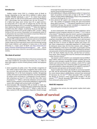
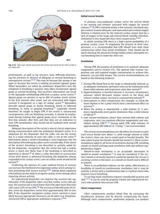
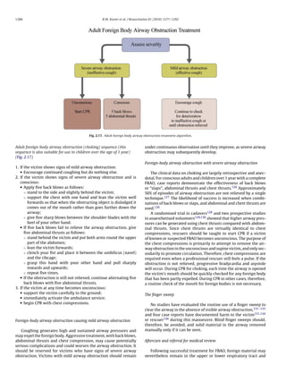
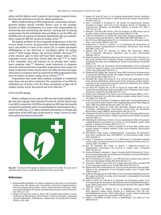
![C.D. Deakin et al. / Resuscitation 81 (2010) 1293–1304 1301
6. Weisfeldt ML, Sitlani CM, Ornato JP, et al. Survival after application of auto- arrhythmia analysis algorithm performance, incorporating new waveforms,
matic external defibrillators before arrival of the emergency medical system: and enhancing safety. A statement for health professionals from the American
evaluation in the resuscitation outcomes consortium population of 21 million. Heart Association Task Force on Automatic External Defibrillation, Subcom-
J Am Coll Cardiol 2010;55:1713–20. mittee on AED Safety and Efficacy. Circulation 1997;95:1677–82.
7. Myerburg RJ, Fenster J, Velez M, et al. Impact of community-wide police 37. Dickey W, Dalzell GW, Anderson JM, Adgey AA. The accuracy of decision-
car deployment of automated external defibrillators on survival from out-of- making of a semi-automatic defibrillator during cardiac arrest. Eur Heart J
hospital cardiac arrest. Circulation 2002;106:1058–64. 1992;13:608–15.
8. Capucci A, Aschieri D, Piepoli MF, Bardy GH, Iconomu E, Arvedi M. Tripling 38. Atkinson E, Mikysa B, Conway JA, et al. Specificity and sensitivity of automated
survival from sudden cardiac arrest via early defibrillation without traditional external defibrillator rhythm analysis in infants and children. Ann Emerg Med
education in cardiopulmonary resuscitation. Circulation 2002;106:1065–70. 2003;42:185–96.
9. van Alem AP, Vrenken RH, de Vos R, Tijssen JG, Koster RW. Use of auto- 39. Cecchin F, Jorgenson DB, Berul CI, et al. Is arrhythmia detection by automatic
mated external defibrillator by first responders in out of hospital cardiac arrest: external defibrillator accurate for children? Sensitivity and specificity of an
prospective controlled trial. BMJ 2003;327:1312. automatic external defibrillator algorithm in 696 pediatric arrhythmias. Circu-
10. Valenzuela TD, Bjerke HS, Clark LL, et al. Rapid defibrillation by nontraditional lation 2001;103:2483–8.
responders: the Casino Project. Acad Emerg Med 1998;5:414–5. 40. van Alem AP, Sanou BT, Koster RW. Interruption of cardiopulmonary resusci-
11. Spearpoint KG, Gruber PC, Brett SJ. Impact of the Immediate Life Support course tation with the use of the automated external defibrillator in out-of-hospital
on the incidence and outcome of in-hospital cardiac arrest calls: an observa- cardiac arrest. Ann Emerg Med 2003;42:449–57.
tional study over 6 years. Resuscitation 2009;80:638–43. 41. Rea TD, Helbock M, Perry S, et al. Increasing use of cardiopulmonary resuscita-
12. Waalewijn RA, Tijssen JG, Koster RW. Bystander initiated actions in out- tion during out-of-hospital ventricular fibrillation arrest: survival implications
of-hospital cardiopulmonary resuscitation: results from the Amsterdam of guideline changes. Circulation 2006;114:2760–5.
Resuscitation Study (ARREST). Resuscitation 2001;50:273–9. 42. Gundersen K, Kvaloy JT, Kramer-Johansen J, Steen PA, Eftestol T. Development
13. Swor RA, Jackson RE, Cynar M, et al. Bystander CPR, ventricular fibrillation, and of the probability of return of spontaneous circulation in intervals without
survival in witnessed, unmonitored out-of-hospital cardiac arrest. Ann Emerg chest compressions during out-of-hospital cardiac arrest: an observational
Med 1995;25:780–4. study. BMC Med 2009;7:6.
14. Holmberg M, Holmberg S, Herlitz J. Effect of bystander cardiopulmonary resus- 43. Lloyd MS, Heeke B, Walter PF, Langberg JJ. Hands-on defibrillation: an analy-
citation in out-of-hospital cardiac arrest patients in Sweden. Resuscitation sis of electrical current flow through rescuers in direct contact with patients
2000;47:59–70. during biphasic external defibrillation. Circulation 2008;117:2510–4.
15. Vaillancourt C, Verma A, Trickett J, et al. Evaluating the effectiveness of 44. Miller PH. Potential fire hazard in defibrillation. JAMA 1972;221:192.
dispatch-assisted cardiopulmonary resuscitation instructions. Acad Emerg 45. Hummel III RS, Ornato JP, Weinberg SM, Clarke AM. Spark-generating proper-
Med 2007;14:877–83. ties of electrode gels used during defibrillation. A potential fire hazard. JAMA
16. O’Neill JF, Deakin CD. Evaluation of telephone CPR advice for adult cardiac arrest 1988;260:3021–4.
patients. Resuscitation 2007;74:63–7. 46. ECRI. Defibrillation in oxygen-enriched environments [hazard]. Health Devices
17. Yang CW, Wang HC, Chiang WC, et al. Interactive video instruction improves 1987;16:113–4.
the quality of dispatcher-assisted chest compression-only cardiopulmonary 47. Lefever J, Smith A. Risk of fire when using defibrillation in an oxygen enriched
resuscitation in simulated cardiac arrests. Crit Care Med 2009;37:490–5. atmosphere. Med Devices Agency Safety Notices 1995;3:1–3.
18. Yang CW, Wang HC, Chiang WC, et al. Impact of adding video communication 48. Ward ME. Risk of fires when using defibrillators in an oxygen enriched atmo-
to dispatch instructions on the quality of rescue breathing in simulated cardiac sphere. Resuscitation 1996;31:173.
arrests—a randomized controlled study. Resuscitation 2008;78:327–32. 49. Theodorou AA, Gutierrez JA, Berg RA. Fire attributable to a defibrillation
19. Koster RW, Baubin MA, Caballero A, et al. European Resuscitation Council attempt in a neonate. Pediatrics 2003;112:677–9.
Guidelines for Resuscitation 2010. Section 2. Adult basic life support and use 50. Robertshaw H, McAnulty G. Ambient oxygen concentrations during simulated
of automated external defibrillators. Resuscitation 2010;81:1277–92. cardiopulmonary resuscitation. Anaesthesia 1998;53:634–7.
20. Berdowski J, Schulten RJ, Tijssen JG, van Alem AP, Koster RW. Delaying a shock 51. Cantello E, Davy TE, Koenig KL. The question of removing a ventilation bag
after takeover from the automated external defibrillator by paramedics is asso- before defibrillation. J Accid Emerg Med 1998;15:286.
ciated with decreased survival. Resuscitation 2010;81:287–92. 52. Deakin CD, Paul V, Fall E, Petley GW, Thompson F. Ambient oxygen concentra-
21. Zafari AM, Zarter SK, Heggen V, et al. A program encouraging early defibril- tions resulting from use of the Lund University Cardiopulmonary Assist System
lation results in improved in-hospital resuscitation efficacy. J Am Coll Cardiol (LUCAS) device during simulated cardiopulmonary resuscitation. Resuscitation
2004;44:846–52. 2007;74:303–9.
22. Destro A, Marzaloni M, Sermasi S, Rossi F. Automatic external defibrillators in 53. Kerber RE, Kouba C, Martins J, et al. Advance prediction of transtho-
the hospital as well? Resuscitation 1996;31:39–43. racic impedance in human defibrillation and cardioversion: importance of
23. Forcina MS, Farhat AY, O’Neil WW, Haines DE. Cardiac arrest survival after impedance in determining the success of low-energy shocks. Circulation
implementation of automated external defibrillator technology in the in- 1984;70:303–8.
hospital setting. Crit Care Med 2009;37:1229–36. 54. Kerber RE, Grayzel J, Hoyt R, Marcus M, Kennedy J. Transthoracic resistance in
24. Domanovits H, Meron G, Sterz F, et al. Successful automatic external defibrilla- human defibrillation. Influence of body weight, chest size, serial shocks, paddle
tor operation by people trained only in basic life support in a simulated cardiac size and paddle contact pressure. Circulation 1981;63:676–82.
arrest situation. Resuscitation 1998;39:47–50. 55. Sado DM, Deakin CD, Petley GW, Clewlow F. Comparison of the effects of
25. Cusnir H, Tongia R, Sheka KP, et al. In hospital cardiac arrest: a role for automatic removal of chest hair with not doing so before external defibrillation on
defibrillation. Resuscitation 2004;63:183–8. transthoracic impedance. Am J Cardiol 2004;93:98–100.
26. Chan PS, Krumholz HM, Nichol G, Nallamothu BK. Delayed time to defibrillation 56. Walsh SJ, McCarty D, McClelland AJ, et al. Impedance compensated biphasic
after in-hospital cardiac arrest. N Engl J Med 2008;358:9–17. waveforms for transthoracic cardioversion of atrial fibrillation: a multi-centre
27. Cummins RO, Eisenberg MS, Litwin PE, Graves JR, Hearne TR, Hallstrom AP. comparison of antero-apical and antero-posterior pad positions. Eur Heart J
Automatic external defibrillators used by emergency medical technicians: a 2005;26:1298–302.
controlled clinical trial. JAMA 1987;257:1605–10. 57. Deakin CD, Sado DM, Petley GW, Clewlow F. Differential contribution of skin
28. Stults KR, Brown DD, Kerber RE. Efficacy of an automated external defibrillator impedance and thoracic volume to transthoracic impedance during external
in the management of out-of-hospital cardiac arrest: validation of the diagnos- defibrillation. Resuscitation 2004;60:171–4.
tic algorithm and initial clinical experience in a rural environment. Circulation 58. Deakin C, Sado D, Petley G, Clewlow F. Determining the optimal paddle force
1986;73:701–9. for external defibrillation. Am J Cardiol 2002;90:812–3.
29. Kramer-Johansen J, Edelson DP, Abella BS, Becker LB, Wik L, Steen PA. Pauses 59. Manegold JC, Israel CW, Ehrlich JR, et al. External cardioversion of atrial fib-
in chest compression and inappropriate shocks: a comparison of manual and rillation in patients with implanted pacemaker or cardioverter-defibrillator
semi-automatic defibrillation attempts. Resuscitation 2007;73:212–20. systems: a randomized comparison of monophasic and biphasic shock energy
30. Pytte M, Pedersen TE, Ottem J, Rokvam AS, Sunde K. Comparison of hands-off application. Eur Heart J 2007;28:1731–8.
time during CPR with manual and semi-automatic defibrillation in a manikin 60. Alferness CA. Pacemaker damage due to external countershock in patients
model. Resuscitation 2007;73:131–6. with implanted cardiac pacemakers. Pacing Clin Electrophysiol 1982;5:
31. Edelson DP, Abella BS, Kramer-Johansen J, et al. Effects of compression depth 457–8.
and pre-shock pauses predict defibrillation failure during cardiac arrest. Resus- 61. Panacek EA, Munger MA, Rutherford WF, Gardner SF. Report of nitropatch
citation 2006;71:137–45. explosions complicating defibrillation. Am J Emerg Med 1992;10:128–9.
32. Eftestol T, Sunde K, Steen PA. Effects of interrupting precordial compressions 62. Wrenn K. The hazards of defibrillation through nitroglycerin patches. Ann
on the calculated probability of defibrillation success during out-of-hospital Emerg Med 1990;19:1327–8.
cardiac arrest. Circulation 2002;105:2270–3. 63. Pagan-Carlo LA, Spencer KT, Robertson CE, Dengler A, Birkett C, Kerber
33. Yu T, Weil MH, Tang W, et al. Adverse outcomes of interrupted precordial RE. Transthoracic defibrillation: importance of avoiding electrode placement
compression during automated defibrillation. Circulation 2002;106:368–72. directly on the female breast. J Am Coll Cardiol 1996;27:449–52.
34. Wik L, Kramer-Johansen J, Myklebust H, et al. Quality of cardiopulmonary 64. Deakin CD, Sado DM, Petley GW, Clewlow F. Is the orientation of the api-
resuscitation during out-of-hospital cardiac arrest. JAMA 2005;293:299–304. cal defibrillation paddle of importance during manual external defibrillation?
35. Abella BS, Alvarado JP, Myklebust H, et al. Quality of cardiopulmonary resusci- Resuscitation 2003;56:15–8.
tation during in-hospital cardiac arrest. JAMA 2005;293:305–10. 65. Kirchhof P, Eckardt L, Loh P, et al. Anterior-posterior versus anterior-lateral
36. Kerber RE, Becker LB, Bourland JD, et al. Automatic external defibrillators for electrode positions for external cardioversion of atrial fibrillation: a ran-
public access defibrillation: recommendations for specifying and reporting domised trial. Lancet 2002;360:1275–9.](https://image.slidesharecdn.com/fullerc2010guidelines-120109122937-phpapp02/85/Full-erc-2010_guidelines-83-320.jpg)
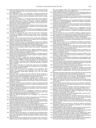
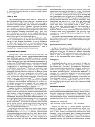
![C.D. Deakin et al. / Resuscitation 81 (2010) 1305–1352 1309
ing no difference in short- or long-term survival.183,189,191,193–199 resuscitation (CPR). For all in-hospital cardiac arrests, ensure that:
in one study, survival of the event was lower when physicians
were part of the resuscitation team.199 Studies indirectly compar- • cardiorespiratory arrest is recognised immediately;
ing resuscitation outcomes between physician-staffed and other • help is summoned using a standard telephone number;
systems are difficult to interpret because of the extremely high • CPR is started immediately using airway adjuncts, e.g., a pocket
variability between systems, independent of physician-staffing.200 mask and, if indicated, defibrillation attempted as rapidly as pos-
Although some studies have documented higher survival rates sible and certainly within 3 min.
after cardiac arrest in EMS systems that include experienced
physicians,186,188,201–203 compared with those that rely on non- The exact sequence of actions after in-hospital cardiac arrest
physician providers,201,202,204,205 other comparisons have found no will depend on many factors, including:
difference in survival between systems using paramedics or physi-
cians as part of the response.206,207 Well-organised non-physician
• location (clinical/non-clinical area; monitored/unmonitored
systems with highly trained paramedics have also reported high
area);
survival rates.200 Given the inconsistent evidence, the inclusion or
• training of the first responders;
exclusion of physicians among prehospital personnel responding
• number of responders;
to cardiac arrests will depend largely on existing local policy.
• equipment available;
• hospital response system to cardiac arrest and medical emergen-
Termination of resuscitation rules cies (e.g., MET, RRT).
One high-quality, prospective study has demonstrated that
Location
application of a ‘basic life support termination of resuscitation rule’
is predictive of death when applied by defibrillation-only emer-
Patients who have monitored arrests are usually diagnosed
gency medical technicians.208 The rule recommends termination
rapidly. Ward patients may have had a period of deterioration and
when there is no return of spontaneous circulation, no shocks are
an unwitnessed arrest.6,8 Ideally, all patients who are at high risk
administered, and the arrest is not witnessed by EMS personnel. Of
of cardiac arrest should be cared for in a monitored area where
776 patients with cardiac arrest for whom the rule recommended
facilities for immediate resuscitation are available.
termination, four survived [0.5% (95% CI 0.2–0.9)]. Implementation
of the rule would reduce the transportation rate by almost two
thirds. Four studies have shown external generalisability of this Training of first responders
rule.209–212
Additional studies have shown associations with futility of cer- All healthcare professionals should be able to recognise cardiac
tain variables such as no ROSC at scene; non-shockable rhythm; arrest, call for help and start CPR. Staff should do what they have
unwitnessed arrest; no bystander CPR, call response time and been trained to do. For example, staff in critical care and emer-
patient demographics.213–218 gency medicine will have more advanced resuscitation skills than
Two in-hospital studies and one emergency department study staff who are not involved regularly in resuscitation in their nor-
showed that the reliability of termination of resuscitation rules is mal clinical role. Hospital staff who attend a cardiac arrest may
limited in these settings.219–221 have different levels of skill to manage the airway, breathing and
Prospectively validated termination of resuscitation rules such circulation. Rescuers must undertake only the skills in which they
as the ‘basic life support termination of resuscitation rule’ can be are trained and competent.
used to guide termination of prehospital CPR in adults; however,
these must be validated in an emergency medical services system Number of responders
similar to the one in which implementation is proposed. Other rules
for various provider levels, including in-hospital providers, may be The single responder must ensure that help is coming. If other
helpful to reduce variability in decision-making; however, rules staff are nearby, several actions can be undertaken simultaneously.
should be prospectively validated prior to implementation.
Equipment available
CPR versus defibrillation first
All clinical areas should have immediate access to resuscitation
There is evidence that performing chest compressions while equipment and drugs to facilitate rapid resuscitation of the patient
retrieving and charging a defibrillator improves the probability of in cardiopulmonary arrest. Ideally, the equipment used for CPR
survival.222 EMS personnel should provide good-quality CPR while (including defibrillators) and the layout of equipment and drugs
a defibrillator is retrieved, applied and charged, but routine delivery should be standardised throughout the hospital.224,225
of a pre-specified period of CPR (e.g., 2 or 3 min) before rhythm anal-
ysis and a shock is delivered is not recommended. Some emergency
Resuscitation team
medical services have already fully implemented a pre-specified
period of chest compressions before defibrillation; given the lack
The resuscitation team may take the form of a traditional cardiac
of convincing data either supporting or refuting this strategy, it is
arrest team, which is called only when cardiac arrest is recognised.
reasonable for them to continue this practice (see Section 3).223
Alternatively, hospitals may have strategies to recognise patients
at risk of cardiac arrest and summon a team (e.g., MET or RRT)
4c In-hospital resuscitation before cardiac arrest occurs. The term ‘resuscitation team’ reflects
the range of response teams. In hospital cardiac arrests are rarely
After in-hospital cardiac arrest, the division between basic life sudden or unexpected. A strategy of recognising patients at risk of
support and advanced life support is arbitrary; in practice, the cardiac arrest may enable some of these arrests to be prevented, or
resuscitation process is a continuum and is based on common sense. may prevent futile resuscitation attempts in those who are unlikely
The public expect that clinical staff can undertake cardiopulmonary to benefit from CPR.](https://image.slidesharecdn.com/fullerc2010guidelines-120109122937-phpapp02/85/Full-erc-2010_guidelines-91-320.jpg)
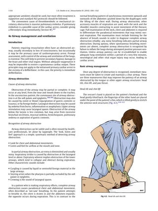
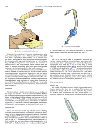
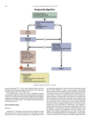
![C.D. Deakin et al. / Resuscitation 81 (2010) 1305–1352 1335
Disability (optimising neurological recovery) with intensive glucose control.688 Another recent study and two
meta-analyses of studies of tight glucose control versus conven-
Cerebral perfusion tional glucose control in critically ill patients showed no significant
difference in mortality but found tight glucose control was associ-
Immediately after ROSC there is a period of cerebral ated with a significantly increased risk of hypoglycaemia.689–691
hyperaemia.670 After asphyxial cardiac arrest, brain oedema may Severe hypoglycaemia is associated with increased mortality in
occur transiently after ROSC but it is rarely associated with clinically critically ill patients,692 and comatose patients are at particular
relevant increases in intracranial pressure.671,672 Autoregulation of risk from unrecognised hypoglycaemia. There is some evidence
cerebral blood flow is impaired for some time after cardiac arrest, that, irrespective of the target range, variability in glucose values is
which means that cerebral perfusion varies with cerebral perfusion associated with mortality.693
pressure instead of being linked to neuronal activity.673,674 As dis- Based on the available data, following ROSC blood glucose
cussed previously, following ROSC, maintain mean arterial pressure should be maintained at ≤10 mmol l−1 (180 mg dl−1 ).694 Hypo-
near the patient’s normal level. glycaemia should be avoided. Strict glucose control should not
be implemented in adult patients with ROSC after cardiac arrest
Sedation because of the increased risk of hypoglycaemia.
Although it has been common practice to sedate and ventilate Temperature control
patients for at least 24 h after ROSC, there are no high-level data to
support a defined period of ventilation, sedation and neuromuscu- Treatment of hyperpyrexia
lar blockade after cardiac arrest. Patients need to be well-sedated A period of hyperthermia (hyperpyrexia) is common in the
during treatment with therapeutic hypothermia, and the duration first 48 h after cardiac arrest.695–697 Several studies document
of sedation and ventilation is therefore influenced by this treat- an association between post-cardiac arrest pyrexia and poor
ment. There are no data to indicate whether or not the choice outcomes.498,695,697–700 There are no randomised controlled trials
of sedation influences outcome, but a combination of opioids evaluating the effect of treatment of pyrexia (defined as ≥37.6 ◦ C)
and hypnotics is usually used. Short-acting drugs (e.g., propofol, compared to no temperature control in patients after cardiac arrest.
alfentanil, remifentanil) will enable earlier neurological assess- Although the effect of elevated temperature on outcome is not
ment. Adequate sedation will reduce oxygen consumption. During proved, it seems prudent to treat any hyperthermia occurring after
hypothermia, optimal sedation can reduce or prevent shivering, cardiac arrest with antipyretics or active cooling.
which enable the target temperature to be achieved more rapidly.
Use of published sedation scales for monitoring these patients (e.g., Therapeutic hypothermia
the Richmond or Ramsay Scales) may be helpful.675,676 Animal and human data indicate that mild hypothermia is
neuroprotective and improves outcome after a period of global
Control of seizures cerebral hypoxia-ischaemia.701,702 Cooling suppresses many of
the pathways leading to delayed cell death, including apopto-
Seizures or myoclonus or both occur in 5–15% of adult sis (programmed cell death). Hypothermia decreases the cerebral
patients who achieve ROSC and 10–40% of those who remain metabolic rate for oxygen (CMRO2 ) by about 6% for each 1 ◦ C reduc-
comatose.498,677–680 Seizures increase cerebral metabolism by up tion in temperature703 and this may reduce the release of excitatory
to 3-fold681 and may cause cerebral injury: treat promptly and amino acids and free radicals.701 Hypothermia blocks the intra-
effectively with benzodiazepines, phenytoin, sodium valproate, cellular consequences of excitotoxin exposure (high calcium and
propofol, or a barbiturate. Myoclonus can be particularly difficult to glutamate concentrations) and reduces the inflammatory response
treat; phenytoin is often ineffective. Clonazepam is the most effec- associated with the post-cardiac arrest syndrome.
tive antimyoclonic drug, but sodium valproate, levetiracetam and
propofol may also be effective.682 Maintenance therapy should be Which post-cardiac arrest patients should be cooled?. All stud-
started after the first event once potential precipitating causes (e.g., ies of post-cardiac arrest therapeutic hypothermia have included
intracranial haemorrhage, electrolyte imbalance) are excluded. No only patients in coma. There is good evidence supporting the use
studies directly address the use of prophylactic anticonvulsant of induced hypothermia in comatose survivors of out-of-hospital
drugs after cardiac arrest in adults. cardiac arrest caused by VF. One randomised trial704 and a pseudo-
randomised trial669 demonstrated improved neurological outcome
Glucose control at hospital discharge or at 6 months in comatose patients after
out-of-hospital VF cardiac arrest. Cooling was initiated within min-
There is a strong association between high blood glucose utes to hours after ROSC and a temperature range of 32–34 ◦ C was
after resuscitation from cardiac arrest and poor neurological maintained for 12–24 h. Two studies with historical control groups
outcome.498–501,504,634,683,684 Although one randomised controlled showed improvement in neurological outcome after therapeutic
trial in a cardiac surgical intensive care unit showed that tight con- hypothermia for comatose survivors of VF cardiac arrest.705–707
trol of blood glucose (4.4–6.1 mmol l−1 or 80–110 mg dl−1 ) using Extrapolation of these data to other cardiac arrests (e.g., other initial
insulin reduced hospital mortality in critically ill adults,685 a sec- rhythms, in-hospital arrests, paediatric patients) seems reasonable
ond study by the same group in medical ICU patients showed but is supported by only lower level data.
no mortality benefit from tight glucose control.686 In one ran- One small, randomised trial showed reduced plasma lactate
domised trial of patients resuscitated from OHCA with ventricular values and oxygen extraction ratios in a group of comatose sur-
fibrillation, strict glucose control (72–108 mg dl−1 , 4–6 mmol l−1 ]) vivors after cardiac arrest with asystole or PEA who were cooled
gave no survival benefit compared with moderate glucose control with a cooling cap.708 Six studies with historical control groups
(108–144 mg dl−1 , 6–8 mmol l−1 ) and there were more episodes of have shown benefit using therapeutic hypothermia in comatose
hypoglycaemia in the strict glucose control group.687 A large ran- survivors of OHCA after all rhythm arrests.629,632,709–712 Two non-
domised trial of intensive glucose control (4.5–6.0 mmol l−1 ) versus randomised studies with concurrent controls indicate possible
conventional glucose control (10 mmol l−1 or less) in general ICU benefit of hypothermia following cardiac arrest from other initial
patients reported increased 90-day mortality in patients treated rhythms in- and out-of-hospital.713,714](https://image.slidesharecdn.com/fullerc2010guidelines-120109122937-phpapp02/85/Full-erc-2010_guidelines-117-320.jpg)
![C.D. Deakin et al. / Resuscitation 81 (2010) 1305–1352 1337
and there is evidence that this therapy makes these tests less reli- hypoxemia, it is reasonable to use unprocessed EEG interpretation
able. (specifically identifying generalized suppression to less than 20 V,
burst suppression pattern with generalized epileptic activity, or dif-
Clinical examination fuse periodic complexes on a flat background) observed between
24 and 72 h after ROSC to assist the prediction of a poor outcome
There are no clinical neurological signs that reliably predict poor (FPR 3%, 95% CI 0.9–11%) in comatose survivors of cardiac arrest
outcome (cerebral performance category [CPC] 3 or 4, or death) less not treated with hypothermia.774 There is inadequate evidence to
than 24 h after cardiac arrest. In adult patients who are comatose support the routine use of other electrophysiological studies (e.g.,
after cardiac arrest, and who have not been treated with hypother- abnormal brainstem auditory evoked potentials) for prognostica-
mia and who do not have confounding factors (such as hypotension, tion of poor outcome in comatose cardiac arrest survivors.606
sedatives or muscle relaxants), the absence of both pupillary light
and corneal reflex at ≥72 h reliably predicts poor outcome (FPR Imaging studies
0%; 95% CI 0–9%).680 Absence of vestibulo-ocular reflexes at ≥24 h
(FPR 0%; 95% CI 0–14%)764,765 and a GCS motor score of 2 or less at Many imaging modalities (magnetic resonance imaging [MRI],
≥72 h (FPR 5%; 95% CI 2–9%)680 are less reliable. Other clinical signs, computed tomography [CT], single photon emission computed
including myoclonus, are not recommended for predicting poor tomography [SPECT], cerebral angiography, transcranial Doppler,
outcome. The presence of myoclonus status in adults is strongly nuclear medicine, near infra-red spectroscopy [NIRS]) have been
associated with poor outcome,679,680,766–768 but rare cases of good studied to determine their utility for prediction of outcome in adult
neurological recovery have been described and accurate diagnosis cardiac arrest survivors.606 There are no level one or level two stud-
is problematic.769–773 ies that support the use of any imaging modality to predict outcome
of comatose cardiac arrest survivors. Overall, those imaging stud-
Biochemical markers ies that have been undertaken were limited by small sample sizes,
variable time of imaging (many very late in the course), lack of com-
Serum neuronal specific enolase elevations are associ- parison with a standardised method of prognostication, and early
ated with poor outcome for comatose patients after cardiac withdrawal of care. Despite tremendous potential, neuroimaging
arrest.680,748,774–792 Although specific cut-off values with a false has yet to be proven as an independently accurate modality for
positive rate of 0% have been reported, clinical application is prediction of outcome in individual comatose cardiac arrest sur-
limited due to variability in the 0% FPR cut-off values reported vivors and, at this time, its routine use for this purpose is not
among various studies. recommended.
Serum S100 elevations are associated with
poor outcome for comatose patients after cardiac Impact of therapeutic hypothermia on prognostication
arrest.680,774–776,782,784,785,787,788,791,793–798
Many other serum markers measured after sustained return There is inadequate evidence to recommend a specific approach
of spontaneous circulation have been associated with poor out- to prognosticating poor outcome in post-cardiac arrest patients
come after cardiac arrest, including BNP,799 vWF,800 ICAM-1,800 treated with therapeutic hypothermia. There are no clinical neuro-
procalcitonin,794 IL-1ra, RANTES, sTNFRII, IL-6, IL-8 and IL-10.645 logical signs, electrophysiological studies, biomarkers, or imaging
However, other studies found no relationship between outcome modalities that can reliably predict neurological outcome in the
and serum IL-8,793 and procalcitonin and sTREM-1.801 first 24 h after cardiac arrest. Based on limited available evidence,
Worse outcomes for comatose survivors of cardiac arrest potentially reliable prognosticators of poor outcome in patients
are also associated with increased levels of cerebrospinal fluid treated with therapeutic hypothermia after cardiac arrest include
(CSF)-CK802,803 and cerebrospinal fluid-CKBB.774,775,777,789,803–807 bilateral absence of N20 peak on SSEP ≥ 24 h after cardiac arrest
However, one study found no relationship between cerebrospinal (FPR 0%, 95% CI 0–69%) and the absence of both corneal and pupil-
fluid-CKBB and prognosis.808 lary reflexes 3 or more days after cardiac arrest (FPR 0%, 95%
Outcomes are also associated with increased cerebrospinal fluid CI 0–48%).766,810 Limited available evidence also suggests that a
levels of other markers including NSE,775,784,789 ), S100,784 LDH, Glasgow Motor Score of 2 or less at 3 days post-ROSC (FPR 14%
GOT,777,803 neurofilament,809 acid phosphatase and lactate.803 [95% CI 3–44%])766 and the presence of status epilepticus (FPR of
Cerebrospinal fluid levels of beta-d-N-acetylglucosaminidase, and 7% [95% CI 1–25%] to 11.5% [95% CI 3–31%])811,812 are potentially
pyruvate were not associated with the prognosis of cardiac unreliable prognosticators of poor outcome in post-cardiac arrest
arrest.803 patients treated with therapeutic hypothermia. One study of 111
In summary, evidence does not support the use of serum or post-cardiac arrest patients treated with therapeutic hypothermia
CSF biomarkers alone as predictors of poor outcomes in comatose attempted to validate prognostic criteria proposed by the Amer-
patients after cardiac arrest with or without treatment with ther- ican Academy of Neurology.774,813 This study demonstrated that
apeutic hypothermia (TH). Limitations included small numbers of clinical exam findings at 36–72 h were unreliable predictors of
patients and/or inconsistency in cut-off values for predicting poor poor neurological outcome while bilaterally absent N20 peak on
outcome. somatosensory evoked potentials (false positive rate 0%, 95% CI
0–13%) and unreactive electroencephalogram background (false
Electrophysiological studies positive rate 0%, 95% CI 0–13%) were the most reliable. A decision
rule derived using this dataset demonstrated that the presence of
No electrophysiological study reliably predicts outcome of a two independent predictors of poor neurological outcome (incom-
comatose patient within the first 24 h after cardiac arrest. If plete recovery brainstem reflexes, early myoclonus, unreactive
somatosensory evoked potentials (SSEP) are measured after 24 h electroencephalogram and bilaterally absent cortical SSEPs) pre-
in comatose cardiac arrest survivors not treated with therapeu- dicted poor neurological outcome with a false positive rate of 0%
tic hypothermia, bilateral absence of the N20 cortical response to (95% CI 0–14%). Serum biomarkers such as NSE are potentially valu-
median nerve stimulation predicts poor outcome (death or CPC 3 or able as adjunctive studies in prognostication of poor outcome in
4) with a FPR of 0.7% (95% CI 0.1–3.7).774 In the absence of confound- patients treated with hypothermia, but their reliability is limited
ing circumstances such as sedatives, hypotension, hypothermia or because few patients have been studied and the assay is not well](https://image.slidesharecdn.com/fullerc2010guidelines-120109122937-phpapp02/85/Full-erc-2010_guidelines-119-320.jpg)
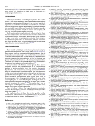
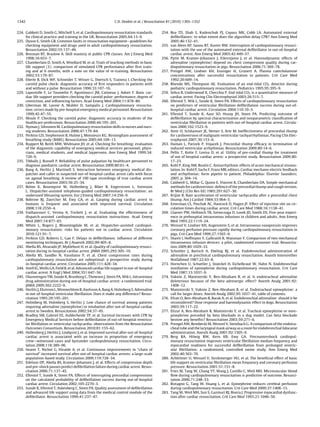
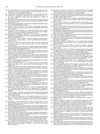
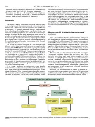
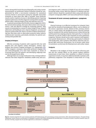
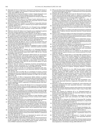
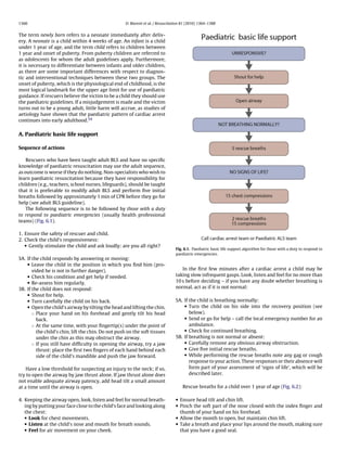
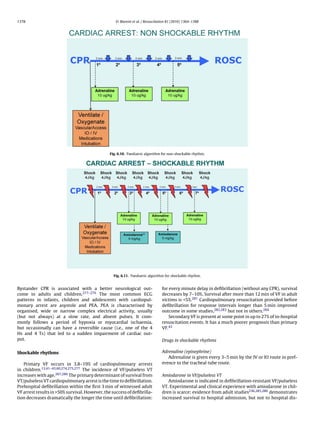
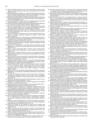
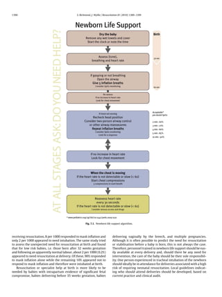

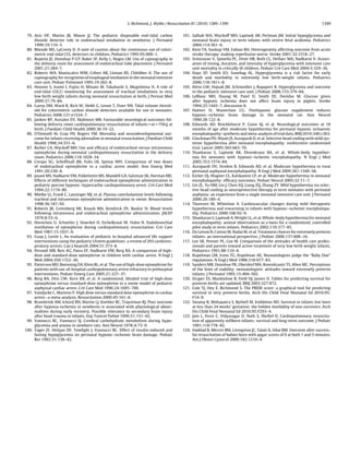
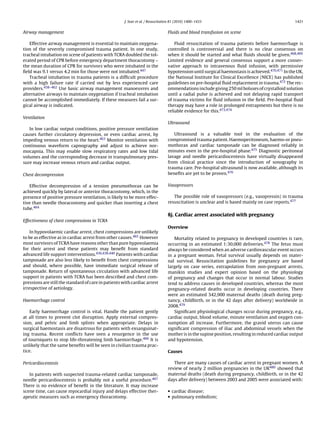
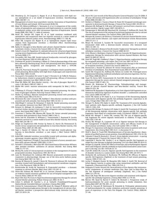
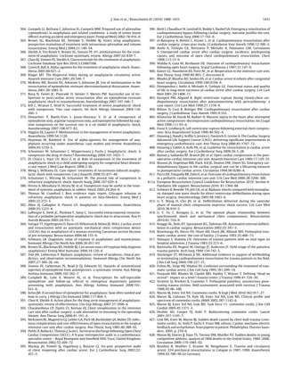


The document provides guidelines for cardiopulmonary resuscitation (CPR) published by the European Resuscitation Council in 2010. It highlights several main changes from the 2005 guidelines, including increased emphasis on high-quality, uninterrupted chest compressions; minimizing interruptions during defibrillation; and recognition of the potential harm of hyperoxaemia after return of spontaneous circulation. Other changes covered include updated treatment recommendations for acute coronary syndromes, greater use of capnography and therapeutic hypothermia, and revised pediatric CPR guidelines focusing on compression quality and automated external defibrillator use in children.


![ONFH[AVN HIP] -TRIPLE REGIME -A NOVAL SURGICAL CONCEPT .pptx](https://cdn.slidesharecdn.com/ss_thumbnails/onfhavnhip2026koaconcalicutdrgokuldevdrmashraf-260210064517-213ec005-thumbnail.jpg?width=640&height=640&fit=bounds)


![PERI-PROSTHETIC FRACTURE NAIL-PLATE CONSTRUCT [NPC].pptx](https://cdn.slidesharecdn.com/ss_thumbnails/drarunkumardrmohamedashrafperiprostheticfrasturenail-plateconstructnpc-260209164459-7e9d15a1-thumbnail.jpg?width=640&height=640&fit=bounds)

![CTEV [ clubfoot] DR ARUN LAL ,DR MOHAMED ASHRAF travancore medical college k...](https://cdn.slidesharecdn.com/ss_thumbnails/ctevclubfootdrarunlaldrmohamedashraftravancoremedicalcollegekollamkeralaindia-260208063247-18fc466c-thumbnail.jpg?width=640&height=640&fit=bounds)