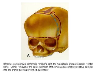
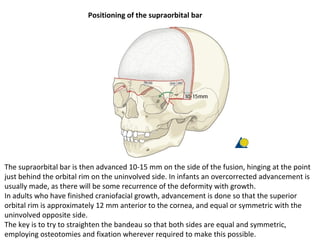
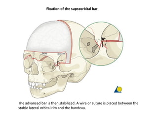
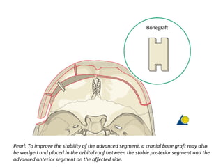
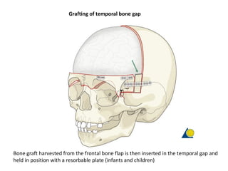
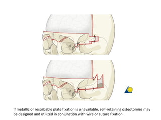
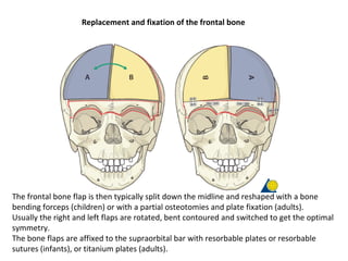
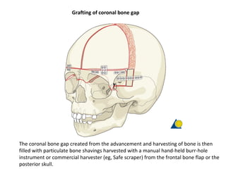
Frontoorbital Advancement
Bifrontal craniotomy is performed removing both the hypoplastic and protuberant frontal
bone. Further removal of the basal extension of the involved coronal suture (blue dashes)
into the cranial base is performed by rongeur
Positioning of the supraorbital bar
The supraorbital bar is then advanced 10-15 mm on the side of the fusion, hinging at the point
just behind the orbital rim on the uninvolved side. In infants an overcorrected advancement is
usually made, as there will be some recurrence of the deformity with growth.
In adults who have finished craniofacial growth, advancement is done so that the superior
orbital rim is approximately 12 mm anterior to the cornea, and equal or symmetric with the
uninvolved opposite side.
The key is to try to straighten the bandeau so that both sides are equal and symmetric,
employing osteotomies and fixation wherever required to make this possible.
Fixation of the supraorbital bar
The advanced bar is then stabilized. A wire or suture is placed between the
stable lateral orbital rim and the bandeau.
Pearl: To improve the stability of the advanced segment, a cranial bone graft may also
be wedged and placed in the orbital roof between the stable posterior segment and the
advanced anterior segment on the affected side.
Grafting of temporal bone gap
Bone graft harvested from the frontal bone flap is then inserted in the temporal gap and
held in position with a resorbable plate (infants and children)
If metallic or resorbable plate fixation is unavailable, self-retaining osteotomies may
be designed and utilized in conjunction with wire or suture fixation.
Replacement and fixation of the frontal bone
flap
The frontal bone flap is then typically split down the midline and reshaped with a bone
bending forceps (children) or with a partial osteotomies and plate fixation (adults).
Usually the right and left flaps are rotated, bent contoured and switched to get the optimal
symmetry.
The bone flaps are affixed to the supraorbital bar with resorbable plates or resorbable
sutures (infants), or titanium plates (adults).
Grafting of coronal bone gap
The coronal bone gap created from the advancement and harvesting of bone is then
filled with particulate bone shavings harvested with a manual hand-held burr-hole
instrument or commercial harvester (eg, Safe scraper) from the frontal bone flap or the
posterior skull.
Grafting of coronal bone gap
The coronal bone gap created from the advancement and harvesting of bone is then
filled with particulate bone shavings harvested with a manual hand-held burr-hole
instrument or commercial harvester (eg, Safe scraper) from the frontal bone flap or the
posterior skull.

Frontoorbital advancement

  • 1.
  • 2.
    Bifrontal craniotomy isperformed removing both the hypoplastic and protuberant frontal bone. Further removal of the basal extension of the involved coronal suture (blue dashes) into the cranial base is performed by rongeur
  • 3.
    Positioning of thesupraorbital bar The supraorbital bar is then advanced 10-15 mm on the side of the fusion, hinging at the point just behind the orbital rim on the uninvolved side. In infants an overcorrected advancement is usually made, as there will be some recurrence of the deformity with growth. In adults who have finished craniofacial growth, advancement is done so that the superior orbital rim is approximately 12 mm anterior to the cornea, and equal or symmetric with the uninvolved opposite side. The key is to try to straighten the bandeau so that both sides are equal and symmetric, employing osteotomies and fixation wherever required to make this possible.
  • 4.
    Fixation of thesupraorbital bar The advanced bar is then stabilized. A wire or suture is placed between the stable lateral orbital rim and the bandeau.
  • 5.
    Pearl: To improvethe stability of the advanced segment, a cranial bone graft may also be wedged and placed in the orbital roof between the stable posterior segment and the advanced anterior segment on the affected side.
  • 6.
    Grafting of temporalbone gap Bone graft harvested from the frontal bone flap is then inserted in the temporal gap and held in position with a resorbable plate (infants and children)
  • 7.
    If metallic orresorbable plate fixation is unavailable, self-retaining osteotomies may be designed and utilized in conjunction with wire or suture fixation.
  • 8.
    Replacement and fixationof the frontal bone flap The frontal bone flap is then typically split down the midline and reshaped with a bone bending forceps (children) or with a partial osteotomies and plate fixation (adults). Usually the right and left flaps are rotated, bent contoured and switched to get the optimal symmetry. The bone flaps are affixed to the supraorbital bar with resorbable plates or resorbable sutures (infants), or titanium plates (adults).
  • 9.
    Grafting of coronalbone gap The coronal bone gap created from the advancement and harvesting of bone is then filled with particulate bone shavings harvested with a manual hand-held burr-hole instrument or commercial harvester (eg, Safe scraper) from the frontal bone flap or the posterior skull.
  • 10.
    Grafting of coronalbone gap The coronal bone gap created from the advancement and harvesting of bone is then filled with particulate bone shavings harvested with a manual hand-held burr-hole instrument or commercial harvester (eg, Safe scraper) from the frontal bone flap or the posterior skull.