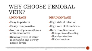
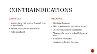
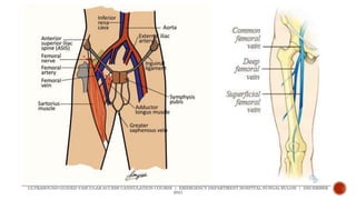
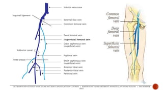
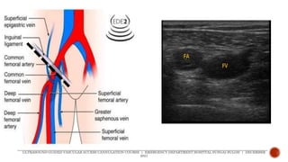
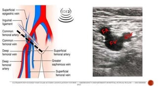
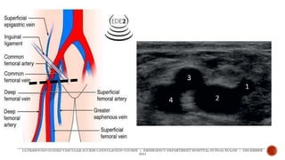
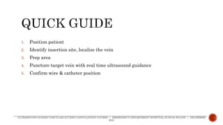
3. ADVANTAGE
Easy to perform
Easily compressible
No risk of pneumothorax
or haemothorax
Relatively free of other
monitoring and airway
access device
DISADVANTAGE
High risk of infection
High rate of thrombosis
Rare complications:
Retroperitoneal bleeding
Bowel penetration
Bladder rupture
ULTRASOUND-GUIDED VASCULAR ACCESS CANNULATION COURSE | EMERGENCY DEPARTMENT HOSPITAL SUNGAI BULOH | DECEMBER
2021
4. ABSOLUTE
Venous injury at level of femoral vein
or proximally
Known or suspected thrombosis
Patient refusal
RELATIVE
Bleeding disorders
Skin infection over the site of access
Obscure anatomical landmarks
Absence of a clearly palpable femoral
artery
History of vasculitis
Previous radiation therapy
9. Common femoral artery
midpoint of inguinal
ligament
Common femoral vein
medial to common
femoral artery
ULTRASOUND-GUIDED VASCULAR ACCESS CANNULATION COURSE | EMERGENCY DEPARTMENT HOSPITAL SUNGAI BULOH | DECEMBER
2021
13. 1. Position patient
2. Identify insertion site, localize the vein
3. Prep area
4. Puncture target vein with real time ultrasound guidance
5. Confirm wire & catheter position
ULTRASOUND-GUIDED VASCULAR ACCESS CANNULATION COURSE | EMERGENCY DEPARTMENT HOSPITAL SUNGAI BULOH | DECEMBER
2021
14. 1. Position patient
2. Identify insertion site, localize the vein
3. Prep area
4. Puncture target vein with real time ultrasound guidance
5. Confirm wire & catheter position
ULTRASOUND-GUIDED VASCULAR ACCESS CANNULATION COURSE | EMERGENCY DEPARTMENT HOSPITAL SUNGAI BULOH | DECEMBER
2021
Slight abduction +
external rotation
Reverse
Trendelenburg
15. 1. Position patient
2. Identify insertion site, localize the vein
3. Prep area
4. Puncture target vein with real time ultrasound guidance
5. Confirm wire & catheter position
ULTRASOUND-GUIDED VASCULAR ACCESS CANNULATION COURSE | EMERGENCY DEPARTMENT HOSPITAL SUNGAI BULOH | DECEMBER
2021
Vein medial to
artery
View short axis &
long axis
Compressible vein,
no thrombus
16. 1. Position patient
2. Identify insertion site, localize the vein
3. Prep area
4. Puncture target vein with real time ultrasound guidance
5. Confirm wire & catheter position
ULTRASOUND-GUIDED VASCULAR ACCESS CANNULATION COURSE | EMERGENCY DEPARTMENT HOSPITAL SUNGAI BULOH | DECEMBER
2021
Aseptic technique
Local anaesthetic
17. 1. Position patient
2. Identify insertion site, localize the vein
3. Prep area
4. Puncture target vein with real time
ultrasound guidance
5. Confirm wire & catheter position
ULTRASOUND-GUIDED VASCULAR ACCESS CANNULATION COURSE | EMERGENCY DEPARTMENT HOSPITAL SUNGAI BULOH | DECEMBER
2021
Short axis OR long
axis
Identify & confirm
needle tip position
18. 1. Position patient
2. Identify insertion site, localize the vein
3. Prep area
4. Puncture target vein with real time ultrasound guidance
5. Confirm wire & catheter position
ULTRASOUND-GUIDED VASCULAR ACCESS CANNULATION COURSE | EMERGENCY DEPARTMENT HOSPITAL SUNGAI BULOH | DECEMBER
2021
The main advantage of FV cannulation is that it is easy to perform due to the
presence of predictable anatomic landmarks. The structures are superficially located,
and easily compressible if bleeding occurs. There is no risk of pneumothorax or
haemothorax. Cannulation of FV is favoured during resuscitation.(30)
The patient is positioned with the hip in the neutral position or with slight hip
abduction and external rotation. This will minimize the overlap of the femoral artery and
vein, keeping the vein on the medial side of the artery. The reverse Trendelenburg
position will increase the CFV cross-sectional area by >50%.