A summary for learning the muscles of the shoulder including their attachments, innervation, etc., without having to have too many books open. Resources: "Grey’s anatomy", "Taschenatlas Anatomie", "McMinn’s Clinical Atlas of Human Anatomy" and Wikipedia. Awaiting further proof-reading!
The thalamus acts as a relay center and plays a key role in sensory and motor functions. It is divided into nuclear groups including anterior, medial, lateral, and intralaminar. Specific nuclei relay information from particular sensory pathways to corresponding cortical areas, while nonspecific nuclei have broader connections. The thalamus receives subcortical inputs and sends outputs to widespread areas of cortex, regulating cortical activity and integrating sensory, motor, and limbic functions. Damage to different thalamic regions can cause sensory deficits, involuntary movements, or pain syndromes on the opposite side of the body.
This document lists various anatomical structures and their relationships to specific vertebral levels in the body. Some key points include:
- The bifurcation of the common carotid artery occurs at C4, while the beginning of the esophagus and trachea occur at C6.
- The brachiocephalic trunk bifurcates at the sternoclavicular joint.
- The aortic arch begins at T3/4 and ends at T4/5.
- The thoracic duct crosses from right to left at T5.
- The inferior vena cava and right phrenic nerve enter the diaphragm at T8.
- The abdominal aorta bifurcates into the common i
A summary for learning the muscles of the hip including their attachments, innervation, etc., without having to have too many books open. Resources: "Grey's anatomy", "Taschenatlas Anatomie", "McMinn's Clinical Atlas of Human Anatomy" and Wikipedia. Awaiting further proof-reading!
The anterior and posterior muscles of the thigh are summarized in 3 sentences:
Anterior: The quadriceps femoris muscle is located on the anterior thigh and extends the knee. The sartorius muscle runs obliquely across the anterior thigh. The gracilis and semitendinosus muscles make up the pes anserinus on the medial thigh.
Posterior: The hamstring muscles - the biceps femoris, semitendinosus, and semimembranosus muscles - are located on the posterior thigh and flex the knee. The gluteal muscles originating from the pelvis insert on the posterior thigh and extend and laterally rotate the thigh.
The document discusses the anatomy of the upper limb, including its bones, joints, muscles, nerves, and vasculature. The upper limb contains the scapula, humerus, radius, ulna, and carpals that form its skeleton. It discusses the brachial plexus formed from spinal nerves that provides innervation. Muscles are categorized based on their action, innervation, and location in the anterior or posterior compartments of the arm and forearm. The radial, median, musculocutaneous, and ulnar nerves provide motor and sensory function. Blood is supplied to the upper limb primarily from the axillary and brachial arteries.
The shoulder joint is a ball and socket joint formed by the humerus, scapula, and clavicle. It has the greatest range of motion of any joint. The glenohumeral joint allows the arm to move in many directions but is less stable due to its shallow socket. A series of muscles including the rotator cuff provide dynamic stability. The shoulder complex also includes the acromioclavicular, sternoclavicular, and scapulothoracic joints. The bones, ligaments, muscles, and nerves of the shoulder are described in detail in the document.
The document describes the anatomy and structures of the spinal cord. It discusses:
- The spinal cord's position in the vertebral canal and its extensions above into the medulla oblongata and below into the conus medullaris and filum terminale.
- The external features including two enlargements known as the cervical and lumbosacral enlargements.
- Internal structures such as the gray matter containing the anterior, posterior and lateral horns, and the white matter containing ascending, descending and proprioceptive tracts that transmit sensory and motor signals.
- Ascending tracts that transmit sensory information to the brain and descending tracts that convey motor commands from the brain.
-
This document summarizes the anatomy and functions of different areas of the cerebral cortex. It describes the allocortex which makes up 10% of the cortex, and the neocortex which is the remaining 90%. It then discusses the six layers of the neocortex and different cell types. It provides details on agranular and granular cortices and their characteristics. Specific areas of the cortex are then described in more detail such as the frontal, parietal, and polar cortices. The document outlines different classification schemes for cortical areas and focuses on the 52 areas described by Brodmann. It provides in-depth descriptions of the primary motor, premotor, frontal eye field, supplementary motor, and prefrontal cortical areas.
A summary for learning the muscles of the upper limb including their attachments, innervation, etc., without having to have too many books open. Resources: "Gray’s Anatomy", "Taschenatlas der Anatomie" and Wikipedia. Awaiting further proof-reading!
This document summarizes the bones of the upper limb, including the clavicle and scapula. It describes the morphological features, attachments, ossification centers, and joints related to the clavicle and scapula. Specifically, it outlines that the clavicle has two ends, convex and concave sections, and attaches to muscles like the trapezius and deltoid. It ossifies from two centers and its joints include the sternoclavicular and acromioclavicular joints. For the scapula, it notes the scapula's angles, borders, processes, surfaces, and rotator cuff muscles that attach to it.
This diagram labels the major nerves and vessels of the lower leg, including the anterior tibial nerve and vessels on the front of the leg, the superficial peroneal nerve on the side of the leg, and branches of the posterior tibial nerve like the saphenous nerve and sural nerve in the back of the leg.
Imaging of atlanto occipital and atlantoaxial traumatic injuriesSumiya Arshad
This document discusses imaging of injuries to the craniocervical junction (CCJ). It begins by reviewing the anatomy of the CCJ, including bones and ligaments. It then describes classifications of CCJ injuries and how CT and MR imaging can identify relevant injuries and clinical effects. Specific injuries covered include atlanto-occipital dissociation, occipital condyle fractures, fractures of C1 with transverse ligament rupture, and atlantoaxial distraction or rotatory deformity from alar ligament tears. Thin-slice CT is recommended for initial evaluation, while MR helps evaluate soft tissues and rule out spinal cord injury. Proper classification of CCJ injuries guides management of unstable or complex cases.
This document describes the origin, insertion, action, and nerve innervation of 55 muscles of the head, neck, back, abdomen, and upper limbs. The muscles control functions like facial expression, eye movement, swallowing, breathing, posture, and shoulder and arm movement. They originate on bones like the skull, vertebrae, ribs, and clavicle, and insert on structures like the eyebrows, eyelids, mouth, hyoid bone, scapula, and humerus. Each muscle has a specific action, such as raising the eyebrows, depressing the eyeballs, or extending the vertebral column. They are innervated by cranial nerves like the facial nerve or spinal nerves.
Young Partner Nap - 2012. május 10.
Bádonyi Szabolcs (Excel-Networking) előadása a díjnyertes brit gyártó újdonságaival.
Egyrészt bemutatta a párhuzamos optikát használó MTP optikai rendszereket, melyek már az adatközpontok számára készültek, és 40-100 GigE teljesítményig támogatják azt. 25 év rendszergarancia a biztosíték ezekre az új típusú optikai eszközökre (kazetták, optikai panelek, gerinckábelek, stb.) és rendszerekre. Másrészt bemutatott olyan újdonságokat, mint a 24 portos V-alakú ún. "szög" panel, mellyel hely spórolható a rack szekrényben, mivel nincs szükség rendezősávokra, és elkerülhető a patch kábel RJ45-ös végződésének terhelése és a kábel feszítéséből adódó kontakthiba.
Betekintést nyerünk, a nonverbális kommunikáció értelmezésébe.A testmozgás világába: a gesztusok, a test, a végtagok, a fej, a kéz és a láb mozdulatait mutatja be.
Graphisoft Park Egészségnap - Munkahelyi gyógytorna Andras Kiss
2013. május 30.
Graphisoft Park
http://bhc.hu/egeszsegnap
Fehér Katalin: Munkahelyi gyógytorna
Az ülőmunkából adódó mozgásszervi panaszok megelőzési lehetőségei. Könnyen elsajátítható módszerek, hasznos tanácsok gyakorlati bemutatóval.
This document discusses various types of spinal tumors, including their location, characteristics, and MRI appearances. It covers primary spinal tumors such as schwannomas, neurofibromas, meningiomas, ependymomas, astrocytomas, and hemangioblastomas. It describes these tumors' demographics, symptoms, classification as interdural exteramedullary or interdural intramedullary, and features on T1-, T2-, and contrast-enhanced MRI sequences. Rare tumors like lipomas, paragangliomas, and subependymomas are also outlined briefly.
The document discusses lower back pain, including common causes such as disc injuries, degeneration, muscle strains, and nerve root compression. It explains that discs can degenerate naturally over time and have poor blood supply, making them prone to injury and slow to heal. While muscles heal quickly, they can weaken or tighten, contributing to back pain. The document outlines how acute lower back pain lasts up to 6 weeks as tissues heal, while chronic pain lasts over 3 months even though tissues may have healed. It describes how pain pathways become sensitized, causing pain to be felt more often and without real tissue threat. Management strategies discussed include exercise, posture, lifting correctly, and pacing activities to avoid reinforcing pain behaviors.
This document provides an overview of spinal cord tumors, including:
- They constitute 3-4% of all CNS tumors and 6% of pediatric CNS tumors.
- The most common types are astrocytomas and ependymomas.
- Treatment involves surgical resection followed by radiation therapy if needed.
- Prognostic factors include tumor type/grade, extent, and patient age/function.
This document provides an overview of osteochondroma, which is a benign bone tumor characterized by a bony projection with a cartilage cap. It summarizes the definition, epidemiology, pathogenesis, clinical presentation, imaging features, complications, and risk of malignant transformation. Osteochondroma most commonly involves the long bones such as the femur and humerus during childhood. Imaging plays an important role in the diagnosis and surveillance for complications or malignant degeneration.
The document provides an introductory overview of spine anatomy, including:
1) It describes the basic vertebral structures such as the vertebral body, pedicles, lamina, processes and foramen.
2) It explains the sagittal curves of the spine including the primary thoracic and sacral kyphosis and secondary cervical and lumbar lordosis.
3) It outlines the four regions of the spine - cervical, thoracic, lumbar, and sacrococcygeal - and provides some key details about typical vertebrae in each region.
Spinal tumors can be classified as extradural, intradural/extramedullary, or intramedullary based on their location. Extradural tumors are most common and often metastatic, while intradural tumors are rare and usually involve nerve sheath tumors or meningiomas. Primary spinal tumors can be benign or malignant, with benign tumors not invading other tissues and malignant tumors potentially spreading. Symptoms depend on tumor location and can include weakness, pain, numbness, and loss of bowel/bladder control. Treatment involves surgery to remove the tumor when possible along with radiation and chemotherapy for malignant tumors. Many benign and malignant tumor types are described in the document.
Presentation1.pptx, radiological vascular anatomy of the chest and abdomen.Abdellah Nazeer
This document provides an overview of the radiological vascular anatomy of the chest and abdomen. It describes the anatomy and branches of the major arteries and veins in these regions, including the coronary arteries, abdominal aorta, celiac axis, superior mesenteric artery, inferior mesenteric artery, renal arteries, inferior vena cava, portal vein system, and iliac arteries. Diagrams and CT images are included to illustrate the typical anatomy and branching patterns of these important blood vessels.
Presentation1.pptx, radiological vascular anatomy of the upper and lower limbs.Abdellah Nazeer
The document describes the arterial and venous anatomy of the upper and lower limbs. It begins by outlining the arterial supply to the upper limb, starting from the subclavian artery and its branches. It then discusses the arteries of the forearm, hand, and veins of the upper limb. For the lower limb, it discusses the femoral artery and its branches that supply the thigh as well as the arteries of the leg. It includes diagrams to illustrate the key structures and their relationships. CT, MR, and conventional angiography images are also provided to demonstrate the vascular anatomy.
Describes cross sectional anatomy of the mediastinum , and lobar and segmental anatomy of the lung with teaching points and radiological guidelines and multiple examples of lobar and segmental pathologies and how we localize these pathologies .Also the types of chest CT images and indications of chest CT.
Week 5. Basics and clinical uses of MR spectroscopy.Dr. Jakab András
The document provides information about an upcoming course, including:
1. Upcoming lecture topics and dates, including MR Spectroscopy on October 30th.
2. Details about an upcoming final test on basic imaging techniques and spectroscopy.
3. Where to find study materials for the test, including lecture materials in PDF format.
This document provides information about an upcoming "Multimodal Imaging in Neurosciences" course, including:
1) Dates and topics for upcoming lectures, as well as details about a final test on basic imaging techniques.
2) An overview of various neuroimaging modalities like CT, MRI, PET, and their applications.
3) A brief history of the development of high-intensity focused ultrasound (HIFU) technology from the 1880s to present.
Week 3. Neurosurgical planning with multimodal imagingDr. Jakab András
The document discusses the use of multimodal imaging in neurosurgery. It describes how multimodal imaging can provide maximum information beyond just anatomical structures, including blood supply, function, and spatial visualization to help with surgical planning and navigation. It outlines some of the key indications for neurosurgery like tumors, arteriovenous malformations, epilepsy, and discusses how clinicians can utilize different imaging modalities like MRI, DTI, fMRI, and PET to obtain information on anatomy, vessels, eloquent tracts, function and laterality, tumor characterization and metabolism, and localization for stereotactic planning.
Week 2. Diffusion magnetic resonance imaging, tractography, mapping the brain...Dr. Jakab András
The document summarizes key points about multimodal neuroimaging techniques, with a focus on diffusion magnetic resonance imaging (MRI) and fiber tracking. It discusses how diffusion MRI can be used to measure and visualize water diffusion in the brain, which provides information about tissue microstructure and white matter pathways. Specifically, it describes diffusion tensor imaging (DTI) and how it is used to quantify diffusion anisotropy and direction. The clinical and research applications of DTI and fiber tractography are also summarized, such as characterizing white matter integrity and disorders, assessing brain tumors, and mapping brain connectivity and development.
Week 1. Basics of multimodal imaging and image processing. Functional magneti...Dr. Jakab András
This document discusses multimodal neuroimaging. It provides an introduction to combining multiple imaging modalities such as CT, MRI, PET, and EEG to gain complementary information. Key benefits of multimodal imaging include anatomical alignment of images and fusion of structural and functional data. Examples of hybrid imaging devices that facilitate multimodal approaches are PET-CT and PET-MRI scanners. The document also gives an overview of functional MRI techniques for mapping brain activity and networks involved in sensory, motor, cognitive and resting state functions.
A review of recent evidences for macroscopic reorganisation from in vivo imaging studies. This presentation focuses on the neuroplastic changes of white matter and the possible mechanisms behind this.
Charting the human thalamus - basic contepts and recent developmentsDr. Jakab András
This document summarizes a study on developing a probabilistic tractography and segmentation method to chart the human thalamus. The study used diffusion tensor imaging and probabilistic tractography to visualize cortico-thalamic connections. It then developed a statistical shape model of the mean thalamus atlas incorporating these connectivity maps. The method was able to align the atlas to individual subjects' geometry with sub-millimeter accuracy, outperforming conventional alignment methods. This individualized target mapping method could help guide image-guided neurosurgery of the thalamus.
Connectivity-augmented Surgical Targeting: Individualization of a 3D Atlas of...Dr. Jakab András
This study developed a tool to generate individualized target maps of the thalamus for image-guided neurosurgery. Researchers aligned a 3D probabilistic atlas of the thalamus to patient MRI scans using statistical shape models, refined by diffusion tensor tractography data on corticothalamic connections. Comparison to conventional alignment methods showed the new technique provided superior matching accuracy of less than 1mm. Evaluation using post-mortem high-resolution MRI confirmed the spatial accuracy for identifying intrathalamic landmarks. The individualized thalamic maps incorporating structural and connectivity data have potential for direct image-guided targeting in neurosurgical procedures.
Fejezetek a keresztmetszeti anatómiából - 2. A gerinc anatómiája
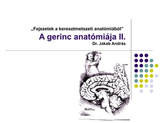
1. „ Fejezetek a keresztmetszeti anatómiából” A gerinc anatómiája II. Dr. Jakab András
2.
3.
4.
5. A gerincvelő és a gerincoszlop viszonya (C7) processus spinosus arcus vertebrae processus transversus periosteum sp. epidurale dura mater spinalis sp. subarachnoideum arachnoidea mater pia mater radix posterior radix anterior ganglion spinale n. spinalis; r. posterior n. spinalis; r. anterior
![Gerincvelő – makroszkópos anatómia ,[object Object],[object Object],[object Object],[object Object],[object Object],[object Object],[object Object],[object Object],[object Object]](data:image/gif;base64,R0lGODlhAQABAIAAAAAAAP///yH5BAEAAAAALAAAAAABAAEAAAIBRAA7)