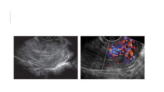
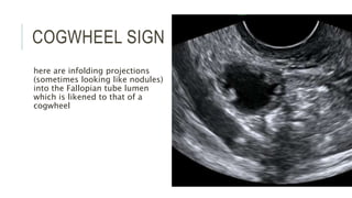
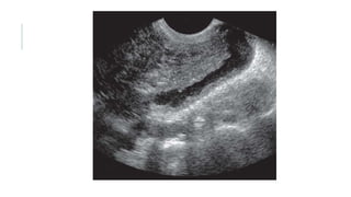
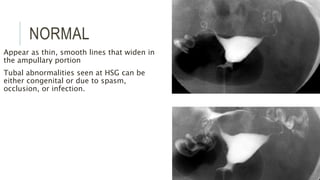
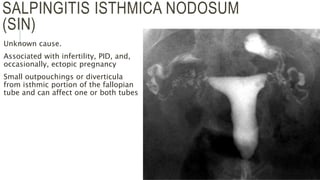
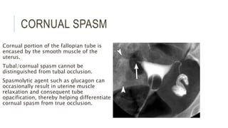
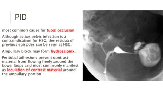
FALLOPIAN TUBE -
IMAGING
20181204
Dr. Abdul Razak C A
FALLOPIAN TUBE
The normal fallopian tube is difficult to identify by transabdominal or
transvaginal sonography unless it is dilated or surrounded by fluid.
Undulating echogenic structure approximately 8 to 10 mm in width,
running posterolaterally from the uterus to lie within the posterior
cul-de-sac near the ovary.
Lumen is not seen unless it is fluid filled.
Developmental abnormalities of the fallopian tube are rare.
Abnormalities of tube include pregnancy, infection, and neoplasm
PELVIC INFLAMMATORY DISEASE
most often associated with gonorrhea and chlamydia.
 by ascent from cervix and endometrium.
direct extension from appendiceal, diverticular, or postsurgical abscesses
Hematogenous spread from tuberculosis.
Bilateral, except when caused by direct extension
IUCD increases the risk of PID
pain, fever, pelvic tenderness, and vaginal discharge. A pelvic mass may be
palpated.
Chronic pelvic pain, infertility, and increased risk of ectopic pregnancy.
cervicitis
endometritis,
acute salpingitis, and pyosalpinx
finally the region of both ovaries and the peritoneum.
Hydrosalpinx
Pyosalpinx
Thickened tubal wall (≥5 mm) is indicative of acute disease.
appearances of tubal wall structure:
 (1) cogwheel sign, seen mainly in acute disease;
 (2) “beads on a string” sign, seen only in chronic disease;
 (3) incomplete septa,
 (4) diametrically opposed indentations in the wall (“waist sign”) had the highest
likelihood ratio in discriminating hydrosalpinx from other adnexal masses
COGWHEEL SIGN
here are infolding projections
(sometimes looking like nodules)
into the Fallopian tube lumen
which is likened to that of a
cogwheel
“beads on a string” sign
WAIST SIGN
Tubo-ovarian complex
Tubo-ovarian abscess - complex multiloculated mass with variable
septations, irregular margins, and scattered internal echoes. There is
usually posterior acoustic enhancement, and a fluid-debris level or
gas may occasionally be seen within the mass.
sonographically guided transvaginal aspiration and drainage.
Chronic PID, extensive fibrosis and adhesions may obscure the
margins of the pelvic organs, which blend into a large, poorly defined
mass.
Transvaginal image shows a very
large ovary surrounded by a rim
of highly echogenic and inflamed
fat (arrows). There are complex
fluid collections within the ovary.
There is no normal architecture.
tube (T) is distended and
elongated and filled with debris
representing pus. The ovary (O)
is similarly filled with pus, with
indistinct borders, showing a
tubo-ovarian complex.
CARCINOMA
least common (0.3%) of all gynecologic malignancies, with
adenocarcinoma being the most common histologic type.
sixth decade
pain, vaginal bleeding, and a pelvic mass
profuse watery discharge, known as hydrops tubae profluens
usually involves the distal end
sausage-shaped, solid, or cystic mass with papillary projections.
HSG
10–12 cm in length and course along the superior aspect of the broad ligament
Three segments radiographically
Interstitial or cornual region
Isthmic portion between the interstitial and ampullary regions.
Ampullary portion is the widened region near the ovary. Fimbriated part is is not
usually seen at HSG.
NORMAL
Appear as thin, smooth lines that widen in
the ampullary portion
Tubal abnormalities seen at HSG can be
either congenital or due to spasm,
occlusion, or infection.
SALPINGITIS ISTHMICA NODOSUM
(SIN)
Unknown cause.
Associated with infertility, PID, and,
occasionally, ectopic pregnancy
Small outpouchings or diverticula
from isthmic portion of the fallopian
tube and can affect one or both tubes
CORNUAL SPASM
Cornual portion of the fallopian tube is
encased by the smooth muscle of the
uterus.
Tubal/cornual spasm cannot be
distinguished from tubal occlusion.
Spasmolytic agent such as glucagon can
occasionally result in uterine muscle
relaxation and consequent tube
opacification, thereby helping differentiate
cornual spasm from true occlusion.
PID
most common cause for tubal occlusion
Although active pelvic infection is a
contraindication for HSG, the residua of
previous episodes can be seen at HSG.
Ampullary block may form hydrosalpinx.
Peritubal adhesions prevent contrast
material from flowing freely around the
bowel loops and most commonly manifest
as loculation of contrast material around
the ampullary portion
Postoperative evaluation of the
fallopian tubes
documentation of tubal occlusion
following tubal ligation.
abrupt termination of the tube at
the surgical site or mild bulbous
expansion of the tube with cutoff
May also demonstrate tubal
patency without extravasation of
contrast material after reversal of
a ligation procedure.
Essure –
 microinsert placed hysteroscopically
into each fallopian tube
 induces scar formation around itself
 HSG is usually performed to evaluate
for tubal occlusion
TUBAL POLYPS
Ectopic endometrial tissue
located in the interstitial portion
of the tube.
Smooth, rounded filling defects,
without concomitant dilatation or
tubal occlusion.
Ectopic pregnancy
 tubal filling defect

Fallopian tube Imaging Radiology

  • 1.
  • 2.
    FALLOPIAN TUBE The normalfallopian tube is difficult to identify by transabdominal or transvaginal sonography unless it is dilated or surrounded by fluid. Undulating echogenic structure approximately 8 to 10 mm in width, running posterolaterally from the uterus to lie within the posterior cul-de-sac near the ovary. Lumen is not seen unless it is fluid filled. Developmental abnormalities of the fallopian tube are rare. Abnormalities of tube include pregnancy, infection, and neoplasm
  • 4.
    PELVIC INFLAMMATORY DISEASE mostoften associated with gonorrhea and chlamydia.  by ascent from cervix and endometrium. direct extension from appendiceal, diverticular, or postsurgical abscesses Hematogenous spread from tuberculosis. Bilateral, except when caused by direct extension IUCD increases the risk of PID pain, fever, pelvic tenderness, and vaginal discharge. A pelvic mass may be palpated. Chronic pelvic pain, infertility, and increased risk of ectopic pregnancy.
  • 5.
    cervicitis endometritis, acute salpingitis, andpyosalpinx finally the region of both ovaries and the peritoneum.
  • 9.
  • 10.
    Pyosalpinx Thickened tubal wall(≥5 mm) is indicative of acute disease. appearances of tubal wall structure:  (1) cogwheel sign, seen mainly in acute disease;  (2) “beads on a string” sign, seen only in chronic disease;  (3) incomplete septa,  (4) diametrically opposed indentations in the wall (“waist sign”) had the highest likelihood ratio in discriminating hydrosalpinx from other adnexal masses
  • 11.
    COGWHEEL SIGN here areinfolding projections (sometimes looking like nodules) into the Fallopian tube lumen which is likened to that of a cogwheel
  • 12.
    “beads on astring” sign
  • 13.
  • 16.
    Tubo-ovarian complex Tubo-ovarian abscess- complex multiloculated mass with variable septations, irregular margins, and scattered internal echoes. There is usually posterior acoustic enhancement, and a fluid-debris level or gas may occasionally be seen within the mass. sonographically guided transvaginal aspiration and drainage. Chronic PID, extensive fibrosis and adhesions may obscure the margins of the pelvic organs, which blend into a large, poorly defined mass.
  • 17.
    Transvaginal image showsa very large ovary surrounded by a rim of highly echogenic and inflamed fat (arrows). There are complex fluid collections within the ovary. There is no normal architecture.
  • 18.
    tube (T) isdistended and elongated and filled with debris representing pus. The ovary (O) is similarly filled with pus, with indistinct borders, showing a tubo-ovarian complex.
  • 19.
    CARCINOMA least common (0.3%)of all gynecologic malignancies, with adenocarcinoma being the most common histologic type. sixth decade pain, vaginal bleeding, and a pelvic mass profuse watery discharge, known as hydrops tubae profluens usually involves the distal end sausage-shaped, solid, or cystic mass with papillary projections.
  • 20.
    HSG 10–12 cm inlength and course along the superior aspect of the broad ligament Three segments radiographically Interstitial or cornual region Isthmic portion between the interstitial and ampullary regions. Ampullary portion is the widened region near the ovary. Fimbriated part is is not usually seen at HSG.
  • 21.
    NORMAL Appear as thin,smooth lines that widen in the ampullary portion Tubal abnormalities seen at HSG can be either congenital or due to spasm, occlusion, or infection.
  • 22.
    SALPINGITIS ISTHMICA NODOSUM (SIN) Unknowncause. Associated with infertility, PID, and, occasionally, ectopic pregnancy Small outpouchings or diverticula from isthmic portion of the fallopian tube and can affect one or both tubes
  • 23.
    CORNUAL SPASM Cornual portionof the fallopian tube is encased by the smooth muscle of the uterus. Tubal/cornual spasm cannot be distinguished from tubal occlusion. Spasmolytic agent such as glucagon can occasionally result in uterine muscle relaxation and consequent tube opacification, thereby helping differentiate cornual spasm from true occlusion.
  • 24.
    PID most common causefor tubal occlusion Although active pelvic infection is a contraindication for HSG, the residua of previous episodes can be seen at HSG. Ampullary block may form hydrosalpinx. Peritubal adhesions prevent contrast material from flowing freely around the bowel loops and most commonly manifest as loculation of contrast material around the ampullary portion
  • 25.
    Postoperative evaluation ofthe fallopian tubes documentation of tubal occlusion following tubal ligation. abrupt termination of the tube at the surgical site or mild bulbous expansion of the tube with cutoff
  • 26.
    May also demonstratetubal patency without extravasation of contrast material after reversal of a ligation procedure. Essure –  microinsert placed hysteroscopically into each fallopian tube  induces scar formation around itself  HSG is usually performed to evaluate for tubal occlusion
  • 27.
    TUBAL POLYPS Ectopic endometrialtissue located in the interstitial portion of the tube. Smooth, rounded filling defects, without concomitant dilatation or tubal occlusion.
  • 28.