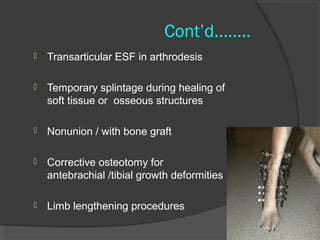
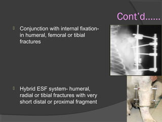
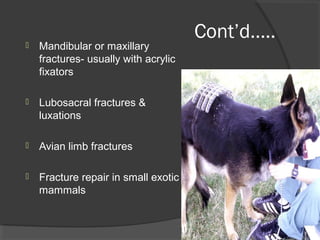
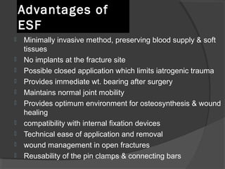
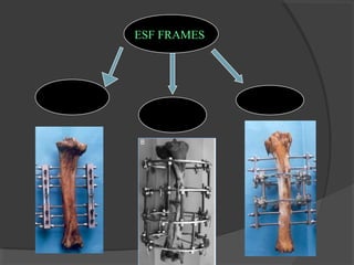
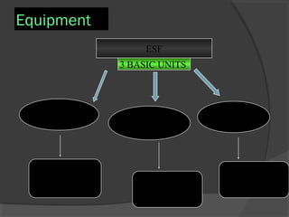
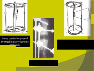
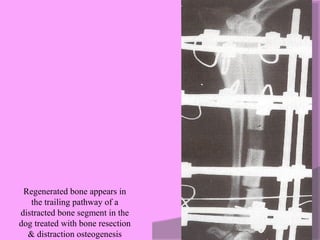
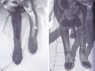
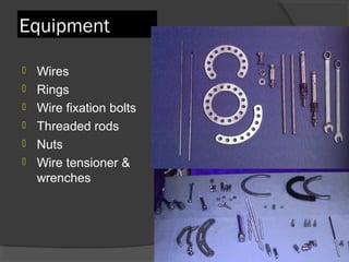
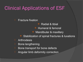
External skeletal  fixation techniques Shahnawaz ahmad bhat  4857
Definition  A method of fracture treatment ,that involves the percutaneous placement of a series of transcortical pins or wires , which are externally connected to a rigid frame
Indications for use   Long bone fractures Open fractures  Comminuted fractures that cannot be anatomically reconstructed Osteomyelitis High-energy fractures with soft- tissue injuries and vascular compromise
Cont’d........ Transarticular ESF in arthrodesis  Temporary splintage during healing of soft tissue or  osseous structures  Nonunion  / with bone graft  Corrective osteotomy for antebrachial /tibial growth deformities Limb lengthening procedures
Cont’d…… Conjunction with internal fixation- in humeral, femoral or tibial fractures  Hybrid ESF system- humeral, radial or tibial fractures with very short distal or proximal fragment
Cont’d….. Mandibular or maxillary  fractures- usually with acrylic fixators Lubosacral fractures & luxations  Avian limb fractures Fracture repair in small exotic mammals
Advantages of ESF Minimally invasive method, preserving blood supply & soft tissues No implants at the fracture site  Possible closed application which limits iatrogenic trauma Provides immediate wt. bearing after surgery  Maintains normal joint mobility  Provides optimum environment for osteosynthesis & wound healing  compatibility with internal fixation devices Technical ease of application and removal wound management in open fractures  Reusability of the pin clamps & connecting bars
Disadvantages of ESF   Device must be cleaned and monitored regularly Care to prevent additional damage to animal/device Aftercare is more labor intensive  More rigid type II and III frames cannot be used for fractures of femur & humerus Difficult to apply and more pain in areas of increased muscle mass Extremely high cost
ESF FRAMES   LINEAR CIRCULAR HYBRID
Equipment   ESF 3 BASIC UNITS  FIXATION PINS   EXTERNAL  CONNECTORS  LINKAGE  DEVICES   Inserted into bone  To hold major Fragments   Support fractured  Bones  Attach fixation  Pins & external  Connectors
FIXATION PINS : classification  on basis of implantation method  HALF PIN   penetrate only one skin  surface but both bone cortices FULL PIN Panetrate one skin  surface then both  cortices ,and then  the opposite Skin  Surface
On basis of structural design Smooth pin  : Rely on friction to remain stable in the bone Threaded pins :  Positive profile and negative profile Centrally threaded and end threaded
Positive & negative profile threaded pins   Pins in which core diameter of the threaded section is smaller than the diameter of the smooth section have negative thread profile  If the core diameter is consistent b/w smooth & threaded regions ,thread profile is positive
Positive profile transfixation pins used in ESF
Centrally threaded & End threaded pins   Centrally threaded pins are used as full pins with type  ÌÌ or ÌÌÌ external fixator frames  Central threads engage bone & smooth pin ends extend beyond the skin surface  End threaded pins are described a/c to no. of cortices engaged by threads (one cortex & two cortex end threaded pins)  End treaded pin   Centrally threaded  pin
External connectors  Made up of stainless steel, titanium alloy , carbon fiber,aluminium or acrylic  External fixator & linkage devices may be fashioned from acrylic for ACRYLIC -PIN EXTERNAL FIXATOR (APEF) APEF system contains +ve profile threaded fixation pins ,packaged acrylic & sterilized acrylic column molding tubes
SK double clamp Secur-U clamp S K single   clamp Kirschner double clamp Kirschner single  clamp Kirschner type linkage devices for joining fixation  Pins to external connecting bars & external connecting bars to each other.Note the larger holes for the external  connecting bars & smaller holes in the bolts for  Fixation pins Linkage devices
Linear ESF Configurations   Type  Ì ,ÌÌ or ÌÌÌ configuration  TYPE Ì Configuration   Utilize half pin splintage fixation pins  Connecting frame  placed on only one side of the limb  Unilateral constructs  Classified as  * type Іa (unilateral and uniplanar) * type Іb (unilateral and biplanar)
Type Ǐ a (unilateral –uniplanar ) fixators All the fixation pins & connecting frame occupy a single plane (uniplanar) Usually applied to cranial medial surface of radius/ tibia and lateral surface of femur/ humerus  Fixation pins are half pins
Type Ǐb (unilateral -biplanar )  fixators  Applied most often to radius & tibia  For radius &  tibia–one ext. bar is placed on craniomedial surface & 2 nd  bar on craniolateral surface
Type ǏǏ (bilateral-uniplanar )   fixators  Utilize full pin splintage fixation pins  Cannot be placed on femur/ humerus because of adjacent body wall Applied only to radius/tibia in mediolateral plane  Maximal type ІІa & minimal type ІІb Maximal type  ІІa Minimal type  ІІb
Type ǏǏǏ(Bilateral-biplanar)fixator Combination of  type І and type ІІ Type І and type ІІ systems placed approx. 90 ° to each other  Can’t be applied to femur or humerus  Indicated in very large dogs
TIE-IN CONFIGURATION  Combination of ESF (type Іa or Іb) with intramedullary pin  Increases rigidity of fracture fixation in humerus & femur as type ІІ &Type ІІІ frames cannot be applied  No. of fixation pins is limited to 2 or 3 pins above & below the fracture
Tie –in configuration
Biomechanics Fixator rigidity depends on: Type of configuration-  Type ІІІ  >  type ІІ  >  type І  biplanar type Іb more resistant to cranial/caudal shear forces compared with type ІІ.  Number of used pins: at least 2 & up to 4 pins/bone segment Diameter and design of the pins – the diameter  ≤  25 % of the bone’s diameter The fixator rigidity  by widening pin spacing within pin groups & by  the distance between pin groups The most central pin should be 1-2 cm away from the fracture line
Cont’d….. Moving the connecting bar closer to the bone makes the pins more resistant to bending Using a “tie-in”  configuration increase significant in bending strength
Fixation pin application  Preoperative planning  Patient fracture assessment Most important principle -asceptic surgery  Injured limb suspended from hook in the ceiling  Patient Draping choose a surgical approach Delimitation of safe corridors  for insertion of pins
Cont’d…….. Most proximal and distal pins inserted after a stab skin incision  Predrilling a pilot hole with slightly smaller diameter drill bit  Transfixating pins inserted by using low-speed power drill  Fracture reduced & connecting bar connected to proximal and distal pins by clamps Clamps placed 1-2 cm away from the skin surface  Central pins are inserted above and below the fracture Rest of needed pins inserted & bolts tightened
Postoperative management Postoperative analgesia  Pin-skin interface cleaned with antiseptic solution  Incision around pins released/extended in case of skin tension  Sterile gauze sponges placed around & b/w fixation pins  Limb wrapped with vetrap (bandage material ) Activity restricted to least walking & physical rehabilitation
Circular External Skeletal Fixators(CESF)    ( Ilizarov technique) Most recent innovation in ESF technique in dogs and cats & also in large animals. Developed by the Russian physician Gavriil A. Ilizarov  CESF consist of a series of complete and/or incomplete external rings that are interconnected by a series of threaded rods
Cont’d….. These are modular systems which  can be assembled in numerous  configurations to  stabilize fractures and arthrodeses, perform bone lengthening and transport  correct angular, translational and rotational deformities Each ring is secured in position along the rod by placing a nut on either side of the ring
Cont’d…. Elongation of the frame allows for  distraction osteogenesis  in which regenerate bone is produced within the gap created when an osteotomy is distracted slowly and sequentially “ Discontinuity of a skeletal segment necessarily triggers the repair process which will continue as long as integrity of both osteogenic tissue & its vascular supply is maintained”  Traditional CFs use small diameter wires, rather than pins, as fixation elements
Standard circular fixator frame for fracture  management
Bones can be lengthened by streching a corticotomy 1mm a day   The regenerate is composed  of  Microcolumns originating from  both bone ends  Radiograph of distraction  Callus at 3 wks
Regenerated bone appears in  the trailing pathway of a  distracted bone segment in the  dog treated with bone resection  & distraction osteogenesis
 
Equipment Wires  Rings Wire fixation bolts Threaded rods  Nuts  Wire tensioner & wrenches
Wires : 1.0,1.2 or1.5mm diameter used in cats/ dogs Olive wires- wires with a bead positioned midway along the wire  Provide interfragmentary compression & increase stability of frame construct  more the no. of fixation wires-  more  stability of fixation  Wire angles  < 45°should be avoided
Olive wires
Rings   Partial ring & full ring  Five–eighth/stretch ring are used when rings would limit joint motion (elbow/stifle) Partial rings -versatile  Full rings -more available holes for rods  & wire, not versatile  ring diameter ,  axial stiffness, torsional & bending stiffness  Smallest ring allowing  ≥2 cm distance b/w skin & inner circumference of ring selected
Wire fixation bolts  Cannulated wire fixation bolts –allow wire passage via a concentrically placed hole at base of bolt head  Slotted wire fixation bolts have an eccentric slot located under the bolt head & parallel to its long axis  Wire must be fixed without deformation
Frame construction  Threaded  rods used to connect rings & form frame  Telescopic rods- hollow rods used as supports & connecting elements of ring  Frames are constructed so that one ring & its wires are placed at proximal end and another ring &  its wires are placed at distal end of long bone  2 additional rings placed so that their wires panetrate proximal & distal bone segments close to the fracture
Wire tensioner
Hybrid External Fixator Combination of a ring & a linear fixator  Indicated for fractures with juxtaarticular bone segments  May be applied to radius,tibia,femur & humerus Can also be used to stabilize corrective osteotomies for angular limb deformities
Larger transfixation wires  can be combined with the  use of rings & wires in  a Hybrid frame
Clinical Applications of ESF Fracture fixation  Radial & tibial  Humeral & femoral  Mandibular & maxillary  Stabilization of spinal fractures & luxations  Arthrodesis  Bone lengthening Bone transport for bone defects  Angular limb deformity correction
Fixation of spinal fractures & luxations Spinal arch external fixator components developed  for  spinal stabilization  Fluoroscopically guided percutaneous pin placement can be performed when applying external skeletal spinal fixation
Bone transport (limb salvage) Method by which one or two segments of normal bone adjacent to a defect are slowly transported into the defect while new bone forms in the distraction pathway  Used in dogs with radial/tibial neoplasia or severe osteomyelitis with bone loss  A segment of bone is created with a corticotomy made 2-3 cm from one end of one of the main  fragment
 
Acrylic external fixation   Acrylics are commonly used for the repair of fractures  The acrylic column acts as both the connecting rod and transfixation pin-gripping device Dental acrylic or hoof repair acrylic are suitable  It can be moulded over pins after curing to a dough stage Acrylic ESF offers the advantage of reduced cost , improved versatility & simplified application technique when compared with kirschner ESF
Advantages of acrylic system Ability to contour the connecting bar to match any fracture configuration  Fixation pins of any diameter may be used Fixation pins do not have to be in the same longitudinal plane Light in weight encouraging earlier return to function
cont’d…… Placing the positive profile threaded pin without the difficulties of passing them through clamps Inexpensive system
 
Uses of acrylic fixators  Fixation of mandibular & maxillary fractures  Fixation of long bone fractures  Primary fixation device or intra-oral splint for mandibular & maxillary frcatures
Complications of ESF  Pin tract infection  Focal osteomyelitis  Ring sequestrum Premature pin loosening Instability at the fracture site  Pin breaking  Pin tract osteolysis Pressure necrosis of skin Iatrogenic bone fracture soft tissue impalement
thank you

External skeletal fixators

  • 1.
    External skeletal fixation techniques Shahnawaz ahmad bhat 4857
  • 2.
    Definition Amethod of fracture treatment ,that involves the percutaneous placement of a series of transcortical pins or wires , which are externally connected to a rigid frame
  • 3.
    Indications for use Long bone fractures Open fractures Comminuted fractures that cannot be anatomically reconstructed Osteomyelitis High-energy fractures with soft- tissue injuries and vascular compromise
  • 4.
    Cont’d........ Transarticular ESFin arthrodesis Temporary splintage during healing of soft tissue or osseous structures Nonunion / with bone graft Corrective osteotomy for antebrachial /tibial growth deformities Limb lengthening procedures
  • 5.
    Cont’d…… Conjunction withinternal fixation- in humeral, femoral or tibial fractures Hybrid ESF system- humeral, radial or tibial fractures with very short distal or proximal fragment
  • 6.
    Cont’d….. Mandibular ormaxillary fractures- usually with acrylic fixators Lubosacral fractures & luxations Avian limb fractures Fracture repair in small exotic mammals
  • 7.
    Advantages of ESFMinimally invasive method, preserving blood supply & soft tissues No implants at the fracture site Possible closed application which limits iatrogenic trauma Provides immediate wt. bearing after surgery Maintains normal joint mobility Provides optimum environment for osteosynthesis & wound healing compatibility with internal fixation devices Technical ease of application and removal wound management in open fractures Reusability of the pin clamps & connecting bars
  • 8.
    Disadvantages of ESF Device must be cleaned and monitored regularly Care to prevent additional damage to animal/device Aftercare is more labor intensive More rigid type II and III frames cannot be used for fractures of femur & humerus Difficult to apply and more pain in areas of increased muscle mass Extremely high cost
  • 9.
    ESF FRAMES LINEAR CIRCULAR HYBRID
  • 10.
    Equipment ESF 3 BASIC UNITS FIXATION PINS EXTERNAL CONNECTORS LINKAGE DEVICES Inserted into bone To hold major Fragments Support fractured Bones Attach fixation Pins & external Connectors
  • 11.
    FIXATION PINS :classification on basis of implantation method HALF PIN penetrate only one skin surface but both bone cortices FULL PIN Panetrate one skin surface then both cortices ,and then the opposite Skin Surface
  • 12.
    On basis ofstructural design Smooth pin : Rely on friction to remain stable in the bone Threaded pins : Positive profile and negative profile Centrally threaded and end threaded
  • 13.
    Positive & negativeprofile threaded pins Pins in which core diameter of the threaded section is smaller than the diameter of the smooth section have negative thread profile If the core diameter is consistent b/w smooth & threaded regions ,thread profile is positive
  • 14.
  • 15.
    Centrally threaded &End threaded pins Centrally threaded pins are used as full pins with type ÌÌ or ÌÌÌ external fixator frames Central threads engage bone & smooth pin ends extend beyond the skin surface End threaded pins are described a/c to no. of cortices engaged by threads (one cortex & two cortex end threaded pins) End treaded pin Centrally threaded pin
  • 16.
    External connectors Made up of stainless steel, titanium alloy , carbon fiber,aluminium or acrylic External fixator & linkage devices may be fashioned from acrylic for ACRYLIC -PIN EXTERNAL FIXATOR (APEF) APEF system contains +ve profile threaded fixation pins ,packaged acrylic & sterilized acrylic column molding tubes
  • 17.
    SK double clampSecur-U clamp S K single clamp Kirschner double clamp Kirschner single clamp Kirschner type linkage devices for joining fixation Pins to external connecting bars & external connecting bars to each other.Note the larger holes for the external connecting bars & smaller holes in the bolts for Fixation pins Linkage devices
  • 18.
    Linear ESF Configurations Type Ì ,ÌÌ or ÌÌÌ configuration TYPE Ì Configuration Utilize half pin splintage fixation pins Connecting frame placed on only one side of the limb Unilateral constructs Classified as * type Іa (unilateral and uniplanar) * type Іb (unilateral and biplanar)
  • 19.
    Type Ǐ a(unilateral –uniplanar ) fixators All the fixation pins & connecting frame occupy a single plane (uniplanar) Usually applied to cranial medial surface of radius/ tibia and lateral surface of femur/ humerus Fixation pins are half pins
  • 20.
    Type Ǐb (unilateral-biplanar ) fixators Applied most often to radius & tibia For radius & tibia–one ext. bar is placed on craniomedial surface & 2 nd bar on craniolateral surface
  • 21.
    Type ǏǏ (bilateral-uniplanar) fixators Utilize full pin splintage fixation pins Cannot be placed on femur/ humerus because of adjacent body wall Applied only to radius/tibia in mediolateral plane Maximal type ІІa & minimal type ІІb Maximal type ІІa Minimal type ІІb
  • 22.
    Type ǏǏǏ(Bilateral-biplanar)fixator Combinationof type І and type ІІ Type І and type ІІ systems placed approx. 90 ° to each other Can’t be applied to femur or humerus Indicated in very large dogs
  • 23.
    TIE-IN CONFIGURATION Combination of ESF (type Іa or Іb) with intramedullary pin Increases rigidity of fracture fixation in humerus & femur as type ІІ &Type ІІІ frames cannot be applied No. of fixation pins is limited to 2 or 3 pins above & below the fracture
  • 24.
  • 25.
    Biomechanics Fixator rigiditydepends on: Type of configuration- Type ІІІ > type ІІ > type І biplanar type Іb more resistant to cranial/caudal shear forces compared with type ІІ. Number of used pins: at least 2 & up to 4 pins/bone segment Diameter and design of the pins – the diameter ≤ 25 % of the bone’s diameter The fixator rigidity by widening pin spacing within pin groups & by the distance between pin groups The most central pin should be 1-2 cm away from the fracture line
  • 26.
    Cont’d….. Moving theconnecting bar closer to the bone makes the pins more resistant to bending Using a “tie-in” configuration increase significant in bending strength
  • 27.
    Fixation pin application Preoperative planning Patient fracture assessment Most important principle -asceptic surgery Injured limb suspended from hook in the ceiling Patient Draping choose a surgical approach Delimitation of safe corridors for insertion of pins
  • 28.
    Cont’d…….. Most proximaland distal pins inserted after a stab skin incision Predrilling a pilot hole with slightly smaller diameter drill bit Transfixating pins inserted by using low-speed power drill Fracture reduced & connecting bar connected to proximal and distal pins by clamps Clamps placed 1-2 cm away from the skin surface Central pins are inserted above and below the fracture Rest of needed pins inserted & bolts tightened
  • 29.
    Postoperative management Postoperativeanalgesia Pin-skin interface cleaned with antiseptic solution Incision around pins released/extended in case of skin tension Sterile gauze sponges placed around & b/w fixation pins Limb wrapped with vetrap (bandage material ) Activity restricted to least walking & physical rehabilitation
  • 30.
    Circular External SkeletalFixators(CESF) ( Ilizarov technique) Most recent innovation in ESF technique in dogs and cats & also in large animals. Developed by the Russian physician Gavriil A. Ilizarov CESF consist of a series of complete and/or incomplete external rings that are interconnected by a series of threaded rods
  • 31.
    Cont’d….. These aremodular systems which can be assembled in numerous configurations to stabilize fractures and arthrodeses, perform bone lengthening and transport correct angular, translational and rotational deformities Each ring is secured in position along the rod by placing a nut on either side of the ring
  • 32.
    Cont’d…. Elongation ofthe frame allows for distraction osteogenesis in which regenerate bone is produced within the gap created when an osteotomy is distracted slowly and sequentially “ Discontinuity of a skeletal segment necessarily triggers the repair process which will continue as long as integrity of both osteogenic tissue & its vascular supply is maintained” Traditional CFs use small diameter wires, rather than pins, as fixation elements
  • 33.
    Standard circular fixatorframe for fracture management
  • 34.
    Bones can belengthened by streching a corticotomy 1mm a day The regenerate is composed of Microcolumns originating from both bone ends Radiograph of distraction Callus at 3 wks
  • 35.
    Regenerated bone appearsin the trailing pathway of a distracted bone segment in the dog treated with bone resection & distraction osteogenesis
  • 36.
  • 37.
    Equipment Wires Rings Wire fixation bolts Threaded rods Nuts Wire tensioner & wrenches
  • 38.
    Wires : 1.0,1.2or1.5mm diameter used in cats/ dogs Olive wires- wires with a bead positioned midway along the wire Provide interfragmentary compression & increase stability of frame construct more the no. of fixation wires- more stability of fixation Wire angles < 45°should be avoided
  • 39.
  • 40.
    Rings Partial ring & full ring Five–eighth/stretch ring are used when rings would limit joint motion (elbow/stifle) Partial rings -versatile Full rings -more available holes for rods & wire, not versatile ring diameter , axial stiffness, torsional & bending stiffness Smallest ring allowing ≥2 cm distance b/w skin & inner circumference of ring selected
  • 41.
    Wire fixation bolts Cannulated wire fixation bolts –allow wire passage via a concentrically placed hole at base of bolt head Slotted wire fixation bolts have an eccentric slot located under the bolt head & parallel to its long axis Wire must be fixed without deformation
  • 42.
    Frame construction Threaded rods used to connect rings & form frame Telescopic rods- hollow rods used as supports & connecting elements of ring Frames are constructed so that one ring & its wires are placed at proximal end and another ring & its wires are placed at distal end of long bone 2 additional rings placed so that their wires panetrate proximal & distal bone segments close to the fracture
  • 43.
  • 44.
    Hybrid External FixatorCombination of a ring & a linear fixator Indicated for fractures with juxtaarticular bone segments May be applied to radius,tibia,femur & humerus Can also be used to stabilize corrective osteotomies for angular limb deformities
  • 45.
    Larger transfixation wires can be combined with the use of rings & wires in a Hybrid frame
  • 46.
    Clinical Applications ofESF Fracture fixation Radial & tibial Humeral & femoral Mandibular & maxillary Stabilization of spinal fractures & luxations Arthrodesis Bone lengthening Bone transport for bone defects Angular limb deformity correction
  • 47.
    Fixation of spinalfractures & luxations Spinal arch external fixator components developed for spinal stabilization Fluoroscopically guided percutaneous pin placement can be performed when applying external skeletal spinal fixation
  • 48.
    Bone transport (limbsalvage) Method by which one or two segments of normal bone adjacent to a defect are slowly transported into the defect while new bone forms in the distraction pathway Used in dogs with radial/tibial neoplasia or severe osteomyelitis with bone loss A segment of bone is created with a corticotomy made 2-3 cm from one end of one of the main fragment
  • 49.
  • 50.
    Acrylic external fixation Acrylics are commonly used for the repair of fractures The acrylic column acts as both the connecting rod and transfixation pin-gripping device Dental acrylic or hoof repair acrylic are suitable It can be moulded over pins after curing to a dough stage Acrylic ESF offers the advantage of reduced cost , improved versatility & simplified application technique when compared with kirschner ESF
  • 51.
    Advantages of acrylicsystem Ability to contour the connecting bar to match any fracture configuration Fixation pins of any diameter may be used Fixation pins do not have to be in the same longitudinal plane Light in weight encouraging earlier return to function
  • 52.
    cont’d…… Placing thepositive profile threaded pin without the difficulties of passing them through clamps Inexpensive system
  • 53.
  • 54.
    Uses of acrylicfixators Fixation of mandibular & maxillary fractures Fixation of long bone fractures Primary fixation device or intra-oral splint for mandibular & maxillary frcatures
  • 55.
    Complications of ESF Pin tract infection Focal osteomyelitis Ring sequestrum Premature pin loosening Instability at the fracture site Pin breaking Pin tract osteolysis Pressure necrosis of skin Iatrogenic bone fracture soft tissue impalement
  • 56.