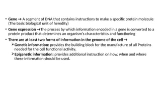
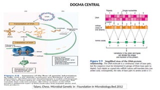
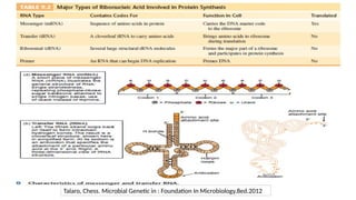
• Gene →A segment of DNA that contains instructions to make a specific protein molecule
(The basic biological unit of heredity)
• Gene expression →The process by which information encoded in a gene is converted to a
protein product that determines an organism’s characteristics and functioning
• There are at least two forms of information in the genome of the cell →
Genetic information: provides the building block for the manufacture of all Proteins
needed for the cell functional activity.
Epigenetic information: provides additional instruction on how, when and where
these information should be used.
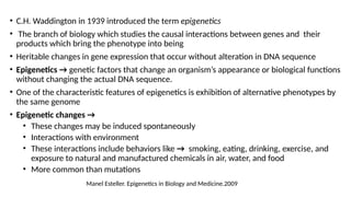
• C.H. Waddingtonin 1939 introduced the term epigenetics
• The branch of biology which studies the causal interactions between genes and their
products which bring the phenotype into being
• Heritable changes in gene expression that occur without alteration in DNA sequence
• Epigenetics → genetic factors that change an organism’s appearance or biological functions
without changing the actual DNA sequence.
• One of the characteristic features of epigenetics is exhibition of alternative phenotypes by
the same genome
• Epigenetic changes →
• These changes may be induced spontaneously
• Interactions with environment
• These interactions include behaviors like → smoking, eating, drinking, exercise, and
exposure to natural and manufactured chemicals in air, water, and food
• More common than mutations
Manel Esteller. Epigenetics in Biology and Medicine.2009
26.
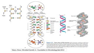
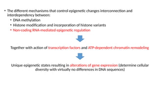
• The differentmechanisms that control epigenetic changes interconnection and
interdependency between:
• DNA methylation
• Histone modification and incorporation of histone variants
• Non-coding RNA-mediated epigenetic regulation
Together with action of transcription factors and ATP-dependent chromatin remodeling
Unique epigenetic states resulting in alterations of gene expression (determine cellular
diversity with virtually no differences in DNA sequences)
27.
repositioning of nucleosomes
incorporationof histone variants
methylation,
acetylation,
phosphorylation,
ubiquitination,
sumoylation, etc…
Long ncRNAs
small RNAs
Cytosine methylation
Adenine methylation
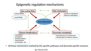
Epigenetic regulation mechanisms
Gene Expression
Non-coding RNAs
Histone Modifications Chromatin remodeling
DNA Methylation
• All these mechanisms realized by the specific pathways and demands specific enzymes
small RNAs
Igor Yakovlev.2016
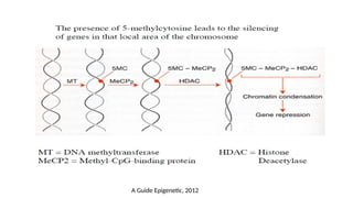
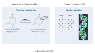
1. DNA methylation
•The addition of a methyl-group to one of the bases in the deoxyribonucleic acid chain
• Does not change the primary DNA sequence
• Generally repressive to transcription
• Constituting an important mechanism for :
• Gene silencing in embryonic development
• Inactivation of defined tumor suppressor genes in human cancers
• Cytosine methylation is the most studied modification
• Adenine has been found to be methylated in prokaryotes and plants
32.
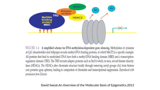
• In mammals,DNA methylation occurs mainly on the fifth carbon of the cytosine base→
5-methylcytosine or 5-methylcytidine (5-mC)
• Almost exclusively found at CpG dinucleotides ( CpG : 100-2,000 bp)
• 5-mC is a potent epigenetic marker and regulator of gene expression (<1% of nucleotides)
• Methylated CpG clusters → named CpG islands (28 million of CpG regions in the genome
and unmethylated across cell types)
• CpG islands – at gene promoters have been associated with gene inactivation
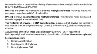
• DNA methylationis catalyzed by a family of enzymes → DNA methyltransferase (includes
DNMT1, DNMT3a and DNMT3b)
• DNMT3a and DNMT3b are known as de novo methyltransferases → able to methylate
previously unmethylated CpG dinucleotides
• DNMT1 is known as a maintenance methyltransferase → methylates hemi-methylated
DNA during replication and repair DNA
• The Tet family of enzymes → DNA demethylation –a process that involve the successive
oxidation of 5-mC to 5-hydroxymethyl- (5-hmC), 5-formyl- (5-fC), and 5-carboxy- (5-caC)
cytosine
• A glycosylase of the BER (Base Excision Repair) pathway, TDG → repair the 5-
hydroxymethyluracil which can result from deamination of 5-hmC (DNA demethylation )
• Methylation occur :
1. De Novo methylation
2. Maintenance Methylation
3. Demethylation of DNA
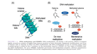
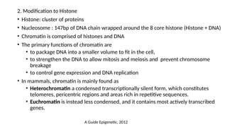
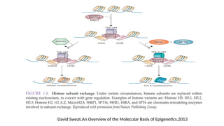
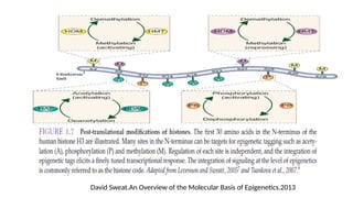
2. Modification toHistone
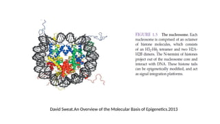
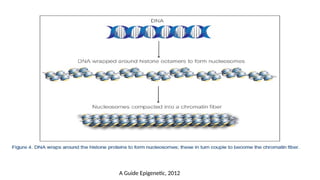
• Histone: cluster of proteins
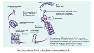
• Nucleosome : 147bp of DNA chain wrapped around the 8 core histone (Histone + DNA)
• Chromatin is comprised of histones and DNA
• The primary functions of chromatin are
• to package DNA into a smaller volume to fit in the cell,
• to strengthen the DNA to allow mitosis and meiosis and prevent chromosome
breakage
• to control gene expression and DNA replication
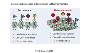
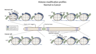
• In mammals, chromatin is mainly found as
• Heterochromatin a condensed transcriptionally silent form, which constitutes
telomeres, pericentric regions and areas rich in repetitive sequences.
• Euchromatin is instead less condensed, and it contains most actively transcribed
genes.
A Guide Epigenetic, 2012
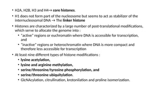
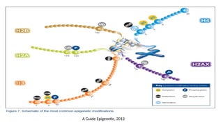
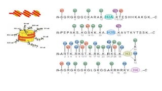
• H2A, H2B,H3 and H4→ core histones.
• H1 does not form part of the nucleosome but seems to act as stabilizer of the
internucleosomal DNA → The linker histone
• Histones are characterized by a large number of post-translational modifications,
which serve to allocate the genome into :
• “active” regions or euchromatin where DNA is accessible for transcription,
and
• “inactive” regions or heterochromatin where DNA is more compact and
therefore less accessible for transcription
• At least nine different types of histone modifications :
• lysine acetylation,
• lysine and arginine methylation,
• serine/threonine/tyrosine phosphorylation, and
• serine/threonine ubiquitylation.
• GlcNAcylation, citrullination, krotonilation and proline isomerization.
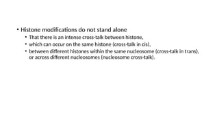
• Histone modificationsdo not stand alone
• That there is an intense cross-talk between histone,
• which can occur on the same histone (cross-talk in cis),
• between different histones within the same nucleosome (cross-talk in trans),
or across different nucleosomes (nucleosome cross-talk).
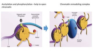
a. Histone Acetylation
•All histones can be acetylated
• The enzymes responsible for regulating the acetylation of histone tails are
histone acetyltransferases (HAT) and deacetylases (HDAC)
• Promoter-localized acetylation → Histone acetylation is largely targeted to
promoter regions
• Acetylation on lysine residues leads to → relaxation of the chromatin structure
and allows the binding of transcription factors → significantly increases gene
expression
50.
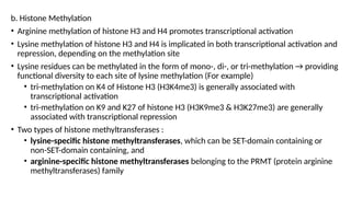
b. Histone Methylation
•Arginine methylation of histone H3 and H4 promotes transcriptional activation
• Lysine methylation of histone H3 and H4 is implicated in both transcriptional activation and
repression, depending on the methylation site
• Lysine residues can be methylated in the form of mono-, di-, or tri-methylation → providing
functional diversity to each site of lysine methylation (For example)
• tri-methylation on K4 of Histone H3 (H3K4me3) is generally associated with
transcriptional activation
• tri-methylation on K9 and K27 of histone H3 (H3K9me3 & H3K27me3) are generally
associated with transcriptional repression
• Two types of histone methyltransferases :
• lysine-specific histone methyltransferases, which can be SET-domain containing or
non-SET-domain containing, and
• arginine-specific histone methyltransferases belonging to the PRMT (protein arginine
methyltransferases) family
51.
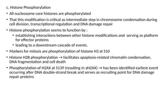
c. Histone Phosphorylation
•All nucleosome core histones are phosphorylated
• That this modification is critical as intermediate step in chromosome condensation during
cell division, transcriptional regulation and DNA damage repair
• Histone phosphorylation seems to function by :
• establishing interactions between other histone modifications and serving as platform
for effector proteins
• leading to a downstream cascade of events.
• Markers for mitosis are phosphorylation of histone H3 at S10
• Histone H2B phosphorylation → facilitates apoptosis-related chromatin condensation,
DNA fragmentation and cell death
• Phosphorylation of H2AX at S139 (resulting in γH2AX) → has been identified earliest event
occurring after DNA double-strand break and serves as recruiting point for DNA damage
repair proteins
52.
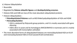
d. Histone Ubiquitylation
•Reversible
• Regulated by histone ubiquitin ligases and deubiquitylating enzymes.
• Histone H2A and H2B are two of the most abundant ubiquitylated proteins
• Histone Ubiquitylation :
• Polyubiquitylated histones such as K63-linked polyubiquitylation of H2A and H2AX
• Monoubiquitylation
• H2A is catalyzed by Polycomb group proteins, and it is mostly associated with gene
silencing
• H2B is Bre1 in yeast and its homologues RNF20/RNF40 in mammals and it is mainly
associated with transcription activation
• The most abundant forms of ubiquitylated histones are monoubiquitylated H2A on K119
and monoubiquitylated H2B on K123 (yeast)/K120 (vertebrates)
53.
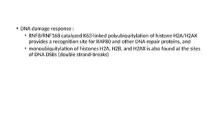
• DNA damageresponse :
• RNF8/RNF168 catalyzed K63-linked polyubiquitylation of histone H2A/H2AX
provides a recognition site for RAP80 and other DNA repair proteins, and
• monoubiquitylation of histones H2A, H2B, and H2AX is also found at the sites
of DNA DSBs (double strand-breaks)
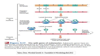
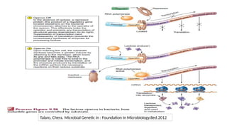
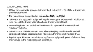
3. NON-CODING RNAs
•90% of the eukaryotic genome is transcribed but only 1 – 2% of these transcripts
encode for proteins
• The majority are transcribed as non-coding RNAs (ncRNAs)
• ncRNAs play a big part in epigenetic regulation of gene expression in addition to
their roles at the transcriptional and post-transcriptional level.
• Non-coding RNAs can be divided into two main types→ infrastructural and
regulatory ncRNAs.
• Infrastructural ncRNAs seem to have a housekeeping role in translation and
splicing and include species such as ribosomal, transfer, small nuclear RNAs.
• Regulatory ncRNAs are more interesting from an epigenetic point of view as they
are involved in the modification of other RNAs
57.
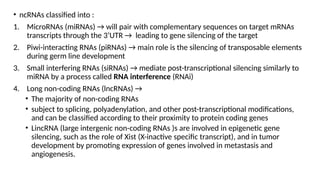
• ncRNAs classifiedinto :
1. MicroRNAs (miRNAs) → will pair with complementary sequences on target mRNAs
transcripts through the 3’UTR → leading to gene silencing of the target
2. Piwi-interacting RNAs (piRNAs) → main role is the silencing of transposable elements
during germ line development
3. Small interfering RNAs (siRNAs) → mediate post-transcriptional silencing similarly to
miRNA by a process called RNA interference (RNAi)
4. Long non-coding RNAs (lncRNAs) →
• The majority of non-coding RNAs
• subject to splicing, polyadenylation, and other post-transcriptional modifications,
and can be classified according to their proximity to protein coding genes
• LincRNA (large intergenic non-coding RNAs )s are involved in epigenetic gene
silencing, such as the role of Xist (X-inactive specific transcript), and in tumor
development by promoting expression of genes involved in metastasis and
angiogenesis.
58.
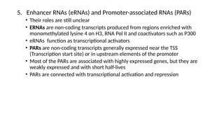
5. Enhancer RNAs(eRNAs) and Promoter-associated RNAs (PARs)
• Their roles are still unclear
• ERNAs are non-coding transcripts produced from regions enriched with
monomethylated lysine 4 on H3, RNA Pol II and coactivators such as P300
• eRNAs function as transcriptional activators
• PARs are non-coding transcripts generally expressed near the TSS
(Transcription start site) or in upstream elements of the promoter
• Most of the PARs are associated with highly expressed genes, but they are
weakly expressed and with short half-lives
• PARs are connected with transcriptional activation and repression
59.
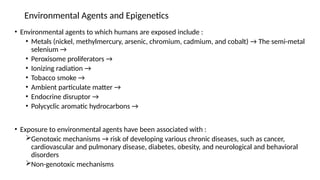
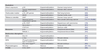
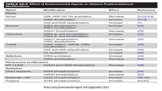
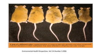
Environmental Agents andEpigenetics
• Environmental agents to which humans are exposed include :
• Metals (nickel, methylmercury, arsenic, chromium, cadmium, and cobalt) → The semi-metal
selenium →
• Peroxisome proliferators →
• Ionizing radiation →
• Tobacco smoke →
• Ambient particulate matter →
• Endocrine disruptor →
• Polycyclic aromatic hydrocarbons →
• Exposure to environmental agents have been associated with :
Genotoxic mechanisms → risk of developing various chronic diseases, such as cancer,
cardiovascular and pulmonary disease, diabetes, obesity, and neurological and behavioral
disorders
Non-genotoxic mechanisms
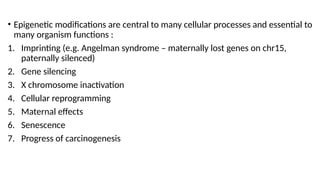
• Epigenetic modificationsare central to many cellular processes and essential to
many organism functions :
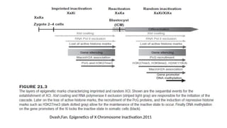
1. Imprinting (e.g. Angelman syndrome – maternally lost genes on chr15,
paternally silenced)
2. Gene silencing
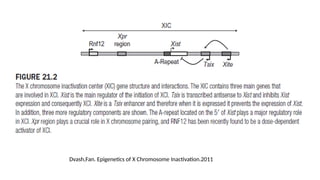
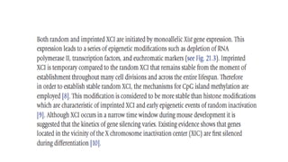
3. X chromosome inactivation
4. Cellular reprogramming
5. Maternal effects
6. Senescence
7. Progress of carcinogenesis
67.
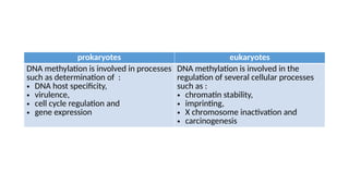
prokaryotes eukaryotes
DNA methylationis involved in processes
such as determination of :
• DNA host specificity,
• virulence,
• cell cycle regulation and
• gene expression
DNA methylation is involved in the
regulation of several cellular processes
such as :
• chromatin stability,
• imprinting,
• X chromosome inactivation and
• carcinogenesis
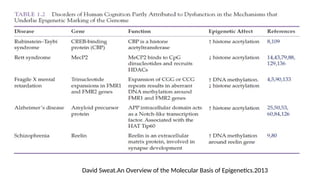
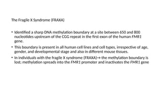
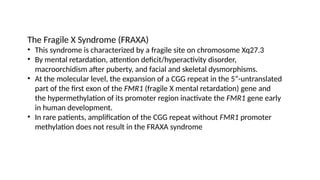
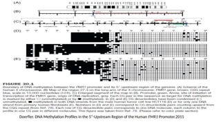
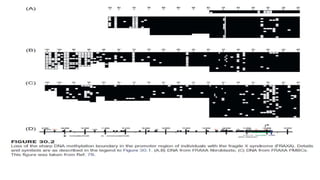
The Fragile XSyndrome (FRAXA)
• Identified a sharp DNA methylation boundary at a site between 650 and 800
nucleotides upstream of the CGG repeat in the first exon of the human FMR1
gene.
• This boundary is present in all human cell lines and cell types, irrespective of age,
gender, and developmental stage and also in different mouse tissues.
• In individuals with the fragile X syndrome (FRAXA)→ the methylation boundary is
lost; methylation spreads into the FMR1 promoter and inactivates the FMR1 gene
70.
The Fragile XSyndrome (FRAXA)
• This syndrome is characterized by a fragile site on chromosome Xq27.3
• By mental retardation, attention deficit/hyperactivity disorder,
macroorchidism after puberty, and facial and skeletal dysmorphisms.
• At the molecular level, the expansion of a CGG repeat in the 5”-untranslated
part of the first exon of the FMR1 (fragile X mental retardation) gene and
the hypermethylation of its promoter region inactivate the FMR1 gene early
in human development.
• In rare patients, amplification of the CGG repeat without FMR1 promoter
methylation does not result in the FRAXA syndrome
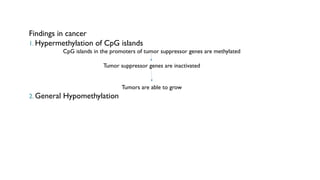
Findings in cancer
1.Hypermethylation of CpG islands
CpG islands in the promoters of tumor suppressor genes are methylated
Tumor suppressor genes are inactivated
Tumors are able to grow
2. General Hypomethylation
77.
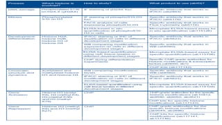
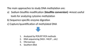
The main approachesto study DNA methylation are:
a) Sodium bisulfite modification (bisulfite conversion) →most useful
tools for analyzing cytosine methylation
b) Sequence-specific enzyme digestion
c) Capture/quantification of methylated DNA
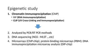
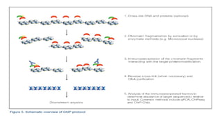
1. Analyzed by PCR/RT PCR methods
2. DNA sequencing (NGS : MLST ,..etc)
3. Microarrays
4. Southern Blot
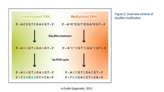
a) Sodium bisulfitemodification
• Sodium bisulfite modification, also known as bisulfite conversion, is one of the most
useful tools for analyzing cytosine methylation. This method is based on treating DNA
with sodium bisulfite in order to determine its methylation pattern.
• Treatment of DNA with sodium bisulfite leads to the deamination of cytosine residues
and converts them to uracil, while 5-mC residues remain the same.
• The treatment will generate specific changes in the DNA sequence that will depend on
the methylation status of individual cytosine residues, potentially providing single-
nucleotide resolution information about the methylation status of a DNA region:
cytosine residues that have been converted to uracil will be detected as thymidine
residues whereas methylated cytosine residues will be detected as cytosines
• Bisulfite-modified DNA can be analyzed by PCR methods that can discriminate the
methylation state of cytosines in specific genomic regions. Alternatively, bisulfite
treatment can be coupled to next generation sequencing to achieve single nucleotide
resolution mapping of cytosine methylation across the whole genome
87.
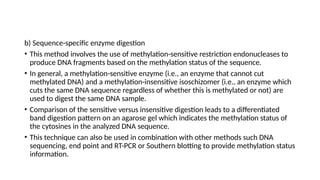
b) Sequence-specific enzymedigestion
• This method involves the use of methylation-sensitive restriction endonucleases to
produce DNA fragments based on the methylation status of the sequence.
• In general, a methylation-sensitive enzyme (i.e., an enzyme that cannot cut
methylated DNA) and a methylation-insensitive isoschizomer (i.e., an enzyme which
cuts the same DNA sequence regardless of whether this is methylated or not) are
used to digest the same DNA sample.
• Comparison of the sensitive versus insensitive digestion leads to a differentiated
band digestion pattern on an agarose gel which indicates the methylation status of
the cytosines in the analyzed DNA sequence.
• This technique can also be used in combination with other methods such DNA
sequencing, end point and RT-PCR or Southern blotting to provide methylation status
information.
88.
c) Capture/quantification ofmethylated DNA
• The use of methylated DNA-binding proteins or antibodies that specifically
recognize methylated DNA is another common method for studying methylated
DNA.
• Antibodies can be used in an ELISA-type assay to specifically detect the
percentage of methylated or hydroxymethylated cytosine residues in the
sample: the higher the signal, the higher percentage of methylated DNA.
• Antibodies can be also used to immunoprecipitate methylated DNA, thereby
enriching a DNA sample in methylated DNA. This technique is also known as
MeDIP (Methylated DNA Immunoprecipitation) and
• it can be coupled to locus specific PCR or to whole genome approaches such as
microarrays or next generation sequencing.
• Similar techniques have been developed using methylated DNA-binding
proteins instead of antibodies.
Editor's Notes
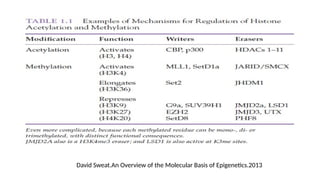
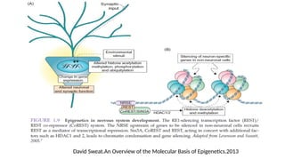
#27 Epigenetic effects can be accomplished by several self-reinforcing and inter-related covalent modifications on DNA and/or chromosomal proteins, such as DNA methylation and histone modifications, and by chromatin remodeling, such as repositioning of nucleosomes. These heritable modifications are collectively termed
“epigenetic codes”


![ONFH[AVN HIP] -TRIPLE REGIME -A NOVAL SURGICAL CONCEPT .pptx](https://cdn.slidesharecdn.com/ss_thumbnails/onfhavnhip2026koaconcalicutdrgokuldevdrmashraf-260210064517-213ec005-thumbnail.jpg?width=640&height=640&fit=bounds)

![PERI-PROSTHETIC FRACTURE NAIL-PLATE CONSTRUCT [NPC].pptx](https://cdn.slidesharecdn.com/ss_thumbnails/drarunkumardrmohamedashrafperiprostheticfrasturenail-plateconstructnpc-260209164459-7e9d15a1-thumbnail.jpg?width=640&height=640&fit=bounds)

