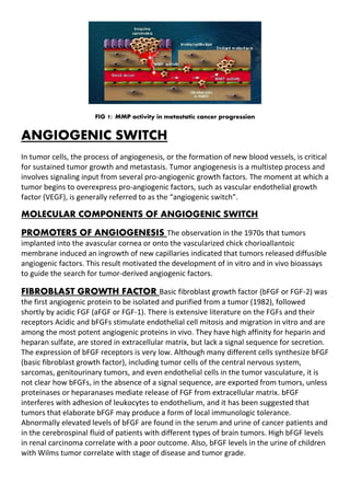
SlideShare a Scribd company logo
1 of 16
Download to read offline
ANGIOGENESIS IN CANCER
FORMATION
SUBMITTED BY
POOJA VERMA
M.SC BIOTECH 3rd SEMESTER
ROLL NO: MSB/14/120
ENROLEMENT NO: A0500214029
ANGIOGENESIS
Angiogenesis is the formation of new blood vessels. This process involves the migration,
growth, and differentiation of endothelial cells, which line the inside wall of blood vessels.
The process of angiogenesis is controlled by chemical signals in the body. These signals can
stimulate both the repair of damaged blood vessels and the formation of new blood
vessels. Other chemical signals, called angiogenesis inhibitors, interfere with blood vessel
formation. Normally, the stimulating and inhibiting effects of these chemical signals are
balanced so that blood vessels form only when and where they are needed.
ANGIOGENESIS AND TUMOR GROWTH
Angiogenesis is the process through which new blood vessels form and grow. Angiogenesis
is a natural physiological function which can be subverted by cancer cells to satisfy their
increasing need for nutrients and oxygen as tumors grow untamed. The angiogenic process,
as currently understood, can be summarized as follows: a cell activated by a lack of oxygen
(or a mutation) releases, among other things, angiogenic factors that attract inflammatory
and endothelial cells and promote their proliferation. In the course of their migration,
inflammatory cells secrete additional substances that intensify the angiogenic call. The
endothelial cells that form existing blood vessels respond to angiogenic signals in their
vicinity by proliferating and secreting proteases, which break open the blood-vessel wall to
enable them to migrate toward the site of the angiogenic stimuli. Proliferating endothelial
cells then organize themselves into new capillary tubes by altering the arrangement of their
adherence-membrane proteins. Finally, through the process of anastomosis, the capillaries
emanating from the arterioles and the venules join to provide a continuous blood flow that
sustains tumor cell metabolism and sets up escaping avenues for metastatic tumor cells.
Epidermal growth factor domain–like 7 (EGFL7) is an extracellular matrix protein that
supports endothelial cell adhesion, promotes cell survival under stress, and forms
perivascular tracks that regulate blood vessel formation. EGFL7 is selectively expressed in
nascent blood vessels in tumors and other proliferating tissues, but is absent or expressed
at low levels in healthy quiescent vessels. Preclinical studies also report that EGFL7 may
promote tumor escape from immunity.
A variety of different growth factors and cytokines can promote angiogenesis, but one of
the most important is vascular endothelial growth factor (VEGF). VEGF is a soluble protein
secreted by most types of cells, excepted by endothelial cells themselves. On the other
hand, endothelial cells express receptors for this growth factor. Hypoxic conditions, as seen
in cancer growth, are a potent trigger of VEGF expression. Interaction of VEGF with its
receptor induces an intracellular signaling cascade that favors endothelial cell survival,
proliferation and migration and increases vascular permeability. VEGF also up-regulates the
expression of other proteins involved in angiogenesis. High serum levels of VEGF correlates
with poor prognosis in cancer patients. VEGF relies on the complementary action of matrix
metalloproteinases (MMPs) for the process of angiogenesis. MMPs are key proteins in the
extension of the vascular bed. MMPs form a family of structurally related, zinc-dependent
endopeptidases secreted by various cell types, including endothelial cells. MMPs
collectively have the potential to degrade all of the protein and proteoglycan components
of the extracellular matrix (ECM) surrounding cells. MMPs also influence important cellular
processes and immune cell functions through proteolytic processing and shedding of
bioactive molecules such as cytokines and growth factors. In the context of tumor
angiogenesis, MMPs digest the blood-vessel walls enabling endothelial cells to make their
way toward the tumor and providing cancer cells with breaches through which they can
spread to distant organs.
FIG 1: MMP activity in metastatic cancer progression
ANGIOGENIC SWITCH
In tumor cells, the process of angiogenesis, or the formation of new blood vessels, is critical
for sustained tumor growth and metastasis. Tumor angiogenesis is a multistep process and
involves signaling input from several pro-angiogenic growth factors. The moment at which a
tumor begins to overexpress pro-angiogenic factors, such as vascular endothelial growth
factor (VEGF), is generally referred to as the “angiogenic switch”.
MOLECULAR COMPONENTS OF ANGIOGENIC SWITCH
PROMOTERS OF ANGIOGENESIS The observation in the 1970s that tumors
implanted into the avascular cornea or onto the vascularized chick chorioallantoic
membrane induced an ingrowth of new capillaries indicated that tumors released diffusible
angiogenic factors. This result motivated the development of in vitro and in vivo bioassays
to guide the search for tumor-derived angiogenic factors.
FIBROBLAST GROWTH FACTOR Basic fibroblast growth factor (bFGF or FGF-2) was
the first angiogenic protein to be isolated and purified from a tumor (1982), followed
shortly by acidic FGF (aFGF or FGF-1). There is extensive literature on the FGFs and their
receptors Acidic and bFGFs stimulate endothelial cell mitosis and migration in vitro and are
among the most potent angiogenic proteins in vivo. They have high affinity for heparin and
heparan sulfate, are stored in extracellular matrix, but lack a signal sequence for secretion.
The expression of bFGF receptors is very low. Although many different cells synthesize bFGF
(basic fibroblast growth factor), including tumor cells of the central nervous system,
sarcomas, genitourinary tumors, and even endothelial cells in the tumor vasculature, it is
not clear how bFGFs, in the absence of a signal sequence, are exported from tumors, unless
proteinases or heparanases mediate release of FGF from extracellular matrix. bFGF
interferes with adhesion of leukocytes to endothelium, and it has been suggested that
tumors that elaborate bFGF may produce a form of local immunologic tolerance.
Abnormally elevated levels of bFGF are found in the serum and urine of cancer patients and
in the cerebrospinal fluid of patients with different types of brain tumors. High bFGF levels
in renal carcinoma correlate with a poor outcome. Also, bFGF levels in the urine of children
with Wilms tumor correlate with stage of disease and tumor grade.
VASCULAR ENDOTHELIAL GROWTH FACTOR
VEGF is an important regulator of both vasculogenesis and angiogeneses. Several cell types
including fibroblasts, ECs (endothelial cells), and keratinocytes release a small amount of
VEGF throughout life. The loss of a VEGF allele always results in embryonic lethality.
Increased levels of VEGF are seen when angiogenesis is necessary, such as during active
wound healing. There are currently six known monomers of VEGF that arise from alternative
splicing of a single gene with eight exons. The documented isoforms contain 121, 145, 165,
183, 189 or 206 amino acids. Some of these isoforms remain associated with cells or
membranes, while others are released extracellularly. Despite these differences, all of them
have identical biological activities. VEGF interacts with two different receptor tyrosine
kinases, VEGFR-1 (Flt-1) and VEGFR-2 (Flk-1), to alter angiogenesis. VEGFR-1 interacts very
strongly with VEGF, but this interaction plays a minor role in the events of angiogenesis. The
interaction of VEGFR-2 with VEGF is a major contributor to the mitogenic, chemotactic,
angiogenic, and increased permeability effects of VEGF. VEGFR-2 expression has been
observed on both endothelial and hematopoietic precursors. VEGF is also capable of
enhancing EC migration, inhibiting EC apoptosis, and inducing the growth of new capillaries
from preexisting vasculature. VEGF helps induce EC migration and sprouting by upregulating
integrin receptors alphaVbeta 3, alpha1beta 1, and alpha1beta2. VEGF also helps activate
matrix metalloproteinase, an important step in the initial stages of angiogenesis. VEGF is
upregulated during hypoxic conditions via the following mechanism, which involves hypoxia-
inducible factor-1 (HIF-1), a protein released during oxygen stress. Responses to ischemia are
mostly regulated by cells within the ischemic area monitoring oxygen concentrations. After
sensing reduced concentrations, these cells increase their expression of genes encoding
vascular growth factors. At the heart of this pathway is HIF-1, a heterodimeric transcription
factor composed of the constitutively expressed HIF-1beta , and the oxygen-sensitive HIF-
1alpha subunits. Although HIF-1alpha is also constitutively expressed, it is broken down
within adequately oxygenated and perfused tissue. When tissues are deprived of oxygen, the
breakdown of HIF-1 is inhibited. HIF-1alpha begins to build up and eventually dimerizes with
HIF-1beta . This complex then binds to DNA, helps recruit coactivators, and activates
transcription of its target genes. This system induces expression of several vasculogenic and
angiogenic growth factors including VEGF and PLG in response to hypoxia. HIF-1 is also
required for expression of VEGFR-1 on EPCs in the bone marrow and the chemotactic
migration of EPCs towards a VEGF gradient.
Anti VEGF Drugs The use of anti-VEGF drugs has been applied to many different fields
of medicine. The American Academy of Ophthalmology recently published a report stating
that the use of anti-VEGF pharmacotherapy is a safe and effective treatment for neovascular
age-related macular degeneration (AMD). Anti-VEGF signaling pathway drugs have also been
tested in a large number of clinical and laboratory studies aimed at preventing angiogeneses
associated with cancer. Some of these drugs target VEGF (Avastin), while others target the
VEGFRs (Nexavar and Sutent). Although these drugs have seen dramatic results in animal
models, the results in many of the clinical trials have been mixed. There have been clinical
trials which show as many as 94% of invasive carcinomas and 88% of in situ carcinomas having
a complete response. These same patients saw no recurrence during the five-year followup.
However, many other angiogeneses inhibitors targeting VEGF signaling pathways have failed
to produce the same long-term responses in a majority of their patients. A short-term
response of either tumor stasis or increased survival was normally observed in these patients;
however after the initial benefit, most patients experienced tumor growth after several
months. These contradictory results have changed the philosophy on the resistance of
tumors to antiangiogenic treatments, as well as the vascular makeup thought to be
associated with the blood vessels that support tumors.
PLATELET DERIVED GROWTH FACTOR
PDGF is another important signaling molecule with several different roles in angiogenesis.
Although originally purified from platelets, it has also been identified in fibroblasts,
astrocytes, ECs, and several other cell types. There are five different isoforms of PDGF that
activate cellular response through two different receptors. Known ligands include A (PDGFA),
B (PDGFB), C (PDGFC), and D (PDGFD), and an AB heterodimer and receptors alpha (PDGFRA)
and beta (PDGFRB). PDGF has few other members of the family, for example VEGF sub-family.
The receptor for PDGF, PDGFR is classified as a receptor tyrosine kinase (RTK), a type of cell
surface receptor. Two types of PDGFRs have been identified: alpha-type and beta-type
PDGFRs. The alpha type binds to PDGF-AA, PDGF-BB and PDGF-AB, whereas the beta type
PDGFR binds with high affinity to PDGF-BB and PDGF-AB. PDGF binds to the PDGFR ligand
binding pocket located within the second and third immunoglobulin domains. Upon
activation by PDGF, these receptors dimerise, and are "switched on" by auto-
phosphorylation of several sites on their cytosolic domains, which serve to mediate binding
of cofactors and subsequently activate signal transduction, for example, through the PI3K
pathway or through reactive oxygen species (ROS)-mediated activation of the STAT3
pathway. Downstream effects of this include regulation of gene expression and the cell
cycle. The role of PI3K has been investigated by several laboratories. Accumulating data
suggests that, while this molecule is, in general, part of growth signaling complex, it plays a
more profound role in controlling cell migration. The different ligand isoforms have variable
affinities for the receptor isoforms, and the receptor isoforms may variably form hetero- or
homo- dimers. This leads to specificity of downstream signaling. It has been shown that the
sis oncogene is derived from the PDGF B-chain gene. PDGF-BB is the highest-affinity ligand
for the PDGFR-beta; PDGFR-beta is a key marker of hepatic stellate cell activation in the
process of fibrogenesis. PDGFs are mitogenic during early developmental stages, driving the
proliferation of undifferentiated mesenchyme and some progenitor populations. During
later maturation stages, PDGF signalling has been implicated in tissue remodelling and
cellular differentiation, and in inductive events involved in patterning and morphogenesis.
In addition to driving mesenchymal proliferation, PDGFs have been shown to direct the
migration, differentiation and function of a variety of specialised mesenchymal and
migratory cell types, both during development and in the adult animal.[11]
Other growth
factors in this family include vascular endothelial growth factors B and C (VEGF-B, VEGF-C)
which are active in angiogenesis and endothelial cell growth, and placenta growth factor
(PlGF) which is also active in angiogenesis.
PDGF plays a role in embryonic development, cell proliferation, cell migration, and
angiogenesis. Over-expression of PDGF has been linked to several diseases such as
atherosclerosis, fibrotic disorders and malignancies. Synthesis occurs due to external stimuli
such as thrombin, low oxygen tension, or other cytokines and growth factors.
PDGF is a required element in cellular division for fibroblasts, a type of connective tissue
cell that is especially prevalent in wound healing. In essence, the PDGFs allow a cell to skip
the G1 checkpoints in order to divide. It has been shown that in monocytes-macrophages
and fibroblasts, exogenously administered PDGF stimulates chemotaxis, proliferation, and
gene expression and significantly augmented the influx of inflammatory cells and
fibroblasts, accelerating extracellular matrix and collagen formation and thus reducing the
time for the healing process to occur. In terms of osteogenic differentiation of
mesenchymal stem cells, comparing PDGF to epidermal growth factor (EGF), which is also
implicated in stimulating cell growth, proliferation, and differentiation, MSCs were shown
to have stronger osteogenic differentiation into bone-forming cells when stimulated by
epidermal growth factor (EGF) versus PDGF. However, comparing the signaling pathways
between them reveals that the PI3K pathway is exclusively activated by PDGF, with EGF
having no effect. Chemically inhibiting the PI3K pathway in PDGF-stimulated cells negates
the differential effect between the two growth factors, and actually gives PDGF an edge in
osteogenic differentiation. Wortmannin is a PI3K-specific inhibitor, and treatment of cells
with Wortmannin in combination with PDGF resulted in enhanced osteoblast
differentiation compared to just PDGF alone, as well as compared to EGF. These results
indicate that the addition of Wortmannin can significantly increase the response of cells
into an osteogenic lineage in the presence of PDGF, and thus might reduce the need for
higher concentrations of PDGF or other growth factors, making PDGF a more viable growth
factor for osteogenic differentiation than other, more expensive growth factors currently
used in the field such as BMP2.
It has been hypothesized that blocking PDGF from interacting with its receptor will reduce
the stability of the growing capillaries rendering them incapable of delivering nutrients to
the cancer cells. Inhibition of PDGF has been attempted with compounds such as CP-
673,451. Rat glioblastomas treated with CP-673,451 showed a 47% decrease in
microvascular density and a 55% decrease in tumor growth. Although a decrease in tumor
growth and angiogenesis has occurred through the treatment of cancers with CP-673,451
during in vitro and in vivo animal studies, clinical trials have not been performed to
demonstrate its efficacy in humans. Another recent study indicated that lycopene, a
carotenoid found in tomatoes, may inhibit PDGF-BB-induced signaling reducing levels of
unwanted angiogenesis.
Tie receptors
The Tie receptors are a family of tyrosine kinases expressed by ECs that mimic the behaviour
of VEGF receptors. To date, Tie1 and Tie2 have been identified, and their mechanisms of
action studied. Genetically altered mice bred without either receptor underwent normal
vasculogenesis, but their ECs lacked normal integrity causing the mice to die from widespread
edema and hemorrhage due to a lack of adequate angiogenesis. While both Tie1 and Tie2
receptors are important for vascular integrity, only Tie2 appears to be vital to vascular
sprouting and branching occurring during angiogenesis. Tie Receptor inhibitors have recently
been produced in the lab. A research group has developed several Tie inhibitors and tested
them in vitro to find one with the best selectivity, potency, and pharmacokinetic parameters.
Although preliminary studies for Tie2 inhibitors have begun, further animal and possibly
clinical trials still remain.
ANGIOPOIETINS
Angiopoietin is part of a family of vascular growth factors that play a role in embryonic and
postnatal angiogenesis. Angiopoietin signaling most directly corresponds with angiogenesis,
the process by which new arteries and veins form from preexisting blood vessels.
Angiogenesis proceeds through sprouting, endothelial cell migration, proliferation, and
vessel destabilization and stabilization. They are responsible for assembling and
disassembling the endothelial lining of blood vessels. Angiopoietin cytokines are involved
with controlling microvascular permeability, vasodilation, and vasoconstriction by signaling
smooth muscle cells surrounding vessels. There are now four identified angiopoietins:
ANGPT1, ANGPT2, ANGPT4. In addition, there are a number of proteins that are closely
related to angiopoietins (ANGPTL1, ANGPTL2, ANGPTL3, ANGPTL4, ANGPTL5, ANGPTL6,
ANGPTL7).
 Angiopoietin-1 is critical for vessel maturation, adhesion, migration, and survival.
Angiopoietin-2, on the other hand, promotes cell death and disrupts vascularization.
Yet, when it is in conjunction with vascular endothelial growth factors, or VEGF, it can
promote neo-vascularization
 Angiopoietins are protein growth factors that act as ligands for the Tie Receptors on
ECs. There are two important angiopoietins that play a role in angiogenesis, Ang-1
and Ang-2. Ang-1 is a well-characterized regulator of angiogenesis. It is an important
agonist of and ligand for Tie2 receptors. Experiments have shown that mice that lack
Ang-1 or Tie2 receptors will develop normal primary vasculature, but will eventually
die because vascular remodeling was never completed. The interaction between Ang-
1 and Tie2 is important in angiogenesis as it helps recruit pericytes to newly created
blood vessels, increasing the stability of the new vasculature and making it less
permeable. Ang-1 also helps induce formation of capillary sprouts and promote
survival of ECs. Overexpression of Ang-1 in transgenic mice led to a greater number
of blood vessels, with larger diameters, and more vascular branches, illustrating the
important role Ang-1 plays in vascular sprout formation.
 The role of Ang-2 is more complicated than that of Ang-1. It appears that Ang-2 is at
least a partial antagonist of Tie2, resulting in pericyte loss. The result of this pericyte
loss is destabilization of the blood vessel. Under normal conditions this helps prevent
excessive angiogenesis. However, the destabilization also allows newly formed blood
vessels to be more plastic. These destabilized vessels show increased endothelial
sprouting and tube formation in the presence of VEGF. This fact has made Ang-2
blockers an important area of study for tumor angiogenesis inhibition. A recent study
created Ang-2-selective peptide-Fc fusion proteins and antibodies; this enabled a
controlled pharmacological inactivation of endogenous Ang-2, allowing the group to
study Ang-2 inactivation without using the lethal knockout approach. The use of
these inhibitors demonstrated tumor inhibition in mice as well as corneal
angiogenesis inhibition in rats. Although the study demonstrated the properties of
Ang-2 in vivo and gave therapeutic possibilities to Ang-2 inhibitors, many questions
still remain. How Ang-2 inhibition blocks EC proliferation is still not completely
understood. In addition, it is unknown if inhibition of both Ang-1 and Ang-2 would
further reduce angiogenesis or if blocking one will offset the inhibition of the other.
Additionally, these studies have only been completed using animal models, and we
can only hope that a similar effect will be seen in humans.
 Despite the current problems understanding Ang-2, the selective inhibition of Ang-2
could have a very high clinical value. Studies have demonstrated that Ang-2
upregulation is seen in several diseases that involve pathogenic angiogenesis
including cancer, macular degeneration, rheumatoid arthritis, osteoarthritis, and
psoriasis. One recent study found that mRNA levels of Ang-2 in metastatic liver cells
and lymph nodes are between 1.5 and 2 times greater than normal levels.
TRANSFORMING GROWTH FACTOR BETA
Transforming Growth Factor-Betas (TFG-beta ) are a family of homodimeric cytokines that help
control many different processes in the body, including angiogenesis. TGF- beta’s are normally found
in the Extra Cellular Matrix of many different cells types. Within the microvasculature, both ECs and
pericytes produce and display receptors for TGF-beta, illustrating the variety of cells capable TGF-
beta expression. To date, both pro- and antiangiogenic properties have been ascribed to TGF-beta. At
low doses TGF-beta helps initiate the angiogenic switch by upregulating angiogenic factors and
proteinases. However, at high doses TGF-beta inhibits EC growth, promotes basement membrane
reformation, and stimulates Smooth muscle cells differentiation and recruitment. Genetic studies in
mice have shown that the loss of TGF-beta leads to leaky vessels lacking structural integrity leading
to premature death. Stimulation of angiogenesis through TGF-beta is mostly via indirect mechanisms.
TGF-beta signals inflammatory mediators to the site of angiogenesis, where inflammatory cells
release proangiogenic factors such as VEGF, FGF, and PDGF.
ANGIOSTATIN
 Angiostatin is a 38 kDa internal fragment of plasminogen that was purified from serum
and urine of mice bearing a subcutaneous Lewis lung carcinoma that suppressed
growth of its lung metastases by inhibiting their angiogenesis.
 It is an endogenous angiogenesis inhibitor (i.e., it blocks the growth of new blood
vessels), and it is currently undergoing clinical trials for its use in anticancer therapy.
 Angiostatin is a larger protein enclosing three to five contiguous kringle modules. Each
module contains two small beta sheets and three disulfide bonds.
 Angiostatin is produced, for example, by autoproteolytic cleavage of plasminogen,
involving extracellular disulfide bond reduction by phosphoglycerate kinase.
Furthermore, angiostatin can be cleaved from plasminogen by different
metalloproteinases (MMPs), elastase, prostate-specific antigen (PSA), 13 KD serine
protease, or 24KD endopeptidase.
 Angiostatin is known to bind many proteins, especially to angiomotin and endothelial
cell surface ATP synthase but also integrins, annexin II, C-met receptor, NG2
proteoglycan, tissue-type plasminogen activator, chondroitin sulfate proteoglycans,
and CD26.
 Additionally, smaller fragments of angiostatin may bind several other proteins. There
is still considerable uncertainty on its mechanism of action, but it seems to involve
inhibition of endothelial cell migration, proliferation and induction of apoptosis. It has
been proposed that angiostatin activity is related, among other things, to the coupling
of its mechanical and redox properties.
INTEGRINS
 Integrins are heterodimeric cell surface receptors for ECM proteins that also play
a role in cell-cell attachment. They contain various alpha- and beta-subunits,
with over 20 different combinations of subunits known. Integrins are important
regulators for many different cell processes including both vasculogenesis and
angiogenesis.
 Of the integrins, alphaVbeta3 is one of the most extensively studied, and has an
important role in angiogenesis. It binds and activates MMP-2 at the tips of
growing blood vessels to help break down the ECM.
 Integrin alphaVbeta3 shows increased expression in vitro when exposed to VEGF
and bFGF. Cell attachment, spreading, and migration are all regulated by integrin
alphaVbeta3 in vitro.
 Angiogenic blood vessels near granulation tissue showed much greater levels of
alphaVbeta3 than vessels in uninjured normal skin. During wound repair,
alphaVbeta3 is localized to the ECs at the ends of the growing vessels. Anti-
alphaVbeta3 monoclonal antibodies reduce bFGF-stimulated angiogenesis,
demonstrating an important relationship between the two proteins.
 Several other integrins have also been implicated in angiogenesis. Inhibition of
integrin alphaVbeta5 hindered angiogenesis stimulated by VEGF. Abs to collagen
receptor integrins (alpha1beta 1 and alpha2beta1) also reduced VEGF-mediated
angiogenesis. A variety of integrins play important roles in angiogenesis
including EC adhesion to ECM, protease localization, and increased EC survival.
This diversity suggests a number of different integrins; each plays a distinct role
that uniquely contributes to the process of angiogenesis.
 There are currently three classes of integrin inhibitors in preclinical and clinical
trials. Some of these include a synthetic peptide Cilengitide (a
alphaVbeta3/alphaVbeta5 inhibitor), a monoclonal Ab Abergin (a alphaVbeta3
antagonist), and a peptidomimetic compound S247 (a
alphaVbeta3/alphaVbeta5) antagonist.
 Phase I trials using Vitaxin (similar to Abergin) were unsuccessful in reducing
tumor growth. A second generation Phase II trial which altered Vitaxin to give it
greater affinity for alphaVbeta3 also failed to reduce tumor growth. Phase I and
II trials with Cilengitide have also been completed. Although some antitumor
effects in the treatment of gliomas were seen with Cilengitide, the study
concluded that its action appeared to be antitumor cell specific as opposed to
angiostatic. Trials in other cancer patients failed to show any reduction in tumor
load using Cilengitide. The low efficacy of the alphaVbeta3/alphaVbeta5
antagonists demonstrates that the mechanistic understanding of integrins in
angiogenesis is not yet fully understood. At this time, antagonists for
alphaVbeta3 or alphaVbeta5 alone do not appear to prevent angiogenesis, and
other strategies need to be examined. Currently, integrin inhibitors for
alpha2beta1 and alpha5beta1 are being tested in phase I and phase II clinical
trials.
CADHERINS
 Cadherins are a class of calcium-binding transmembrane proteins that
play an important role in cell-cell interactions. Several studies have
underlined the important role of one particular cadherin, the vascular
endothelial (VE) cadherin, in neovascularization.
 VE-cadherins are localized exclusively to the adherens junctions in ECs. It
has been suggested that VE-cadherins are important in regulating the
passage of various molecules across the endothelium. In addition, VE-
cadherin plays an important role in mediating EC growth through contact
inhibition.
 Inhibition of VE-cadherins to prevent angiogenesis has been examined in
several different animal models. Monoclonal Abs designed to recognize
certain sections of extracellular repeats found in active VE-cadherins have
been designed. These Abs prevented EC junctional assembly and induced
the disassembly of already existing EC junctions in vitro, abilities that
could help prevent unwanted angiogenesis. Although these original Abs
were found to have some inhibitory effects on angiogenesis at low doses,
significant vascular permeability was found in the heart and lungs of the
mice at moderate to high doses, illustrating toxic side effects of the Abs.
Since these original studies, other monoclonal Abs have been developed
that do not exhibit the vascular permeability problems of the original
version; these include BV14 and E4G10. It is currently hypothesized that
these second-generation monoclonal Abs will be effective because
angiogenic junctions are weaker and contain different epitopes which are
open to monoclonal Ab targeting. Other groups have focused on the gene
sequence of VE-cadherins, which may allow researchers to produce more
specific monoclonal Abs for VE-cadherins and prevent unwanted
angiogenesis.
 VE-cadherins role in retinal neovascularization was also recently
examined in mice. A group induced retinal neovascularization in newborn
mice by exposure to oxygen. Some of the mice were then treated with a
VE-cadherin antagonist while others were treated with a control peptide.
The mice treated with the VE-cadherin antagonist saw significantly
reduced retinal angiogenesis compared to the control group. In addition,
the group treated with the antagonist had reduced levels of EC migration
and proliferation as well as suppressed tubule formation from ECs. As
studies designed to better comprehend VE-cadherins role in angiogenesis
have been completed, it has become clear that the role of cadherins in
the angiogenic pathway is larger than just their adhesive activity. The
ability of VE-cadherins to interact with various signaling molecules
suggests that it has a role in EC growth, migration, survival, and
morphogenesis. Although VE-cadherin inhibitors alone are not capable of
suppressing angiogenesis at this time, combining a VE-cadherin inhibitor
with other inhibitors of angiogenesis may provide more complete
suppression.
Angiogenesis Inhibitors: Current Research and Clinical
Implications
a)Angiostatin
Angiostatin is a 38 kDa internal fragment of plasminogen that displays inhibitory effects
against tumor angiogenesis. When plasminogen is in the vicinity of an implanted and/or
primary tumor, it is cleaved by an unknown protease; a product of this cleavage is the
antiangiogenic protein angiostatin. Research groups have shown that the endogenous
protein is capable of inhibiting the growth of distant metastasis.
Experiments with angiostatin demonstrated that its activity leads to several different
physiological results. As mentioned, angiostatin was shown to reduce the growth of remote
metastasis. This is accomplished by increasing the rate of apoptosis in metastatic tumors.
Apoptosis is increased because angiostatin attacks the energy system of the metastatic tissue
by inhibiting ATP synthase F1F0, leading to caspase-mediated apoptosis. Another effect of
activated angiostatin is the inhibition of capillary endothelial growth in vitro. Mice with
gliomas and melanomas experienced greatly reduced tumor growth and neovascularization
when they were genetically engineered to express angiostatin. The activity of angiostatin was
also examined on a global level using microarray techniques. A total of 189 genes had their
expression levels altered with treatment of angiostatin. Most of these genes were involved
in growth, apoptosis, and migration of ECs, as well as inflammation, demonstrating the wide
range of effects that angiostatin has within the body.
To date, several clinical trials have tested the efficacy of angiostatin as a treatment for several
forms of cancer. Subcutaneous injections of recombinant human angiostatin showed little to
no toxicity in phase I clinical trials. Phase II trials using angiostatin in combination with both
paclitaxel and carboplatin have been completed in non-small-cell lung cancer (NSCLC)
patients. The response rate of the combinational therapy was higher than previous studies
using the chemotherapy alone. The group reported that the overall response rate to the
combined treatment was 39.1%, another 39.1% of the patients remained stable, while the
last 21.7% of the patients saw their disease progress. Although the group did see improved
rates of treatment, they fell below the expected levels. Several groups are working on
alternate methods of administering angiostatin to increase its success as a treatment.
Experiments using intravenous administration of angiostatin genes complexed to cationic
liposomes are ongoing.
b)Endostatin
1. Endostatin is an angiostatic 20-kD internal fragment of the carboxy terminus of
collagen XVIII, an important proteoglycan in basement membranes. It was originally
discovered in the blood and urine of tumor-bearing mice. Two of the enzymes
responsible for the release of endostatin include elastase and cathepsin L.
Endostatin interacts with many different cell surface proteins including the
integrins ( alpha5beta 1 and to an extent also alphaVbeta3 and alphaVbeta5) and
several glycpians. These interactions result in altered EC adhesion and migration.
2. In vitro, endostatin inhibits EC migration, proliferation, and tube formation, three
key aspects of angiogenesis. Inhibition of angiogenesis via endostatin leads to a
reduction of tumor growth in vivo. It appears that this inhibition is partially
accomplished by reducing the expression of VEGF. Endostatin also has the ability to
block existing VEGF from interacting with its receptor VEGFR-2. Endostatin reduces
EC proliferation by arresting the EC cell cycle through the downregulation of cyclin-
D1 promoter transcriptional activity. As a result, the cell is unable to progress
through the G1/S transition. Recent studies have also shown that endostatin
disturbs the survival/death balance via activation of the proapoptotic pathway
through the induction of caspase-9 activation. These pathways induction is due to
the endostatin-led decrease of the anti-apoptotic proteins Bcl-2, Bcl-Xl, and Bad.
Gene array and proteonomic analysis have given insight into the vast number of
genes that can be affected by endostatin treatment in human dermal microvascular
ECs. Approximately 12% of the 74,834 genes represented on the microarray chip
had altered expression levels when treated with endostatin. Both angiostatin and
endostatin cause apoptotic pathway activation. However, the identity and number
of genes regulated by endostatin differ from angiostatin, suggesting alternative
pathways of action.
3. Endostatin is currently being analyzed for therapeutic potential in several forms of
cancer. Using animal studies, a group recently demonstrated that it may be used as
a possible treatment to boost the post-operative prognosis of osteosarcoma
patients. The study was designed to determine whether antiangiogenic treatment
could help prevent the progression of pulmonary metastasis, a secondary problem
often associated with postoperative osteosarcoma. The group injected an
adenovirus encoding endostatin vector (Ad5CMV-mEnd) two weeks after tumor
inoculation. The group found statistically significant differences in the size and
prevalence of pulmonary metastasis between the control and treatment groups
two weeks after the administration of the vector.
c) Tumstatin
1. Degradation of type IV collagen releases a 28 kDa fragment known as tumstatin, a
compound that also displays antiangiogenic properties. Tumstatin binds the
alphaVbeta3 integrin, which results in G1 arrest and the induction of EC apoptosis.
Mice models have shown that exogenous tumstatin is able to inhibit the growth of
tumors. In addition, tumstatin-deficient mice had a much greater microvessel density
near implanted murine tumors, and the mice had a 300% increase in overall tumor
growth.
2. Animal studies determining the viability of tumstatin as an antiangiogenic-drug have
been and continue to be completed. Tumstatin treated mice with teratocarcinomas
showed over a 90% reduction in tumor size compared to controls. The same group also
examined a combination of anti-VEGF (Avastin) and tumstatin treatment compared to
the tumstatin treatment alone. The animals receiving the combination treatment saw
a statistically significant reduction in tumor growth when compared to the tumstatin
alone. These findings demonstrated strong preclinical evidence for a future treatment
of cancer with an anti-VEGF Ab alongside a tumstatin peptide. Another recently
completed animal study examined gene delivery of a tumstatin fragment into
hepatocellular carcinomas (HCCs). In vivo intratumoral injection of the tumstatin
fragment (pSecTag2B-tum-1) greatly diminished the growth of preestablished human
HCCs. In addition, there was a decrease in the amount of CD-34 positive vessels in the
tumor.
d) Platelet Factor-4
1. Platelet Factor-4 (PF-4) is a chemokine naturally secreted by platelets that normally
promotes blood coagulation. In addition to this role, PF-4 is also known to be an
inhibitor of angiogenesis.
2. PF-4 is secreted from the alpha-granules of activated platelets and binds with high
affinity to heparin-like glycosaminoglycans on the surface on ECs blocking them
from further activity. Studies have also shown that PF-4 blocks the upregulation of
MMP-1 and MMP-3, inhibiting EC migration. Finally, PF-4 is also capable of
inhibiting the EC cell cycle by impairing pRB phosphorylation.
3. Despite the antiangiogenic and antitumoral effects of PF-4 in murine human tumor
implant models, PF-4 has not proven to be an effective treatment for human
cancers. The early failure of PF-4 as a monotherapeutic treatment led researchers
to examine different approaches for the use of PF-4 as an angiostatic agent.
4. Recently, researchers produced a novel peptide containing the active fragment of
PF-4 along with vasostatin, an inhibitor of EC proliferation. This peptide was
examined as a potential angiostatic agent in chick embryos. In vivo neovascular
growth was compared between a group receiving the novel peptide, a group only
receiving vasostatin, another receiving PF-4 alone, as well as a control. The chick
embryos receiving the novel peptide showed a statistically significant reduction in
angiogenesis when compared to the other groups. Although the study was only
completed in animals, it demonstrates another possible drug that may be used to
inhibit tumor angiogenesis.
e) Thrombospondin
Thrombospondin-1 (TSP-1), the first naturally occurring angiostatic protein discovered,
is a multidomain matrix glycoprotein that has been shown to be a natural inhibitor of
neovascularization. Unlike previous angiostatic agents discussed, TSP-1 is a native, full-
length protein. TSP-1 is stored in -granules of platelets, where it is complexed with TGF
beta1. When released from the platelets and free from TGFbeta1, TSP-1 inhibits the
migration of ECs and induces EC apoptosis. To slow migration, TSP-1 binds to EC
surface receptors capable of promoting promigratory signals. The induction of
apoptosis in ECs is associated with TSP-1’s ability to alter the concentrations of several
important apoptotic factors. TSP-1 upregulates Bax, downregulates Bcl-2, and
activates the caspase-3 intrinsic pathway, leading to programmed EC death. Other
experiments have shown that mice depleted of TSP-1 saw a 250%–300% increase in
tumor growth rate when implanted with murine tumors. Microvessel density was also
increased near the tumor in these mice.
f) Interleukins
1. Interleukins (ILs) are a group of cytokines that are released by leukocytes and
control a wide range of biological activities. A few of these ILs have been shown to
affect the growth of blood vessels.
2. The ability to either enhance or suppress angiogenesis is based on a Glu-Leu-Arg
(ELR) motif at the NH2 terminus. IL-8 possesses this sequence, and therefore
enhances angiogenesis, while IL-4 does not contain the motif, and is an inhibitor of
angiogenesis.
3. IL-4 acts as an inhibitor of tumor growth, but its mechanism of action likely varies
with different tumor cells. For example, IL-4 is thought to directly inhibit
proliferation of cells from cancers such as colon tumors, head and neck tumors, and
glioblastomas, while in other cases it is thought to induce a host immune response
against the tumor cells such as in B-cell lymphomas and melanomas.
4. There is also evidence that IL-4 inhibits neovascularization, thus inhibiting tumor
growth. In vitro, IL-4 inhibits migration of ECs towards bFGF. In vivo, IL-4 has been
shown to inhibit neovascularization in rat corneas that should have been induced
by the high concentration of bFGF present.
5. These experiments demonstrate that IL-4-mediated suppression of tumor growth
may be due to IL-4s ability to inhibit angiogenic processes.
6. Other noncancer-related animal studies have shown IL-4’s antiangiogenic
capabilities in vivo. One such study examined rats with adjuvant-induced arthritis.
One set of animals received an adenovirus capable of producing IL-4, another group
received a control virus without the IL-4 producing capabilities, and the last
received a saline injection. The group treated with the IL-4 producing adenovirus
saw a statistically significant reduction in blood vessel growth. The reduction in
angiogenesis from the IL-4 treatment appears to be associated with a change in the
pro- and antiangiogenic cytokine levels. Although the study was designed to study
inflammatory arthritis, knowledge gained about IL-4 could be used for the
treatment of cancer in the future.
g) Interferons (IFNs)
1. Interferons (IFNs) belong to a large family of secreted glycoproteins known as
cytokines. They are produced and secreted by a wide variety of immune-related
cells and IFN-alpha has been shown to inhibit angiogenesis in vivo.
2. It is thought that both IFN-alpha and IFN-beta are able to inhibit angiogenesis by
repressing bFGF mRNA and protein levels. In addition to the downregulation of key
angiogenesis signaling factors, IFN-alpha also inhibits the migration of EC cells in
vivo.
3. Several clinical trials have been performed investigating the administration of IFN-
alpha in combination with chemotherapeutics in different forms of cancer. Many
studies have used PEG IFN-alpha, a form of IFN-alpha that has been modified to
have a longer half-life and thus have a greater opportunity to reduce angiogenesis.
h) Tissue Inhibitors of Metalloproteinases
1. Tissue Inhibitors of Metalloproteinases (TIMPs) are a family of proteases, derived
from cartilage, which inhibit MMPs. As previously mentioned, MMPs play an
integral role in the initiation of angiogenesis. They are responsible for EC basement
membrane degradation and EC remodeling . The newly formed ECM developed by
MMPs during the angiogenic response provides a scaffold for ECs to adhere,
migrate, and form tubes for nutrient delivery. The inhibition of MMPs by TIMPs
reduces the angiogenic capacity of ECs. High levels of TIMP-1 greatly inhibit
migration of ECs through gelatin in vitro. The invasive potential, growth, and
neovascularization of metastatic murine melanoma cells were inhibited in vivo
when transfected with TIMP-2.
2. The role of TIMP-3 in tumor angiogenesis was examined using mice models. The
study found that TIMP-3 treatment of mice with lung cancer led to reduced
angiogenesis in vivo through the inhibition of the VEGF-VEGFR-2 signaling pathway
and the induction of EC-apoptosis.
3. The inhibition of this pathway and the EC associated death are in part due to the
inhibition of MMP-2 by TIMP-3.
4. Another study investigated the use of TIMP-1 gene transfer through an adenovirus
as a way to treat established gastric cancer in nude mice. Compared to controls,
mice transfected with TIMP-1 gene saw a significant reduction in the mean number
of tumor vessels. Although the exact mechanism of TIMPs inhibition of cancer
progression remains unclear, TIMPs should still be considered for therapeutic trials
because of their success in animal models.

More Related Content

What's hot

In vitro and in vivo models of angiogenesis
In vitro and in vivo models of angiogenesisIn vitro and in vivo models of angiogenesis
In vitro and in vivo models of angiogenesisVijay Avin BR
 
Angiogenesis, Introduction to Understand the Art.
Angiogenesis, Introduction to Understand the Art.Angiogenesis, Introduction to Understand the Art.
Angiogenesis, Introduction to Understand the Art.Khaled Elmasry
 
Topic of the month.... Antiangiogenic Therapy in Brain Tumors
Topic of the month.... Antiangiogenic Therapy in Brain TumorsTopic of the month.... Antiangiogenic Therapy in Brain Tumors
Topic of the month.... Antiangiogenic Therapy in Brain TumorsProfessor Yasser Metwally
 
VEGF (Vascular endothelial growth factors )
VEGF (Vascular endothelial growth factors )VEGF (Vascular endothelial growth factors )
VEGF (Vascular endothelial growth factors )bhawna singh
 
A systemic review on in vivo & in vitro models of angiogenesis & preliminary ...
A systemic review on in vivo & in vitro models of angiogenesis & preliminary ...A systemic review on in vivo & in vitro models of angiogenesis & preliminary ...
A systemic review on in vivo & in vitro models of angiogenesis & preliminary ...Abhijeet Mihir
 
vasculogenesis and angiogenesis
vasculogenesis and angiogenesisvasculogenesis and angiogenesis
vasculogenesis and angiogenesisAyesha Sadiqa
 
Tumor And Microenvironment
Tumor And MicroenvironmentTumor And Microenvironment
Tumor And Microenvironmentabhitux
 
Vascular endothelial growth factor signaling pathwys in cancer
Vascular endothelial growth factor signaling pathwys in cancerVascular endothelial growth factor signaling pathwys in cancer
Vascular endothelial growth factor signaling pathwys in cancerKarim El-sayed
 
Tumor microenvironment in the body
Tumor microenvironment in the bodyTumor microenvironment in the body
Tumor microenvironment in the bodytinasingh30
 
Haematopoitic growth factors dr. varun
Haematopoitic growth factors dr. varunHaematopoitic growth factors dr. varun
Haematopoitic growth factors dr. varunVarun Goel
 

What's hot (20)

In vitro and in vivo models of angiogenesis
In vitro and in vivo models of angiogenesisIn vitro and in vivo models of angiogenesis
In vitro and in vivo models of angiogenesis
 
Angiogenesis, Introduction to Understand the Art.
Angiogenesis, Introduction to Understand the Art.Angiogenesis, Introduction to Understand the Art.
Angiogenesis, Introduction to Understand the Art.
 
Angiogenesis
AngiogenesisAngiogenesis
Angiogenesis
 
Topic of the month.... Antiangiogenic Therapy in Brain Tumors
Topic of the month.... Antiangiogenic Therapy in Brain TumorsTopic of the month.... Antiangiogenic Therapy in Brain Tumors
Topic of the month.... Antiangiogenic Therapy in Brain Tumors
 
VEGF (Vascular endothelial growth factors )
VEGF (Vascular endothelial growth factors )VEGF (Vascular endothelial growth factors )
VEGF (Vascular endothelial growth factors )
 
Angiogenesis
AngiogenesisAngiogenesis
Angiogenesis
 
Angiogenesis
AngiogenesisAngiogenesis
Angiogenesis
 
A systemic review on in vivo & in vitro models of angiogenesis & preliminary ...
A systemic review on in vivo & in vitro models of angiogenesis & preliminary ...A systemic review on in vivo & in vitro models of angiogenesis & preliminary ...
A systemic review on in vivo & in vitro models of angiogenesis & preliminary ...
 
vasculogenesis and angiogenesis
vasculogenesis and angiogenesisvasculogenesis and angiogenesis
vasculogenesis and angiogenesis
 
Tumor And Microenvironment
Tumor And MicroenvironmentTumor And Microenvironment
Tumor And Microenvironment
 
Vegf signaling
Vegf signalingVegf signaling
Vegf signaling
 
Angiogenisis in tumer
Angiogenisis in tumerAngiogenisis in tumer
Angiogenisis in tumer
 
Vascular endothelial growth factor signaling pathwys in cancer
Vascular endothelial growth factor signaling pathwys in cancerVascular endothelial growth factor signaling pathwys in cancer
Vascular endothelial growth factor signaling pathwys in cancer
 
Angiogenesis
AngiogenesisAngiogenesis
Angiogenesis
 
Tumor induced stroma
Tumor induced stromaTumor induced stroma
Tumor induced stroma
 
Apoptosis
ApoptosisApoptosis
Apoptosis
 
Hematopoiesis
HematopoiesisHematopoiesis
Hematopoiesis
 
Tumor microenvironment in the body
Tumor microenvironment in the bodyTumor microenvironment in the body
Tumor microenvironment in the body
 
Haematopoitic growth factors dr. varun
Haematopoitic growth factors dr. varunHaematopoitic growth factors dr. varun
Haematopoitic growth factors dr. varun
 
Therapeutic Angiogenesis
Therapeutic AngiogenesisTherapeutic Angiogenesis
Therapeutic Angiogenesis
 

Similar to Cancer project

SIGNALING MECHANISM DRIVING ANGIOGENESIS IN HYPOXIC TUMOR MICRO-ENVIRONMENT
SIGNALING MECHANISM DRIVING ANGIOGENESIS IN HYPOXIC TUMOR MICRO-ENVIRONMENTSIGNALING MECHANISM DRIVING ANGIOGENESIS IN HYPOXIC TUMOR MICRO-ENVIRONMENT
SIGNALING MECHANISM DRIVING ANGIOGENESIS IN HYPOXIC TUMOR MICRO-ENVIRONMENTMinali Singh
 
Vascular Endothelial Growth Factor (VEGF): Role, Family, Disease, Challenges ...
Vascular Endothelial Growth Factor (VEGF): Role, Family, Disease, Challenges ...Vascular Endothelial Growth Factor (VEGF): Role, Family, Disease, Challenges ...
Vascular Endothelial Growth Factor (VEGF): Role, Family, Disease, Challenges ...The Lifesciences Magazine
 
The lymphangiogenic growth factors VEGF-C and VEGF-D, Part 1: Fundamentals an...
The lymphangiogenic growth factors VEGF-C and VEGF-D, Part 1: Fundamentals an...The lymphangiogenic growth factors VEGF-C and VEGF-D, Part 1: Fundamentals an...
The lymphangiogenic growth factors VEGF-C and VEGF-D, Part 1: Fundamentals an...belatoth666
 
Molecular Basis of Neoplasia.pptx
Molecular Basis of Neoplasia.pptxMolecular Basis of Neoplasia.pptx
Molecular Basis of Neoplasia.pptxAymanshahzad4
 
Molecular%20Basis%20of%20Neoplasia.pptx
Molecular%20Basis%20of%20Neoplasia.pptxMolecular%20Basis%20of%20Neoplasia.pptx
Molecular%20Basis%20of%20Neoplasia.pptxAymanshahzad4
 
Hall marks of Cancer
Hall marks of CancerHall marks of Cancer
Hall marks of Cancerneralagundi
 
The term angiogenesis is generally applied to the growth of .docx
The term angiogenesis is generally applied to the growth of .docxThe term angiogenesis is generally applied to the growth of .docx
The term angiogenesis is generally applied to the growth of .docxssusera34210
 
angiogenesis..pptx
angiogenesis..pptxangiogenesis..pptx
angiogenesis..pptxsaswati14
 
Tumor suppressor genes
Tumor suppressor genesTumor suppressor genes
Tumor suppressor genesfatmafars
 
PLATELET ACTIVATING FACTOR
PLATELET ACTIVATING FACTORPLATELET ACTIVATING FACTOR
PLATELET ACTIVATING FACTORMulayamSingh10
 
Signalling mechanism in cell growth
Signalling mechanism in cell growthSignalling mechanism in cell growth
Signalling mechanism in cell growthbijitadutta123
 
PLATELET ACTIVATING FACTORS: MECHANISM OF ACTION AND CLINICAL USES
PLATELET ACTIVATING FACTORS: MECHANISM OF ACTION AND CLINICAL USESPLATELET ACTIVATING FACTORS: MECHANISM OF ACTION AND CLINICAL USES
PLATELET ACTIVATING FACTORS: MECHANISM OF ACTION AND CLINICAL USESvikasaagrahari007
 

Similar to Cancer project (20)

Angiogenesis
AngiogenesisAngiogenesis
Angiogenesis
 
Angiogenesis
AngiogenesisAngiogenesis
Angiogenesis
 
SIGNALING MECHANISM DRIVING ANGIOGENESIS IN HYPOXIC TUMOR MICRO-ENVIRONMENT
SIGNALING MECHANISM DRIVING ANGIOGENESIS IN HYPOXIC TUMOR MICRO-ENVIRONMENTSIGNALING MECHANISM DRIVING ANGIOGENESIS IN HYPOXIC TUMOR MICRO-ENVIRONMENT
SIGNALING MECHANISM DRIVING ANGIOGENESIS IN HYPOXIC TUMOR MICRO-ENVIRONMENT
 
Vascular Endothelial Growth Factor (VEGF): Role, Family, Disease, Challenges ...
Vascular Endothelial Growth Factor (VEGF): Role, Family, Disease, Challenges ...Vascular Endothelial Growth Factor (VEGF): Role, Family, Disease, Challenges ...
Vascular Endothelial Growth Factor (VEGF): Role, Family, Disease, Challenges ...
 
The lymphangiogenic growth factors VEGF-C and VEGF-D, Part 1: Fundamentals an...
The lymphangiogenic growth factors VEGF-C and VEGF-D, Part 1: Fundamentals an...The lymphangiogenic growth factors VEGF-C and VEGF-D, Part 1: Fundamentals an...
The lymphangiogenic growth factors VEGF-C and VEGF-D, Part 1: Fundamentals an...
 
Angiogenesis
AngiogenesisAngiogenesis
Angiogenesis
 
Angiogenesis seminar
Angiogenesis seminarAngiogenesis seminar
Angiogenesis seminar
 
Hallmarks of cancer
Hallmarks of cancerHallmarks of cancer
Hallmarks of cancer
 
Molecular Basis of Neoplasia.pptx
Molecular Basis of Neoplasia.pptxMolecular Basis of Neoplasia.pptx
Molecular Basis of Neoplasia.pptx
 
Molecular%20Basis%20of%20Neoplasia.pptx
Molecular%20Basis%20of%20Neoplasia.pptxMolecular%20Basis%20of%20Neoplasia.pptx
Molecular%20Basis%20of%20Neoplasia.pptx
 
Hall marks of Cancer
Hall marks of CancerHall marks of Cancer
Hall marks of Cancer
 
The term angiogenesis is generally applied to the growth of .docx
The term angiogenesis is generally applied to the growth of .docxThe term angiogenesis is generally applied to the growth of .docx
The term angiogenesis is generally applied to the growth of .docx
 
angiogenesis..pptx
angiogenesis..pptxangiogenesis..pptx
angiogenesis..pptx
 
cao2008.pdf
cao2008.pdfcao2008.pdf
cao2008.pdf
 
Tumor suppressor genes
Tumor suppressor genesTumor suppressor genes
Tumor suppressor genes
 
Liver cirrhosis :an update
Liver cirrhosis  :an updateLiver cirrhosis  :an update
Liver cirrhosis :an update
 
PLATELET ACTIVATING FACTOR
PLATELET ACTIVATING FACTORPLATELET ACTIVATING FACTOR
PLATELET ACTIVATING FACTOR
 
Signalling mechanism in cell growth
Signalling mechanism in cell growthSignalling mechanism in cell growth
Signalling mechanism in cell growth
 
Blood
BloodBlood
Blood
 
PLATELET ACTIVATING FACTORS: MECHANISM OF ACTION AND CLINICAL USES
PLATELET ACTIVATING FACTORS: MECHANISM OF ACTION AND CLINICAL USESPLATELET ACTIVATING FACTORS: MECHANISM OF ACTION AND CLINICAL USES
PLATELET ACTIVATING FACTORS: MECHANISM OF ACTION AND CLINICAL USES
 

Recently uploaded

PSYCHIATRIC History collection FORMAT.pptx
PSYCHIATRIC   History collection FORMAT.pptxPSYCHIATRIC   History collection FORMAT.pptx
PSYCHIATRIC History collection FORMAT.pptxPoojaSen20
 
Crayon Activity Handout For the Crayon A
Crayon Activity Handout For the Crayon ACrayon Activity Handout For the Crayon A
Crayon Activity Handout For the Crayon AUnboundStockton
 
Kisan Call Centre - To harness potential of ICT in Agriculture by answer farm...
Kisan Call Centre - To harness potential of ICT in Agriculture by answer farm...Kisan Call Centre - To harness potential of ICT in Agriculture by answer farm...
Kisan Call Centre - To harness potential of ICT in Agriculture by answer farm...Krashi Coaching
 
18-04-UA_REPORT_MEDIALITERAСY_INDEX-DM_23-1-final-eng.pdf
18-04-UA_REPORT_MEDIALITERAСY_INDEX-DM_23-1-final-eng.pdf18-04-UA_REPORT_MEDIALITERAСY_INDEX-DM_23-1-final-eng.pdf
18-04-UA_REPORT_MEDIALITERAСY_INDEX-DM_23-1-final-eng.pdfssuser54595a
 
microwave assisted reaction. General introduction
microwave assisted reaction. General introductionmicrowave assisted reaction. General introduction
microwave assisted reaction. General introductionMaksud Ahmed
 
URLs and Routing in the Odoo 17 Website App
URLs and Routing in the Odoo 17 Website AppURLs and Routing in the Odoo 17 Website App
URLs and Routing in the Odoo 17 Website AppCeline George
 
Alper Gobel In Media Res Media Component
Alper Gobel In Media Res Media ComponentAlper Gobel In Media Res Media Component
Alper Gobel In Media Res Media ComponentInMediaRes1
 
POINT- BIOCHEMISTRY SEM 2 ENZYMES UNIT 5.pptx
POINT- BIOCHEMISTRY SEM 2 ENZYMES UNIT 5.pptxPOINT- BIOCHEMISTRY SEM 2 ENZYMES UNIT 5.pptx
POINT- BIOCHEMISTRY SEM 2 ENZYMES UNIT 5.pptxSayali Powar
 
Solving Puzzles Benefits Everyone (English).pptx
Solving Puzzles Benefits Everyone (English).pptxSolving Puzzles Benefits Everyone (English).pptx
Solving Puzzles Benefits Everyone (English).pptxOH TEIK BIN
 
Sanyam Choudhary Chemistry practical.pdf
Sanyam Choudhary Chemistry practical.pdfSanyam Choudhary Chemistry practical.pdf
Sanyam Choudhary Chemistry practical.pdfsanyamsingh5019
 
APM Welcome, APM North West Network Conference, Synergies Across Sectors
APM Welcome, APM North West Network Conference, Synergies Across SectorsAPM Welcome, APM North West Network Conference, Synergies Across Sectors
APM Welcome, APM North West Network Conference, Synergies Across SectorsAssociation for Project Management
 
Contemporary philippine arts from the regions_PPT_Module_12 [Autosaved] (1).pptx
Contemporary philippine arts from the regions_PPT_Module_12 [Autosaved] (1).pptxContemporary philippine arts from the regions_PPT_Module_12 [Autosaved] (1).pptx
Contemporary philippine arts from the regions_PPT_Module_12 [Autosaved] (1).pptxRoyAbrique
 
Hybridoma Technology ( Production , Purification , and Application )
Hybridoma Technology  ( Production , Purification , and Application  ) Hybridoma Technology  ( Production , Purification , and Application  )
Hybridoma Technology ( Production , Purification , and Application ) Sakshi Ghasle
 
Micromeritics - Fundamental and Derived Properties of Powders
Micromeritics - Fundamental and Derived Properties of PowdersMicromeritics - Fundamental and Derived Properties of Powders
Micromeritics - Fundamental and Derived Properties of PowdersChitralekhaTherkar
 
MENTAL STATUS EXAMINATION format.docx
MENTAL     STATUS EXAMINATION format.docxMENTAL     STATUS EXAMINATION format.docx
MENTAL STATUS EXAMINATION format.docxPoojaSen20
 
SOCIAL AND HISTORICAL CONTEXT - LFTVD.pptx
SOCIAL AND HISTORICAL CONTEXT - LFTVD.pptxSOCIAL AND HISTORICAL CONTEXT - LFTVD.pptx
SOCIAL AND HISTORICAL CONTEXT - LFTVD.pptxiammrhaywood
 
Presentation by Andreas Schleicher Tackling the School Absenteeism Crisis 30 ...
Presentation by Andreas Schleicher Tackling the School Absenteeism Crisis 30 ...Presentation by Andreas Schleicher Tackling the School Absenteeism Crisis 30 ...
Presentation by Andreas Schleicher Tackling the School Absenteeism Crisis 30 ...EduSkills OECD
 
Presiding Officer Training module 2024 lok sabha elections
Presiding Officer Training module 2024 lok sabha electionsPresiding Officer Training module 2024 lok sabha elections
Presiding Officer Training module 2024 lok sabha electionsanshu789521
 
Mastering the Unannounced Regulatory Inspection
Mastering the Unannounced Regulatory InspectionMastering the Unannounced Regulatory Inspection
Mastering the Unannounced Regulatory InspectionSafetyChain Software
 

Recently uploaded (20)

PSYCHIATRIC History collection FORMAT.pptx
PSYCHIATRIC   History collection FORMAT.pptxPSYCHIATRIC   History collection FORMAT.pptx
PSYCHIATRIC History collection FORMAT.pptx
 
Crayon Activity Handout For the Crayon A
Crayon Activity Handout For the Crayon ACrayon Activity Handout For the Crayon A
Crayon Activity Handout For the Crayon A
 
Kisan Call Centre - To harness potential of ICT in Agriculture by answer farm...
Kisan Call Centre - To harness potential of ICT in Agriculture by answer farm...Kisan Call Centre - To harness potential of ICT in Agriculture by answer farm...
Kisan Call Centre - To harness potential of ICT in Agriculture by answer farm...
 
18-04-UA_REPORT_MEDIALITERAСY_INDEX-DM_23-1-final-eng.pdf
18-04-UA_REPORT_MEDIALITERAСY_INDEX-DM_23-1-final-eng.pdf18-04-UA_REPORT_MEDIALITERAСY_INDEX-DM_23-1-final-eng.pdf
18-04-UA_REPORT_MEDIALITERAСY_INDEX-DM_23-1-final-eng.pdf
 
microwave assisted reaction. General introduction
microwave assisted reaction. General introductionmicrowave assisted reaction. General introduction
microwave assisted reaction. General introduction
 
URLs and Routing in the Odoo 17 Website App
URLs and Routing in the Odoo 17 Website AppURLs and Routing in the Odoo 17 Website App
URLs and Routing in the Odoo 17 Website App
 
Alper Gobel In Media Res Media Component
Alper Gobel In Media Res Media ComponentAlper Gobel In Media Res Media Component
Alper Gobel In Media Res Media Component
 
POINT- BIOCHEMISTRY SEM 2 ENZYMES UNIT 5.pptx
POINT- BIOCHEMISTRY SEM 2 ENZYMES UNIT 5.pptxPOINT- BIOCHEMISTRY SEM 2 ENZYMES UNIT 5.pptx
POINT- BIOCHEMISTRY SEM 2 ENZYMES UNIT 5.pptx
 
Solving Puzzles Benefits Everyone (English).pptx
Solving Puzzles Benefits Everyone (English).pptxSolving Puzzles Benefits Everyone (English).pptx
Solving Puzzles Benefits Everyone (English).pptx
 
TataKelola dan KamSiber Kecerdasan Buatan v022.pdf
TataKelola dan KamSiber Kecerdasan Buatan v022.pdfTataKelola dan KamSiber Kecerdasan Buatan v022.pdf
TataKelola dan KamSiber Kecerdasan Buatan v022.pdf
 
Sanyam Choudhary Chemistry practical.pdf
Sanyam Choudhary Chemistry practical.pdfSanyam Choudhary Chemistry practical.pdf
Sanyam Choudhary Chemistry practical.pdf
 
APM Welcome, APM North West Network Conference, Synergies Across Sectors
APM Welcome, APM North West Network Conference, Synergies Across SectorsAPM Welcome, APM North West Network Conference, Synergies Across Sectors
APM Welcome, APM North West Network Conference, Synergies Across Sectors
 
Contemporary philippine arts from the regions_PPT_Module_12 [Autosaved] (1).pptx
Contemporary philippine arts from the regions_PPT_Module_12 [Autosaved] (1).pptxContemporary philippine arts from the regions_PPT_Module_12 [Autosaved] (1).pptx
Contemporary philippine arts from the regions_PPT_Module_12 [Autosaved] (1).pptx
 
Hybridoma Technology ( Production , Purification , and Application )
Hybridoma Technology  ( Production , Purification , and Application  ) Hybridoma Technology  ( Production , Purification , and Application  )
Hybridoma Technology ( Production , Purification , and Application )
 
Micromeritics - Fundamental and Derived Properties of Powders
Micromeritics - Fundamental and Derived Properties of PowdersMicromeritics - Fundamental and Derived Properties of Powders
Micromeritics - Fundamental and Derived Properties of Powders
 
MENTAL STATUS EXAMINATION format.docx
MENTAL     STATUS EXAMINATION format.docxMENTAL     STATUS EXAMINATION format.docx
MENTAL STATUS EXAMINATION format.docx
 
SOCIAL AND HISTORICAL CONTEXT - LFTVD.pptx
SOCIAL AND HISTORICAL CONTEXT - LFTVD.pptxSOCIAL AND HISTORICAL CONTEXT - LFTVD.pptx
SOCIAL AND HISTORICAL CONTEXT - LFTVD.pptx
 
Presentation by Andreas Schleicher Tackling the School Absenteeism Crisis 30 ...
Presentation by Andreas Schleicher Tackling the School Absenteeism Crisis 30 ...Presentation by Andreas Schleicher Tackling the School Absenteeism Crisis 30 ...
Presentation by Andreas Schleicher Tackling the School Absenteeism Crisis 30 ...
 
Presiding Officer Training module 2024 lok sabha elections
Presiding Officer Training module 2024 lok sabha electionsPresiding Officer Training module 2024 lok sabha elections
Presiding Officer Training module 2024 lok sabha elections
 
Mastering the Unannounced Regulatory Inspection
Mastering the Unannounced Regulatory InspectionMastering the Unannounced Regulatory Inspection
Mastering the Unannounced Regulatory Inspection
 

Cancer project

  • 1. ANGIOGENESIS IN CANCER FORMATION SUBMITTED BY POOJA VERMA M.SC BIOTECH 3rd SEMESTER ROLL NO: MSB/14/120 ENROLEMENT NO: A0500214029
  • 2. ANGIOGENESIS Angiogenesis is the formation of new blood vessels. This process involves the migration, growth, and differentiation of endothelial cells, which line the inside wall of blood vessels. The process of angiogenesis is controlled by chemical signals in the body. These signals can stimulate both the repair of damaged blood vessels and the formation of new blood vessels. Other chemical signals, called angiogenesis inhibitors, interfere with blood vessel formation. Normally, the stimulating and inhibiting effects of these chemical signals are balanced so that blood vessels form only when and where they are needed. ANGIOGENESIS AND TUMOR GROWTH Angiogenesis is the process through which new blood vessels form and grow. Angiogenesis is a natural physiological function which can be subverted by cancer cells to satisfy their increasing need for nutrients and oxygen as tumors grow untamed. The angiogenic process, as currently understood, can be summarized as follows: a cell activated by a lack of oxygen (or a mutation) releases, among other things, angiogenic factors that attract inflammatory and endothelial cells and promote their proliferation. In the course of their migration, inflammatory cells secrete additional substances that intensify the angiogenic call. The endothelial cells that form existing blood vessels respond to angiogenic signals in their vicinity by proliferating and secreting proteases, which break open the blood-vessel wall to enable them to migrate toward the site of the angiogenic stimuli. Proliferating endothelial cells then organize themselves into new capillary tubes by altering the arrangement of their adherence-membrane proteins. Finally, through the process of anastomosis, the capillaries emanating from the arterioles and the venules join to provide a continuous blood flow that sustains tumor cell metabolism and sets up escaping avenues for metastatic tumor cells. Epidermal growth factor domain–like 7 (EGFL7) is an extracellular matrix protein that supports endothelial cell adhesion, promotes cell survival under stress, and forms perivascular tracks that regulate blood vessel formation. EGFL7 is selectively expressed in nascent blood vessels in tumors and other proliferating tissues, but is absent or expressed at low levels in healthy quiescent vessels. Preclinical studies also report that EGFL7 may promote tumor escape from immunity.
  • 3. A variety of different growth factors and cytokines can promote angiogenesis, but one of the most important is vascular endothelial growth factor (VEGF). VEGF is a soluble protein secreted by most types of cells, excepted by endothelial cells themselves. On the other hand, endothelial cells express receptors for this growth factor. Hypoxic conditions, as seen in cancer growth, are a potent trigger of VEGF expression. Interaction of VEGF with its receptor induces an intracellular signaling cascade that favors endothelial cell survival, proliferation and migration and increases vascular permeability. VEGF also up-regulates the expression of other proteins involved in angiogenesis. High serum levels of VEGF correlates with poor prognosis in cancer patients. VEGF relies on the complementary action of matrix metalloproteinases (MMPs) for the process of angiogenesis. MMPs are key proteins in the extension of the vascular bed. MMPs form a family of structurally related, zinc-dependent endopeptidases secreted by various cell types, including endothelial cells. MMPs collectively have the potential to degrade all of the protein and proteoglycan components of the extracellular matrix (ECM) surrounding cells. MMPs also influence important cellular processes and immune cell functions through proteolytic processing and shedding of bioactive molecules such as cytokines and growth factors. In the context of tumor angiogenesis, MMPs digest the blood-vessel walls enabling endothelial cells to make their way toward the tumor and providing cancer cells with breaches through which they can spread to distant organs.
  • 4. FIG 1: MMP activity in metastatic cancer progression ANGIOGENIC SWITCH In tumor cells, the process of angiogenesis, or the formation of new blood vessels, is critical for sustained tumor growth and metastasis. Tumor angiogenesis is a multistep process and involves signaling input from several pro-angiogenic growth factors. The moment at which a tumor begins to overexpress pro-angiogenic factors, such as vascular endothelial growth factor (VEGF), is generally referred to as the “angiogenic switch”. MOLECULAR COMPONENTS OF ANGIOGENIC SWITCH PROMOTERS OF ANGIOGENESIS The observation in the 1970s that tumors implanted into the avascular cornea or onto the vascularized chick chorioallantoic membrane induced an ingrowth of new capillaries indicated that tumors released diffusible angiogenic factors. This result motivated the development of in vitro and in vivo bioassays to guide the search for tumor-derived angiogenic factors. FIBROBLAST GROWTH FACTOR Basic fibroblast growth factor (bFGF or FGF-2) was the first angiogenic protein to be isolated and purified from a tumor (1982), followed shortly by acidic FGF (aFGF or FGF-1). There is extensive literature on the FGFs and their receptors Acidic and bFGFs stimulate endothelial cell mitosis and migration in vitro and are among the most potent angiogenic proteins in vivo. They have high affinity for heparin and heparan sulfate, are stored in extracellular matrix, but lack a signal sequence for secretion. The expression of bFGF receptors is very low. Although many different cells synthesize bFGF (basic fibroblast growth factor), including tumor cells of the central nervous system, sarcomas, genitourinary tumors, and even endothelial cells in the tumor vasculature, it is not clear how bFGFs, in the absence of a signal sequence, are exported from tumors, unless proteinases or heparanases mediate release of FGF from extracellular matrix. bFGF interferes with adhesion of leukocytes to endothelium, and it has been suggested that tumors that elaborate bFGF may produce a form of local immunologic tolerance. Abnormally elevated levels of bFGF are found in the serum and urine of cancer patients and in the cerebrospinal fluid of patients with different types of brain tumors. High bFGF levels in renal carcinoma correlate with a poor outcome. Also, bFGF levels in the urine of children with Wilms tumor correlate with stage of disease and tumor grade.
  • 5. VASCULAR ENDOTHELIAL GROWTH FACTOR VEGF is an important regulator of both vasculogenesis and angiogeneses. Several cell types including fibroblasts, ECs (endothelial cells), and keratinocytes release a small amount of VEGF throughout life. The loss of a VEGF allele always results in embryonic lethality. Increased levels of VEGF are seen when angiogenesis is necessary, such as during active wound healing. There are currently six known monomers of VEGF that arise from alternative splicing of a single gene with eight exons. The documented isoforms contain 121, 145, 165, 183, 189 or 206 amino acids. Some of these isoforms remain associated with cells or membranes, while others are released extracellularly. Despite these differences, all of them have identical biological activities. VEGF interacts with two different receptor tyrosine kinases, VEGFR-1 (Flt-1) and VEGFR-2 (Flk-1), to alter angiogenesis. VEGFR-1 interacts very strongly with VEGF, but this interaction plays a minor role in the events of angiogenesis. The interaction of VEGFR-2 with VEGF is a major contributor to the mitogenic, chemotactic, angiogenic, and increased permeability effects of VEGF. VEGFR-2 expression has been observed on both endothelial and hematopoietic precursors. VEGF is also capable of enhancing EC migration, inhibiting EC apoptosis, and inducing the growth of new capillaries from preexisting vasculature. VEGF helps induce EC migration and sprouting by upregulating integrin receptors alphaVbeta 3, alpha1beta 1, and alpha1beta2. VEGF also helps activate matrix metalloproteinase, an important step in the initial stages of angiogenesis. VEGF is upregulated during hypoxic conditions via the following mechanism, which involves hypoxia- inducible factor-1 (HIF-1), a protein released during oxygen stress. Responses to ischemia are mostly regulated by cells within the ischemic area monitoring oxygen concentrations. After sensing reduced concentrations, these cells increase their expression of genes encoding vascular growth factors. At the heart of this pathway is HIF-1, a heterodimeric transcription factor composed of the constitutively expressed HIF-1beta , and the oxygen-sensitive HIF- 1alpha subunits. Although HIF-1alpha is also constitutively expressed, it is broken down within adequately oxygenated and perfused tissue. When tissues are deprived of oxygen, the breakdown of HIF-1 is inhibited. HIF-1alpha begins to build up and eventually dimerizes with HIF-1beta . This complex then binds to DNA, helps recruit coactivators, and activates transcription of its target genes. This system induces expression of several vasculogenic and angiogenic growth factors including VEGF and PLG in response to hypoxia. HIF-1 is also required for expression of VEGFR-1 on EPCs in the bone marrow and the chemotactic migration of EPCs towards a VEGF gradient. Anti VEGF Drugs The use of anti-VEGF drugs has been applied to many different fields of medicine. The American Academy of Ophthalmology recently published a report stating that the use of anti-VEGF pharmacotherapy is a safe and effective treatment for neovascular age-related macular degeneration (AMD). Anti-VEGF signaling pathway drugs have also been tested in a large number of clinical and laboratory studies aimed at preventing angiogeneses associated with cancer. Some of these drugs target VEGF (Avastin), while others target the VEGFRs (Nexavar and Sutent). Although these drugs have seen dramatic results in animal models, the results in many of the clinical trials have been mixed. There have been clinical trials which show as many as 94% of invasive carcinomas and 88% of in situ carcinomas having a complete response. These same patients saw no recurrence during the five-year followup.
  • 6. However, many other angiogeneses inhibitors targeting VEGF signaling pathways have failed to produce the same long-term responses in a majority of their patients. A short-term response of either tumor stasis or increased survival was normally observed in these patients; however after the initial benefit, most patients experienced tumor growth after several months. These contradictory results have changed the philosophy on the resistance of tumors to antiangiogenic treatments, as well as the vascular makeup thought to be associated with the blood vessels that support tumors. PLATELET DERIVED GROWTH FACTOR PDGF is another important signaling molecule with several different roles in angiogenesis. Although originally purified from platelets, it has also been identified in fibroblasts, astrocytes, ECs, and several other cell types. There are five different isoforms of PDGF that activate cellular response through two different receptors. Known ligands include A (PDGFA), B (PDGFB), C (PDGFC), and D (PDGFD), and an AB heterodimer and receptors alpha (PDGFRA) and beta (PDGFRB). PDGF has few other members of the family, for example VEGF sub-family. The receptor for PDGF, PDGFR is classified as a receptor tyrosine kinase (RTK), a type of cell surface receptor. Two types of PDGFRs have been identified: alpha-type and beta-type PDGFRs. The alpha type binds to PDGF-AA, PDGF-BB and PDGF-AB, whereas the beta type PDGFR binds with high affinity to PDGF-BB and PDGF-AB. PDGF binds to the PDGFR ligand binding pocket located within the second and third immunoglobulin domains. Upon activation by PDGF, these receptors dimerise, and are "switched on" by auto- phosphorylation of several sites on their cytosolic domains, which serve to mediate binding of cofactors and subsequently activate signal transduction, for example, through the PI3K pathway or through reactive oxygen species (ROS)-mediated activation of the STAT3 pathway. Downstream effects of this include regulation of gene expression and the cell cycle. The role of PI3K has been investigated by several laboratories. Accumulating data suggests that, while this molecule is, in general, part of growth signaling complex, it plays a more profound role in controlling cell migration. The different ligand isoforms have variable affinities for the receptor isoforms, and the receptor isoforms may variably form hetero- or homo- dimers. This leads to specificity of downstream signaling. It has been shown that the sis oncogene is derived from the PDGF B-chain gene. PDGF-BB is the highest-affinity ligand for the PDGFR-beta; PDGFR-beta is a key marker of hepatic stellate cell activation in the process of fibrogenesis. PDGFs are mitogenic during early developmental stages, driving the proliferation of undifferentiated mesenchyme and some progenitor populations. During later maturation stages, PDGF signalling has been implicated in tissue remodelling and cellular differentiation, and in inductive events involved in patterning and morphogenesis. In addition to driving mesenchymal proliferation, PDGFs have been shown to direct the migration, differentiation and function of a variety of specialised mesenchymal and migratory cell types, both during development and in the adult animal.[11] Other growth factors in this family include vascular endothelial growth factors B and C (VEGF-B, VEGF-C) which are active in angiogenesis and endothelial cell growth, and placenta growth factor (PlGF) which is also active in angiogenesis.
  • 7. PDGF plays a role in embryonic development, cell proliferation, cell migration, and angiogenesis. Over-expression of PDGF has been linked to several diseases such as atherosclerosis, fibrotic disorders and malignancies. Synthesis occurs due to external stimuli such as thrombin, low oxygen tension, or other cytokines and growth factors. PDGF is a required element in cellular division for fibroblasts, a type of connective tissue cell that is especially prevalent in wound healing. In essence, the PDGFs allow a cell to skip the G1 checkpoints in order to divide. It has been shown that in monocytes-macrophages and fibroblasts, exogenously administered PDGF stimulates chemotaxis, proliferation, and gene expression and significantly augmented the influx of inflammatory cells and fibroblasts, accelerating extracellular matrix and collagen formation and thus reducing the time for the healing process to occur. In terms of osteogenic differentiation of mesenchymal stem cells, comparing PDGF to epidermal growth factor (EGF), which is also implicated in stimulating cell growth, proliferation, and differentiation, MSCs were shown to have stronger osteogenic differentiation into bone-forming cells when stimulated by epidermal growth factor (EGF) versus PDGF. However, comparing the signaling pathways between them reveals that the PI3K pathway is exclusively activated by PDGF, with EGF having no effect. Chemically inhibiting the PI3K pathway in PDGF-stimulated cells negates the differential effect between the two growth factors, and actually gives PDGF an edge in osteogenic differentiation. Wortmannin is a PI3K-specific inhibitor, and treatment of cells with Wortmannin in combination with PDGF resulted in enhanced osteoblast differentiation compared to just PDGF alone, as well as compared to EGF. These results indicate that the addition of Wortmannin can significantly increase the response of cells into an osteogenic lineage in the presence of PDGF, and thus might reduce the need for higher concentrations of PDGF or other growth factors, making PDGF a more viable growth factor for osteogenic differentiation than other, more expensive growth factors currently used in the field such as BMP2. It has been hypothesized that blocking PDGF from interacting with its receptor will reduce the stability of the growing capillaries rendering them incapable of delivering nutrients to the cancer cells. Inhibition of PDGF has been attempted with compounds such as CP- 673,451. Rat glioblastomas treated with CP-673,451 showed a 47% decrease in microvascular density and a 55% decrease in tumor growth. Although a decrease in tumor growth and angiogenesis has occurred through the treatment of cancers with CP-673,451 during in vitro and in vivo animal studies, clinical trials have not been performed to demonstrate its efficacy in humans. Another recent study indicated that lycopene, a carotenoid found in tomatoes, may inhibit PDGF-BB-induced signaling reducing levels of unwanted angiogenesis. Tie receptors The Tie receptors are a family of tyrosine kinases expressed by ECs that mimic the behaviour of VEGF receptors. To date, Tie1 and Tie2 have been identified, and their mechanisms of action studied. Genetically altered mice bred without either receptor underwent normal
  • 8. vasculogenesis, but their ECs lacked normal integrity causing the mice to die from widespread edema and hemorrhage due to a lack of adequate angiogenesis. While both Tie1 and Tie2 receptors are important for vascular integrity, only Tie2 appears to be vital to vascular sprouting and branching occurring during angiogenesis. Tie Receptor inhibitors have recently been produced in the lab. A research group has developed several Tie inhibitors and tested them in vitro to find one with the best selectivity, potency, and pharmacokinetic parameters. Although preliminary studies for Tie2 inhibitors have begun, further animal and possibly clinical trials still remain. ANGIOPOIETINS Angiopoietin is part of a family of vascular growth factors that play a role in embryonic and postnatal angiogenesis. Angiopoietin signaling most directly corresponds with angiogenesis, the process by which new arteries and veins form from preexisting blood vessels. Angiogenesis proceeds through sprouting, endothelial cell migration, proliferation, and vessel destabilization and stabilization. They are responsible for assembling and disassembling the endothelial lining of blood vessels. Angiopoietin cytokines are involved with controlling microvascular permeability, vasodilation, and vasoconstriction by signaling smooth muscle cells surrounding vessels. There are now four identified angiopoietins: ANGPT1, ANGPT2, ANGPT4. In addition, there are a number of proteins that are closely related to angiopoietins (ANGPTL1, ANGPTL2, ANGPTL3, ANGPTL4, ANGPTL5, ANGPTL6, ANGPTL7).  Angiopoietin-1 is critical for vessel maturation, adhesion, migration, and survival. Angiopoietin-2, on the other hand, promotes cell death and disrupts vascularization. Yet, when it is in conjunction with vascular endothelial growth factors, or VEGF, it can promote neo-vascularization  Angiopoietins are protein growth factors that act as ligands for the Tie Receptors on ECs. There are two important angiopoietins that play a role in angiogenesis, Ang-1 and Ang-2. Ang-1 is a well-characterized regulator of angiogenesis. It is an important agonist of and ligand for Tie2 receptors. Experiments have shown that mice that lack Ang-1 or Tie2 receptors will develop normal primary vasculature, but will eventually die because vascular remodeling was never completed. The interaction between Ang- 1 and Tie2 is important in angiogenesis as it helps recruit pericytes to newly created blood vessels, increasing the stability of the new vasculature and making it less permeable. Ang-1 also helps induce formation of capillary sprouts and promote survival of ECs. Overexpression of Ang-1 in transgenic mice led to a greater number of blood vessels, with larger diameters, and more vascular branches, illustrating the important role Ang-1 plays in vascular sprout formation.  The role of Ang-2 is more complicated than that of Ang-1. It appears that Ang-2 is at least a partial antagonist of Tie2, resulting in pericyte loss. The result of this pericyte loss is destabilization of the blood vessel. Under normal conditions this helps prevent excessive angiogenesis. However, the destabilization also allows newly formed blood vessels to be more plastic. These destabilized vessels show increased endothelial sprouting and tube formation in the presence of VEGF. This fact has made Ang-2 blockers an important area of study for tumor angiogenesis inhibition. A recent study
  • 9. created Ang-2-selective peptide-Fc fusion proteins and antibodies; this enabled a controlled pharmacological inactivation of endogenous Ang-2, allowing the group to study Ang-2 inactivation without using the lethal knockout approach. The use of these inhibitors demonstrated tumor inhibition in mice as well as corneal angiogenesis inhibition in rats. Although the study demonstrated the properties of Ang-2 in vivo and gave therapeutic possibilities to Ang-2 inhibitors, many questions still remain. How Ang-2 inhibition blocks EC proliferation is still not completely understood. In addition, it is unknown if inhibition of both Ang-1 and Ang-2 would further reduce angiogenesis or if blocking one will offset the inhibition of the other. Additionally, these studies have only been completed using animal models, and we can only hope that a similar effect will be seen in humans.  Despite the current problems understanding Ang-2, the selective inhibition of Ang-2 could have a very high clinical value. Studies have demonstrated that Ang-2 upregulation is seen in several diseases that involve pathogenic angiogenesis including cancer, macular degeneration, rheumatoid arthritis, osteoarthritis, and psoriasis. One recent study found that mRNA levels of Ang-2 in metastatic liver cells and lymph nodes are between 1.5 and 2 times greater than normal levels. TRANSFORMING GROWTH FACTOR BETA Transforming Growth Factor-Betas (TFG-beta ) are a family of homodimeric cytokines that help control many different processes in the body, including angiogenesis. TGF- beta’s are normally found in the Extra Cellular Matrix of many different cells types. Within the microvasculature, both ECs and pericytes produce and display receptors for TGF-beta, illustrating the variety of cells capable TGF- beta expression. To date, both pro- and antiangiogenic properties have been ascribed to TGF-beta. At low doses TGF-beta helps initiate the angiogenic switch by upregulating angiogenic factors and proteinases. However, at high doses TGF-beta inhibits EC growth, promotes basement membrane reformation, and stimulates Smooth muscle cells differentiation and recruitment. Genetic studies in mice have shown that the loss of TGF-beta leads to leaky vessels lacking structural integrity leading to premature death. Stimulation of angiogenesis through TGF-beta is mostly via indirect mechanisms. TGF-beta signals inflammatory mediators to the site of angiogenesis, where inflammatory cells release proangiogenic factors such as VEGF, FGF, and PDGF. ANGIOSTATIN  Angiostatin is a 38 kDa internal fragment of plasminogen that was purified from serum and urine of mice bearing a subcutaneous Lewis lung carcinoma that suppressed growth of its lung metastases by inhibiting their angiogenesis.  It is an endogenous angiogenesis inhibitor (i.e., it blocks the growth of new blood vessels), and it is currently undergoing clinical trials for its use in anticancer therapy.  Angiostatin is a larger protein enclosing three to five contiguous kringle modules. Each module contains two small beta sheets and three disulfide bonds.  Angiostatin is produced, for example, by autoproteolytic cleavage of plasminogen, involving extracellular disulfide bond reduction by phosphoglycerate kinase. Furthermore, angiostatin can be cleaved from plasminogen by different metalloproteinases (MMPs), elastase, prostate-specific antigen (PSA), 13 KD serine protease, or 24KD endopeptidase.
  • 10.  Angiostatin is known to bind many proteins, especially to angiomotin and endothelial cell surface ATP synthase but also integrins, annexin II, C-met receptor, NG2 proteoglycan, tissue-type plasminogen activator, chondroitin sulfate proteoglycans, and CD26.  Additionally, smaller fragments of angiostatin may bind several other proteins. There is still considerable uncertainty on its mechanism of action, but it seems to involve inhibition of endothelial cell migration, proliferation and induction of apoptosis. It has been proposed that angiostatin activity is related, among other things, to the coupling of its mechanical and redox properties. INTEGRINS  Integrins are heterodimeric cell surface receptors for ECM proteins that also play a role in cell-cell attachment. They contain various alpha- and beta-subunits, with over 20 different combinations of subunits known. Integrins are important regulators for many different cell processes including both vasculogenesis and angiogenesis.  Of the integrins, alphaVbeta3 is one of the most extensively studied, and has an important role in angiogenesis. It binds and activates MMP-2 at the tips of growing blood vessels to help break down the ECM.  Integrin alphaVbeta3 shows increased expression in vitro when exposed to VEGF and bFGF. Cell attachment, spreading, and migration are all regulated by integrin alphaVbeta3 in vitro.  Angiogenic blood vessels near granulation tissue showed much greater levels of alphaVbeta3 than vessels in uninjured normal skin. During wound repair, alphaVbeta3 is localized to the ECs at the ends of the growing vessels. Anti- alphaVbeta3 monoclonal antibodies reduce bFGF-stimulated angiogenesis, demonstrating an important relationship between the two proteins.  Several other integrins have also been implicated in angiogenesis. Inhibition of integrin alphaVbeta5 hindered angiogenesis stimulated by VEGF. Abs to collagen receptor integrins (alpha1beta 1 and alpha2beta1) also reduced VEGF-mediated angiogenesis. A variety of integrins play important roles in angiogenesis including EC adhesion to ECM, protease localization, and increased EC survival. This diversity suggests a number of different integrins; each plays a distinct role that uniquely contributes to the process of angiogenesis.  There are currently three classes of integrin inhibitors in preclinical and clinical trials. Some of these include a synthetic peptide Cilengitide (a alphaVbeta3/alphaVbeta5 inhibitor), a monoclonal Ab Abergin (a alphaVbeta3 antagonist), and a peptidomimetic compound S247 (a alphaVbeta3/alphaVbeta5) antagonist.  Phase I trials using Vitaxin (similar to Abergin) were unsuccessful in reducing tumor growth. A second generation Phase II trial which altered Vitaxin to give it greater affinity for alphaVbeta3 also failed to reduce tumor growth. Phase I and II trials with Cilengitide have also been completed. Although some antitumor effects in the treatment of gliomas were seen with Cilengitide, the study concluded that its action appeared to be antitumor cell specific as opposed to
  • 11. angiostatic. Trials in other cancer patients failed to show any reduction in tumor load using Cilengitide. The low efficacy of the alphaVbeta3/alphaVbeta5 antagonists demonstrates that the mechanistic understanding of integrins in angiogenesis is not yet fully understood. At this time, antagonists for alphaVbeta3 or alphaVbeta5 alone do not appear to prevent angiogenesis, and other strategies need to be examined. Currently, integrin inhibitors for alpha2beta1 and alpha5beta1 are being tested in phase I and phase II clinical trials. CADHERINS  Cadherins are a class of calcium-binding transmembrane proteins that play an important role in cell-cell interactions. Several studies have underlined the important role of one particular cadherin, the vascular endothelial (VE) cadherin, in neovascularization.  VE-cadherins are localized exclusively to the adherens junctions in ECs. It has been suggested that VE-cadherins are important in regulating the passage of various molecules across the endothelium. In addition, VE- cadherin plays an important role in mediating EC growth through contact inhibition.  Inhibition of VE-cadherins to prevent angiogenesis has been examined in several different animal models. Monoclonal Abs designed to recognize certain sections of extracellular repeats found in active VE-cadherins have been designed. These Abs prevented EC junctional assembly and induced the disassembly of already existing EC junctions in vitro, abilities that could help prevent unwanted angiogenesis. Although these original Abs were found to have some inhibitory effects on angiogenesis at low doses, significant vascular permeability was found in the heart and lungs of the mice at moderate to high doses, illustrating toxic side effects of the Abs. Since these original studies, other monoclonal Abs have been developed that do not exhibit the vascular permeability problems of the original version; these include BV14 and E4G10. It is currently hypothesized that these second-generation monoclonal Abs will be effective because angiogenic junctions are weaker and contain different epitopes which are open to monoclonal Ab targeting. Other groups have focused on the gene sequence of VE-cadherins, which may allow researchers to produce more specific monoclonal Abs for VE-cadherins and prevent unwanted angiogenesis.  VE-cadherins role in retinal neovascularization was also recently examined in mice. A group induced retinal neovascularization in newborn mice by exposure to oxygen. Some of the mice were then treated with a VE-cadherin antagonist while others were treated with a control peptide. The mice treated with the VE-cadherin antagonist saw significantly reduced retinal angiogenesis compared to the control group. In addition, the group treated with the antagonist had reduced levels of EC migration and proliferation as well as suppressed tubule formation from ECs. As
  • 12. studies designed to better comprehend VE-cadherins role in angiogenesis have been completed, it has become clear that the role of cadherins in the angiogenic pathway is larger than just their adhesive activity. The ability of VE-cadherins to interact with various signaling molecules suggests that it has a role in EC growth, migration, survival, and morphogenesis. Although VE-cadherin inhibitors alone are not capable of suppressing angiogenesis at this time, combining a VE-cadherin inhibitor with other inhibitors of angiogenesis may provide more complete suppression. Angiogenesis Inhibitors: Current Research and Clinical Implications a)Angiostatin Angiostatin is a 38 kDa internal fragment of plasminogen that displays inhibitory effects against tumor angiogenesis. When plasminogen is in the vicinity of an implanted and/or primary tumor, it is cleaved by an unknown protease; a product of this cleavage is the antiangiogenic protein angiostatin. Research groups have shown that the endogenous protein is capable of inhibiting the growth of distant metastasis. Experiments with angiostatin demonstrated that its activity leads to several different physiological results. As mentioned, angiostatin was shown to reduce the growth of remote metastasis. This is accomplished by increasing the rate of apoptosis in metastatic tumors. Apoptosis is increased because angiostatin attacks the energy system of the metastatic tissue by inhibiting ATP synthase F1F0, leading to caspase-mediated apoptosis. Another effect of activated angiostatin is the inhibition of capillary endothelial growth in vitro. Mice with gliomas and melanomas experienced greatly reduced tumor growth and neovascularization when they were genetically engineered to express angiostatin. The activity of angiostatin was also examined on a global level using microarray techniques. A total of 189 genes had their expression levels altered with treatment of angiostatin. Most of these genes were involved in growth, apoptosis, and migration of ECs, as well as inflammation, demonstrating the wide range of effects that angiostatin has within the body. To date, several clinical trials have tested the efficacy of angiostatin as a treatment for several forms of cancer. Subcutaneous injections of recombinant human angiostatin showed little to no toxicity in phase I clinical trials. Phase II trials using angiostatin in combination with both paclitaxel and carboplatin have been completed in non-small-cell lung cancer (NSCLC) patients. The response rate of the combinational therapy was higher than previous studies using the chemotherapy alone. The group reported that the overall response rate to the combined treatment was 39.1%, another 39.1% of the patients remained stable, while the last 21.7% of the patients saw their disease progress. Although the group did see improved rates of treatment, they fell below the expected levels. Several groups are working on alternate methods of administering angiostatin to increase its success as a treatment. Experiments using intravenous administration of angiostatin genes complexed to cationic liposomes are ongoing.
  • 13. b)Endostatin 1. Endostatin is an angiostatic 20-kD internal fragment of the carboxy terminus of collagen XVIII, an important proteoglycan in basement membranes. It was originally discovered in the blood and urine of tumor-bearing mice. Two of the enzymes responsible for the release of endostatin include elastase and cathepsin L. Endostatin interacts with many different cell surface proteins including the integrins ( alpha5beta 1 and to an extent also alphaVbeta3 and alphaVbeta5) and several glycpians. These interactions result in altered EC adhesion and migration. 2. In vitro, endostatin inhibits EC migration, proliferation, and tube formation, three key aspects of angiogenesis. Inhibition of angiogenesis via endostatin leads to a reduction of tumor growth in vivo. It appears that this inhibition is partially accomplished by reducing the expression of VEGF. Endostatin also has the ability to block existing VEGF from interacting with its receptor VEGFR-2. Endostatin reduces EC proliferation by arresting the EC cell cycle through the downregulation of cyclin- D1 promoter transcriptional activity. As a result, the cell is unable to progress through the G1/S transition. Recent studies have also shown that endostatin disturbs the survival/death balance via activation of the proapoptotic pathway through the induction of caspase-9 activation. These pathways induction is due to the endostatin-led decrease of the anti-apoptotic proteins Bcl-2, Bcl-Xl, and Bad. Gene array and proteonomic analysis have given insight into the vast number of genes that can be affected by endostatin treatment in human dermal microvascular ECs. Approximately 12% of the 74,834 genes represented on the microarray chip had altered expression levels when treated with endostatin. Both angiostatin and endostatin cause apoptotic pathway activation. However, the identity and number of genes regulated by endostatin differ from angiostatin, suggesting alternative pathways of action. 3. Endostatin is currently being analyzed for therapeutic potential in several forms of cancer. Using animal studies, a group recently demonstrated that it may be used as a possible treatment to boost the post-operative prognosis of osteosarcoma patients. The study was designed to determine whether antiangiogenic treatment could help prevent the progression of pulmonary metastasis, a secondary problem often associated with postoperative osteosarcoma. The group injected an adenovirus encoding endostatin vector (Ad5CMV-mEnd) two weeks after tumor inoculation. The group found statistically significant differences in the size and prevalence of pulmonary metastasis between the control and treatment groups two weeks after the administration of the vector. c) Tumstatin 1. Degradation of type IV collagen releases a 28 kDa fragment known as tumstatin, a compound that also displays antiangiogenic properties. Tumstatin binds the alphaVbeta3 integrin, which results in G1 arrest and the induction of EC apoptosis. Mice models have shown that exogenous tumstatin is able to inhibit the growth of tumors. In addition, tumstatin-deficient mice had a much greater microvessel density near implanted murine tumors, and the mice had a 300% increase in overall tumor growth.
  • 14. 2. Animal studies determining the viability of tumstatin as an antiangiogenic-drug have been and continue to be completed. Tumstatin treated mice with teratocarcinomas showed over a 90% reduction in tumor size compared to controls. The same group also examined a combination of anti-VEGF (Avastin) and tumstatin treatment compared to the tumstatin treatment alone. The animals receiving the combination treatment saw a statistically significant reduction in tumor growth when compared to the tumstatin alone. These findings demonstrated strong preclinical evidence for a future treatment of cancer with an anti-VEGF Ab alongside a tumstatin peptide. Another recently completed animal study examined gene delivery of a tumstatin fragment into hepatocellular carcinomas (HCCs). In vivo intratumoral injection of the tumstatin fragment (pSecTag2B-tum-1) greatly diminished the growth of preestablished human HCCs. In addition, there was a decrease in the amount of CD-34 positive vessels in the tumor. d) Platelet Factor-4 1. Platelet Factor-4 (PF-4) is a chemokine naturally secreted by platelets that normally promotes blood coagulation. In addition to this role, PF-4 is also known to be an inhibitor of angiogenesis. 2. PF-4 is secreted from the alpha-granules of activated platelets and binds with high affinity to heparin-like glycosaminoglycans on the surface on ECs blocking them from further activity. Studies have also shown that PF-4 blocks the upregulation of MMP-1 and MMP-3, inhibiting EC migration. Finally, PF-4 is also capable of inhibiting the EC cell cycle by impairing pRB phosphorylation. 3. Despite the antiangiogenic and antitumoral effects of PF-4 in murine human tumor implant models, PF-4 has not proven to be an effective treatment for human cancers. The early failure of PF-4 as a monotherapeutic treatment led researchers to examine different approaches for the use of PF-4 as an angiostatic agent. 4. Recently, researchers produced a novel peptide containing the active fragment of PF-4 along with vasostatin, an inhibitor of EC proliferation. This peptide was examined as a potential angiostatic agent in chick embryos. In vivo neovascular growth was compared between a group receiving the novel peptide, a group only receiving vasostatin, another receiving PF-4 alone, as well as a control. The chick embryos receiving the novel peptide showed a statistically significant reduction in angiogenesis when compared to the other groups. Although the study was only completed in animals, it demonstrates another possible drug that may be used to inhibit tumor angiogenesis. e) Thrombospondin Thrombospondin-1 (TSP-1), the first naturally occurring angiostatic protein discovered, is a multidomain matrix glycoprotein that has been shown to be a natural inhibitor of neovascularization. Unlike previous angiostatic agents discussed, TSP-1 is a native, full- length protein. TSP-1 is stored in -granules of platelets, where it is complexed with TGF beta1. When released from the platelets and free from TGFbeta1, TSP-1 inhibits the migration of ECs and induces EC apoptosis. To slow migration, TSP-1 binds to EC surface receptors capable of promoting promigratory signals. The induction of
  • 15. apoptosis in ECs is associated with TSP-1’s ability to alter the concentrations of several important apoptotic factors. TSP-1 upregulates Bax, downregulates Bcl-2, and activates the caspase-3 intrinsic pathway, leading to programmed EC death. Other experiments have shown that mice depleted of TSP-1 saw a 250%–300% increase in tumor growth rate when implanted with murine tumors. Microvessel density was also increased near the tumor in these mice. f) Interleukins 1. Interleukins (ILs) are a group of cytokines that are released by leukocytes and control a wide range of biological activities. A few of these ILs have been shown to affect the growth of blood vessels. 2. The ability to either enhance or suppress angiogenesis is based on a Glu-Leu-Arg (ELR) motif at the NH2 terminus. IL-8 possesses this sequence, and therefore enhances angiogenesis, while IL-4 does not contain the motif, and is an inhibitor of angiogenesis. 3. IL-4 acts as an inhibitor of tumor growth, but its mechanism of action likely varies with different tumor cells. For example, IL-4 is thought to directly inhibit proliferation of cells from cancers such as colon tumors, head and neck tumors, and glioblastomas, while in other cases it is thought to induce a host immune response against the tumor cells such as in B-cell lymphomas and melanomas. 4. There is also evidence that IL-4 inhibits neovascularization, thus inhibiting tumor growth. In vitro, IL-4 inhibits migration of ECs towards bFGF. In vivo, IL-4 has been shown to inhibit neovascularization in rat corneas that should have been induced by the high concentration of bFGF present. 5. These experiments demonstrate that IL-4-mediated suppression of tumor growth may be due to IL-4s ability to inhibit angiogenic processes. 6. Other noncancer-related animal studies have shown IL-4’s antiangiogenic capabilities in vivo. One such study examined rats with adjuvant-induced arthritis. One set of animals received an adenovirus capable of producing IL-4, another group received a control virus without the IL-4 producing capabilities, and the last received a saline injection. The group treated with the IL-4 producing adenovirus saw a statistically significant reduction in blood vessel growth. The reduction in angiogenesis from the IL-4 treatment appears to be associated with a change in the pro- and antiangiogenic cytokine levels. Although the study was designed to study inflammatory arthritis, knowledge gained about IL-4 could be used for the treatment of cancer in the future. g) Interferons (IFNs) 1. Interferons (IFNs) belong to a large family of secreted glycoproteins known as cytokines. They are produced and secreted by a wide variety of immune-related cells and IFN-alpha has been shown to inhibit angiogenesis in vivo. 2. It is thought that both IFN-alpha and IFN-beta are able to inhibit angiogenesis by repressing bFGF mRNA and protein levels. In addition to the downregulation of key angiogenesis signaling factors, IFN-alpha also inhibits the migration of EC cells in vivo.
  • 16. 3. Several clinical trials have been performed investigating the administration of IFN- alpha in combination with chemotherapeutics in different forms of cancer. Many studies have used PEG IFN-alpha, a form of IFN-alpha that has been modified to have a longer half-life and thus have a greater opportunity to reduce angiogenesis. h) Tissue Inhibitors of Metalloproteinases 1. Tissue Inhibitors of Metalloproteinases (TIMPs) are a family of proteases, derived from cartilage, which inhibit MMPs. As previously mentioned, MMPs play an integral role in the initiation of angiogenesis. They are responsible for EC basement membrane degradation and EC remodeling . The newly formed ECM developed by MMPs during the angiogenic response provides a scaffold for ECs to adhere, migrate, and form tubes for nutrient delivery. The inhibition of MMPs by TIMPs reduces the angiogenic capacity of ECs. High levels of TIMP-1 greatly inhibit migration of ECs through gelatin in vitro. The invasive potential, growth, and neovascularization of metastatic murine melanoma cells were inhibited in vivo when transfected with TIMP-2. 2. The role of TIMP-3 in tumor angiogenesis was examined using mice models. The study found that TIMP-3 treatment of mice with lung cancer led to reduced angiogenesis in vivo through the inhibition of the VEGF-VEGFR-2 signaling pathway and the induction of EC-apoptosis. 3. The inhibition of this pathway and the EC associated death are in part due to the inhibition of MMP-2 by TIMP-3. 4. Another study investigated the use of TIMP-1 gene transfer through an adenovirus as a way to treat established gastric cancer in nude mice. Compared to controls, mice transfected with TIMP-1 gene saw a significant reduction in the mean number of tumor vessels. Although the exact mechanism of TIMPs inhibition of cancer progression remains unclear, TIMPs should still be considered for therapeutic trials because of their success in animal models.