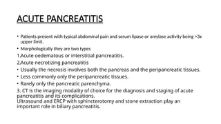
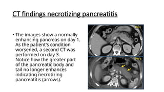
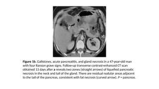
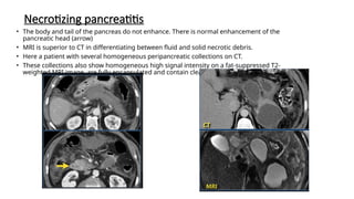
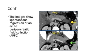
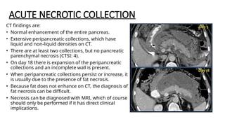
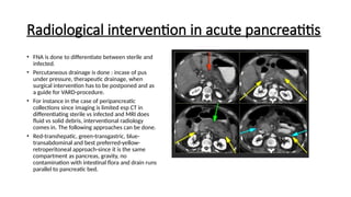
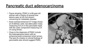
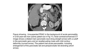
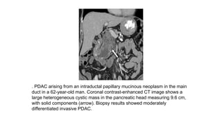
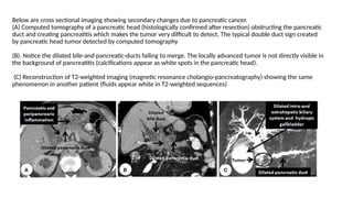
PANCREAS(ANATOMY,
TECHNIQUE AND PATHOLOGY)
LOISE NGURUMI
K/23030006/MMI
YR TWO
ANATOMY
• It is an unpaired mostly retroperitoneal organ.
• Located at L1-L2 vertebral level in the anterior
pararenal space of retroperitenuem with exception
of its tail.
• It is located in the upper abdomen directly behind
the stomach, with the head at next to duodenum.
• In most adults it is 6-10 inches long and 2 inches
wide. In that the diameter of the pancreatic head
does not exceed the transverse diameter of the
adjacent vertebral body.
• It is comprises primarily of a network of tube or
pancreatic duct that releases juices into the
duodenum
• It is divided into five regions
• Anatomical variants such as pancreas divisum
• It has both the endocrine and exocrine functions
hence It is a heterocrine gland
• The pancreas is an important organ in the digestive
system,ie in glucose metabolism and digestion
THE ENDOCRINE PANCREAS
• Consist of nests of cells (islets of Langerhans) that are distributed throughout the
exocrine pancreas.
• Although the cells are in millions, they comprise of only 1% of total pancreas.
• It has a great reserve capacity in that more that 70% of the insulin-seleceting beta
cells must be lost before dysfuntion occurs.
• There are four islet cells types with each producing its producy
• Beta cell-secrets insulin (60%)
• Alpha cell-secrets glucagon (30%)
• Delta cell-somatostatin (<10%)
• The pancreatic polypeptide secreting cell (PP or F cells)(1%)
• Most common disease of the endocrine pancreas is Diabetes mellitus.
Cont’
Arterial supply
• The head is primarily from inferior and superior pancreaticoduodenal arteries.
• The neck, body and tail from branches of the splenic artery such as the dorsal
pancreatic artery, greater pancreatic artery and tranverse pancreatic artery.
Venous drainage
• Venous return is by numerous small veins into the splenic hilum.
• From the head the superior pancreaticoduodenal vein drains into the portal vein
and the inferior drains into the superior mesenteric vein.
Lymphatic drainage
• Follows the blood vessels then collects in splenic, celiac, superior mesenteric and
hepatic nodes.
Functions
Role of imaging
• Imaging endocrine tumors entails combining conventional techniques of morphological imaging
(ultrasound, computed tomography [CT], magnetic resonance imaging [MRI]), endoscopic
explorations and functional imaging using radiopharmaceutical imaging techniques.
• The role of imaging includes the localization of small functioning tumor, differentiation of these
tumors from adenocarcinoma, identification of signs of malignancy and evaluation of extent.
On CT and MRI,
• Most of functioning PNETs are well defined small tumors with intense and homogeneous
enhancement on arterial and portal phases.
• However, some PNETs with a more fibrous content may have a more delayed enhancement that
is best depicted on the delayed phase.
• Other PNETs can present as purely cystic, complex cystic and solid tumors and calcified tumors
• Non-functioning PNETs are larger with less intense and more heterogeneous enhancement
Cont’
• Functional imaging is useful for disease staging, to detect disease recurrence or the
primary and to select patient candidate for peptide receptor radiometabolic treatment.
• Somatostatin receptor scintigraphy (srs) (octreoscan) is still the most available
technique. Gallium 68-SST analogue PET have been demonstrated to be more sensitive
than SRS-SPEC and it will be the future of functional imaging for NET.
• Fdg pet/ct is indicated for more aggressive pnet as defined either by negative srs and
huge tumor burden or ki67 above 10% or poorly differentiated pnec tumors.
• Finally, endoscopic ultrasonography (eus) is particularly suited to detect small size (2 to
5 mm) pancreatic lesions, such as gastrinomas and insulinomas with reported detection
rates from 79% to 94%.
• Due to the proximity of the endoscope, the sensitivity for detection is higher in the head
than in the tail
TECHNIQUE
Pancreas and it
pathologies can be
assessed by.
• Convectional
radiography(A. X-
ray)-chronic
pancreatitis
• Endoscopic
Ultrasound-
abdominal
• CT
• MRI
Unsuspected PDAC in a background of acute pancreatitis in a 63-year-old
man. Because acute pancreatitis of unknown cause was seen at a CT
examination, an MRI examination was performed. (A) Axial T2-weighted fat-
suppressed MR image. (B) MRCP image shows a mass, prompting
endoscopic US. (C) Endoscopic US image shows a mass (arrow) as the
cause of ductal dilatation that was biopsy-proven PDAC
PANCREAS CT PROTOCOL
• Mostly performed as part of other scans such as CT abdomen-pelvis
and CT chest-abdomen-pelvis.
Indications
• Evaluation of;Jaundice, pancreatic tumors and cystic lesions, acute or
chronic pancreatitis, unclear findings on ultrasound or CT abdomen
and pancreatic interventions (e.g CT-guided biopsy, drainage),
identification and characterization of pancreatic calcifications.
Cont’
• Patient position
Supine position, both arms elevated, abdomen centered within the gantry.
• Tube voltage and tube current
<120kVp and automatic exposure control and iterative reconstruction
• Scout-acquired from diaphragm to the iliac crest (or symphysis)
• Scan extent: for arterial phase-mid diaphragm to the iliac crest, venous phase-
above the diaphragm to the iliac crest.
• Scan direction is craniocaudal.
• Scan geometry-FOV: 350, slice thickness <0.625, interval:<0.5mm, reconstruction
algorithm: soft tissue and bone.
• Respiratory; single beath-hold: inspiration.
Cont’-contrast
• Oral contrast-neutral that is 800ml of water 20-30 mins before scan-streak artifacts and two for
clear visualization of the pancreatic borders as the stomach and duodenum will be distended.
• Contrast administration after the non contrasted study, volume of 70-120ml with 30-40 ml
saline chaser at 3-5ml/s
• Arterial phase-minimal scan delay about 20 seconds after injection; this phase in hypervacsular
lesions characterization such as neuroendocrine tumors, vascular lesions.
• Pancreatic phase which is 35-40 sec after CM helps in depiction of hypoattenuating tumors such
as pancreatic ductal adenocarcinoma.
• Portal venous phase -65-70 sec after contrast injection to depict hepatic metastases, venous
thrombosis etc.
• Multiplanar reconstruction entail, axial images, coronal, sagittal, with soft tisse using <2.5mm,
bone window using <2mm.
• For local staging a late arterial and portal venous are the phase to always perform.
MRI TECHNIQUE
• Indication are same as CT
• Patient position.
• Sequences include
• T1 TSE Axial and coronal
• T2 TSE Axial and coronal
• T1 in phase and out of phase in axial-pre Gd and post.
• DWI
• MRCP
• Dual FFE breath hold-coronal-it is preferred instead of out/in phase due to a
short acquisition time.
PATHOLOGIES
They can be classified into
• Acute pancreatitis
• Pancreatic tumors
• Pancreatic cystic lesions
ACUTE PANCREATITIS
• Patients present with typical abdominal pain and serum lipase or amylase activity being >3x
upper limit.
• Morphologically they are two types
1.Acute oedematous or interstitial pancreatitis.
2.Acute necrotizing pancreatitis
• Usually the necrosis involves both the pancreas and the peripancreatic tissues.
• Less commonly only the peripancreatic tissues.
• Rarely only the pancreatic parenchyma.
3. CT is the imaging modality of choice for the diagnosis and staging of acute
pancreatitis and its complications.
Ultrasound and ERCP with sphincterotomy and stone extraction play an
important role in biliary pancreatitis.
CT findings necrotizing pancreatitis
• The images show a normally
enhancing pancreas on day 1.
As the patient's condition
worsened, a second CT was
performed on day 3.
Notice how the greater part
of the pancreatic body and
tail no longer enhances
indicating necrotizing
pancreatitis (arrows).
Figure 1b. Gallstones, acute pancreatitis, and gland necrosis in a 47-year-old man
with four Ranson grave signs. Follow-up transverse contrast-enhanced CT scan
obtained 13 days after a reveals two zones (straight arrows) of liquefied pancreatic
necrosis in the neck and tail of the gland. There are residual nodular areas adjacent
to the tail of the pancreas, consistent with fat necrosis (curved arrow). P = pancreas.
Necrotizing pancreatitis
• The body and tail of the pancreas do not enhance. There is normal enhancement of the
pancreatic head (arrow)
• MRI is superior to CT in differentiating between fluid and solid necrotic debris.
• Here a patient with several homogeneous peripancreatic collections on CT.
• These collections also show homogeneous high signal intensity on a fat-suppressed T2-
weighted MRI image, are fully encapsulated and contain clear fluid (i.e. pseudocysts)
ACUTE PERIPANCREATIC FLUID COLLECTION
• Intraabdominal fluid collections and collections of necrotic tissue are common in
acute pancreatitis.
• These collections develop early in the course of acute pancreatitis.
• They are the result of the release of activated pancreatic enzymes which also
cause necrosis of the surrounding tissues.
• In the early stage, such a collection does not have a wall or capsule.
• About 50% of these collections show spontaneous regression
• Preferred locations of fluid collections are:
Lesser sac
Anterior and posterior pararenal space of the retroperitoneum.
Transverse mesocolon
Small bowel mesentery
Cont’
• The images show
spontaneous
regression of an
acute
peripancreatic
fluid collection
(APFC)
ACUTE NECROTIC COLLECTION
CT findings are:
• Normal enhancement of the entire pancreas.
• Extensive peripancreatic collections, which have
liquid and non-liquid densities on CT.
• There are at least two collections, but no pancreatic
parenchymal necrosis (CTSI: 4).
• On day 18 there is expansion of the peripancreatic
collections and an incomplete wall is present.
• When peripancreatic collections persist or increase, it
is usually due to the presence of fat necrosis.
• Because fat does not enhance on CT, the diagnosis of
fat necrosis can be difficult.
• Necrosis can be diagnosed with MRI, which of course
should only be performed if it has direct clinical
implications.
Radiological intervention in acute pancreatitis
• FNA is done to differentiate between sterile and
infected.
• Percutaneous drainage is done : incase of pus
under pressure, therapeutic drainage, when
surgical intervention has to be postponed and as
a guide for VARD-procedure.
• For instance in the case of peripancreatic
collections since imaging is limited esp CT in
differentiating sterile vs infected and MRI does
fluid vs solid debris, interventional radiology
comes in. The following approaches can be done.
• Red-transhepatic, green-transgastric, blue-
transabdominal and best preferred-yellow-
retroperitoneal approach-since it is the same
compartment as pancreas, gravity, no
contamination with intestinal flora and drain runs
parallel to pancreatic bed.
Figure shows.. Abrupt termination of the duct because of a stone in a 45-year-old man with
chronic pancreatitis. (A) Coronal rapid acquisition with relaxation enhancement MRCP
image shows a dilated pancreatic duct with abrupt termination of the duct in the
pancreatic body (arrow). No associated mass or stone is visible. (B) Axial contrast-enhanced
CT image shows a calcified stone (arrow) in the pancreatic duct, instead of a mass at the
site of ductal termination as seen on the MR image.
CYSTIC PANCREATIC LESIONS
The cause of most pancreatic cysts is unknown. Some are ass with rare
illnesses such as polycystic kidney disease or von Hippel-Lindau disease.
They can be categorized into the following groups:
• Pseudocysts
• Common cystic neoplasms:
• IPMN - intraductal papillary mucinous neoplasm
• SCN - Serous cystic neoplasm
• MCN - Mucinous cystic neoplasm
• Uncommon cystic neoplasms:
• SPEN (solid pseudopapillary epithelial neoplasm)
• Tumors with cystic degeneration:
adenocarcinoma - neuroendocrine tumor
MR versus CT
• CT will depict most pancreatic lesions, but is sometimes unable to depict the cystic
component.
• MR with heavily weighted T2WI and MRCP will better demonstrate the cystic nature
and the internal structure of the cyst and has the advantage of demonstrating the
relationship of the cyst to the pancreatic duct as is seen in IPMN.
• The images show a serous cystic neoplasm (SCN) on a CT.
MRI better shows the central scar.
Pseudocyst
Key CT &MRI findings:
• Unilocular cyst without solid components, central
scar or wall calcification.
• Collection of pancreatic enzymes, blood and
necrotic tissue.
• Debris within a cystic lesion is a specific MR
finding.
• History of pancreatitis or abdominal trauma.
• Cysts develop in 4-6 weeks - usually decrease in
size over time - sometimes enlarge or become
infected.
• Found in any part of the pancreas or anywhere
within the abdomen and sometimes even in the
chest.
• The CT demonstrates a large cyst in the upper
abdomen in a patient who had an acute
pancreatitis
Intraductal papillary mucinous neoplasm
MRI shows a lesion, which consists of multiple small cysts.
This could be a serous cystic neoplasm or a branch-duct
IPMN. The connection of the cystic lesion to the pancreatic
duct indicates that this is a branch-duct IPMN
MRCP image shows a 3.2-cm lesion (arrow)
communicating with the main duct, compatible
with branch-duct IPMN.
Main-duct IPMN in an 88-year-old woman. MRCP image shows
a diffusely dilated 1.7-cm pancreatic duct (arrow) in the setting
of main-duct IPMN.
MCN- Mucinous cystic neoplasm in a 34-
year-old woman. (A) Axial T2-weighted MR
image shows a complex T2-hyperintense
cystic lesion composed of multiple smaller
T2-hyperintense cystic locules (arrow). (B)
Axial noncontrast T1-weighted MR image
shows locules (arrow) and hyperintense
foci (arrowhead) consistent with areas of
hemorrhage. (C) Axial contrast-enhanced
T1-weighted MR image shows enhancing
septa and nodules (arrow). (D) Gross
specimen of the resected malignant MCN
shows a multiloculated cystic mass.
PANCREATIC TUMORS
• Pancreatic adenocarcinoma represent the most common pancreatic cancer, while
pancreatic neuroendocrine tumors (PNETs) also known as pancreatic islet cell tumors the
represent the second most common.
• 65% of pancreatic adenocarcinomas are located within the head, 15% in the corpus and
10% in the tail. The remaining 10% being multifocal or diffuse.
PDAC in a 68-year-old woman. Axial contrast-enhanced CT
image shows an enlarged heterogeneous pancreatic head,
suggesting the presence of an underlying tumor (long arrow)
with intratumoral calcification (short arrow), a rare finding in
PDAC.
Pancreatic duct adenocarcinoma
• Figure showing, PDAC in a 66-year-old
woman with a history of several liver
lesions seen at US (not shown)
concerning for metastatic disease.
• Axial contrast-enhanced CT image shows
an ill-defined, solid, low-attenuation mass
measuring 4.6 cm in the pancreatic head,
with the cystic component measuring 3.4
cm (long arrow).
• Clues to the diagnosis of PDAC include
the heterogeneous lesion with an
enhancing solid component (short arrow).
The lack of a history or clinical findings of
pancreatitis also should suggest PDAC.
Figure showing.. Unsuspected PDAC in the background of acute pancreatitis
in a 63-year-old man (same patient as in Fig 11). Axial contrast-enhanced CT
image shows a dilated main pancreatic duct (long straight arrow) with a cutoff
in the pancreatic genu and an isoattenuating mass with mild contour
deformity (curved arrow). The patient had acute pancreatitis, including
enlargement of the pancreatic tail and peripancreatic fat stranding (short
arrow).
. PDAC arising from an intraductal papillary mucinous neoplasm in the main
duct in a 62-year-old man. Coronal contrast-enhanced CT image shows a
large heterogeneous cystic mass in the pancreatic head measuring 9.6 cm,
with solid components (arrow). Biopsy results showed moderately
differentiated invasive PDAC.
Below are cross sectional imaging showing secondary changes due to pancreatic cancer.
(A) Computed tomography of a pancreatic head (histologically confirmed after resection) obstructing the pancreatic
duct and creating pancreatitis which makes the tumor very difficult to detect. The typical double duct sign created
by pancreatic head tumor detected by computed tomography
(B). Notice the dilated bile-and pancreatic-ducts failing to merge. The locally advanced tumor is not directly visible in
the background of pancreatitis (calcifications appear as white spots in the pancreatic head).
(C) Reconstruction of T2-weighted imaging (magnetic resonance cholangio-pancreatography) showing the same
phenomenon in another patient (fluids appear white in T2-weighted sequences)
Cystic PNET in a 66-year-old man with a history of prostate cancer, and a pancreatic
cyst seen at CT. (A) Axial T2-weighted MR image shows a 4.2-cm cystic lesion
(arrow) in the pancreatic tail, with subtle nodularity (arrowhead) along the cyst wall.
(B) Axial contrast-enhanced T1-weighted MR image shows a thick enhancing wall and
nodularity (arrowhead) of the cystic lesion (arrow). Biopsy yielded World Health
Organization grade 1 (carcinoid) neuroendocrine tumor.
References
• https://radiologyassistant.nl/abdomen/pancreas/pancreas-cystic-lesio
ns
• https://radiologyassistant.nl/abdomen/pancreas/pancreas-carcinoma
-1
• https://radiologyassistant.nl/abdomen/pancreas/acute-pancreatitis
• Radiopedia
• Radiology key
• Textbook-Pathology of disease LANGE 8th
edition.

Endocrine pancreas /computerized Tomography and MRI

  • 1.
  • 2.
    ANATOMY • It isan unpaired mostly retroperitoneal organ. • Located at L1-L2 vertebral level in the anterior pararenal space of retroperitenuem with exception of its tail. • It is located in the upper abdomen directly behind the stomach, with the head at next to duodenum. • In most adults it is 6-10 inches long and 2 inches wide. In that the diameter of the pancreatic head does not exceed the transverse diameter of the adjacent vertebral body. • It is comprises primarily of a network of tube or pancreatic duct that releases juices into the duodenum • It is divided into five regions • Anatomical variants such as pancreas divisum • It has both the endocrine and exocrine functions hence It is a heterocrine gland • The pancreas is an important organ in the digestive system,ie in glucose metabolism and digestion
  • 3.
    THE ENDOCRINE PANCREAS •Consist of nests of cells (islets of Langerhans) that are distributed throughout the exocrine pancreas. • Although the cells are in millions, they comprise of only 1% of total pancreas. • It has a great reserve capacity in that more that 70% of the insulin-seleceting beta cells must be lost before dysfuntion occurs. • There are four islet cells types with each producing its producy • Beta cell-secrets insulin (60%) • Alpha cell-secrets glucagon (30%) • Delta cell-somatostatin (<10%) • The pancreatic polypeptide secreting cell (PP or F cells)(1%) • Most common disease of the endocrine pancreas is Diabetes mellitus.
  • 4.
    Cont’ Arterial supply • Thehead is primarily from inferior and superior pancreaticoduodenal arteries. • The neck, body and tail from branches of the splenic artery such as the dorsal pancreatic artery, greater pancreatic artery and tranverse pancreatic artery. Venous drainage • Venous return is by numerous small veins into the splenic hilum. • From the head the superior pancreaticoduodenal vein drains into the portal vein and the inferior drains into the superior mesenteric vein. Lymphatic drainage • Follows the blood vessels then collects in splenic, celiac, superior mesenteric and hepatic nodes.
  • 5.
  • 6.
    Role of imaging •Imaging endocrine tumors entails combining conventional techniques of morphological imaging (ultrasound, computed tomography [CT], magnetic resonance imaging [MRI]), endoscopic explorations and functional imaging using radiopharmaceutical imaging techniques. • The role of imaging includes the localization of small functioning tumor, differentiation of these tumors from adenocarcinoma, identification of signs of malignancy and evaluation of extent. On CT and MRI, • Most of functioning PNETs are well defined small tumors with intense and homogeneous enhancement on arterial and portal phases. • However, some PNETs with a more fibrous content may have a more delayed enhancement that is best depicted on the delayed phase. • Other PNETs can present as purely cystic, complex cystic and solid tumors and calcified tumors • Non-functioning PNETs are larger with less intense and more heterogeneous enhancement
  • 7.
    Cont’ • Functional imagingis useful for disease staging, to detect disease recurrence or the primary and to select patient candidate for peptide receptor radiometabolic treatment. • Somatostatin receptor scintigraphy (srs) (octreoscan) is still the most available technique. Gallium 68-SST analogue PET have been demonstrated to be more sensitive than SRS-SPEC and it will be the future of functional imaging for NET. • Fdg pet/ct is indicated for more aggressive pnet as defined either by negative srs and huge tumor burden or ki67 above 10% or poorly differentiated pnec tumors. • Finally, endoscopic ultrasonography (eus) is particularly suited to detect small size (2 to 5 mm) pancreatic lesions, such as gastrinomas and insulinomas with reported detection rates from 79% to 94%. • Due to the proximity of the endoscope, the sensitivity for detection is higher in the head than in the tail
  • 8.
    TECHNIQUE Pancreas and it pathologiescan be assessed by. • Convectional radiography(A. X- ray)-chronic pancreatitis • Endoscopic Ultrasound- abdominal • CT • MRI Unsuspected PDAC in a background of acute pancreatitis in a 63-year-old man. Because acute pancreatitis of unknown cause was seen at a CT examination, an MRI examination was performed. (A) Axial T2-weighted fat- suppressed MR image. (B) MRCP image shows a mass, prompting endoscopic US. (C) Endoscopic US image shows a mass (arrow) as the cause of ductal dilatation that was biopsy-proven PDAC
  • 9.
    PANCREAS CT PROTOCOL •Mostly performed as part of other scans such as CT abdomen-pelvis and CT chest-abdomen-pelvis. Indications • Evaluation of;Jaundice, pancreatic tumors and cystic lesions, acute or chronic pancreatitis, unclear findings on ultrasound or CT abdomen and pancreatic interventions (e.g CT-guided biopsy, drainage), identification and characterization of pancreatic calcifications.
  • 10.
    Cont’ • Patient position Supineposition, both arms elevated, abdomen centered within the gantry. • Tube voltage and tube current <120kVp and automatic exposure control and iterative reconstruction • Scout-acquired from diaphragm to the iliac crest (or symphysis) • Scan extent: for arterial phase-mid diaphragm to the iliac crest, venous phase- above the diaphragm to the iliac crest. • Scan direction is craniocaudal. • Scan geometry-FOV: 350, slice thickness <0.625, interval:<0.5mm, reconstruction algorithm: soft tissue and bone. • Respiratory; single beath-hold: inspiration.
  • 11.
    Cont’-contrast • Oral contrast-neutralthat is 800ml of water 20-30 mins before scan-streak artifacts and two for clear visualization of the pancreatic borders as the stomach and duodenum will be distended. • Contrast administration after the non contrasted study, volume of 70-120ml with 30-40 ml saline chaser at 3-5ml/s • Arterial phase-minimal scan delay about 20 seconds after injection; this phase in hypervacsular lesions characterization such as neuroendocrine tumors, vascular lesions. • Pancreatic phase which is 35-40 sec after CM helps in depiction of hypoattenuating tumors such as pancreatic ductal adenocarcinoma. • Portal venous phase -65-70 sec after contrast injection to depict hepatic metastases, venous thrombosis etc. • Multiplanar reconstruction entail, axial images, coronal, sagittal, with soft tisse using <2.5mm, bone window using <2mm. • For local staging a late arterial and portal venous are the phase to always perform.
  • 12.
    MRI TECHNIQUE • Indicationare same as CT • Patient position. • Sequences include • T1 TSE Axial and coronal • T2 TSE Axial and coronal • T1 in phase and out of phase in axial-pre Gd and post. • DWI • MRCP • Dual FFE breath hold-coronal-it is preferred instead of out/in phase due to a short acquisition time.
  • 13.
    PATHOLOGIES They can beclassified into • Acute pancreatitis • Pancreatic tumors • Pancreatic cystic lesions
  • 14.
    ACUTE PANCREATITIS • Patientspresent with typical abdominal pain and serum lipase or amylase activity being >3x upper limit. • Morphologically they are two types 1.Acute oedematous or interstitial pancreatitis. 2.Acute necrotizing pancreatitis • Usually the necrosis involves both the pancreas and the peripancreatic tissues. • Less commonly only the peripancreatic tissues. • Rarely only the pancreatic parenchyma. 3. CT is the imaging modality of choice for the diagnosis and staging of acute pancreatitis and its complications. Ultrasound and ERCP with sphincterotomy and stone extraction play an important role in biliary pancreatitis.
  • 15.
    CT findings necrotizingpancreatitis • The images show a normally enhancing pancreas on day 1. As the patient's condition worsened, a second CT was performed on day 3. Notice how the greater part of the pancreatic body and tail no longer enhances indicating necrotizing pancreatitis (arrows).
  • 16.
    Figure 1b. Gallstones,acute pancreatitis, and gland necrosis in a 47-year-old man with four Ranson grave signs. Follow-up transverse contrast-enhanced CT scan obtained 13 days after a reveals two zones (straight arrows) of liquefied pancreatic necrosis in the neck and tail of the gland. There are residual nodular areas adjacent to the tail of the pancreas, consistent with fat necrosis (curved arrow). P = pancreas.
  • 17.
    Necrotizing pancreatitis • Thebody and tail of the pancreas do not enhance. There is normal enhancement of the pancreatic head (arrow) • MRI is superior to CT in differentiating between fluid and solid necrotic debris. • Here a patient with several homogeneous peripancreatic collections on CT. • These collections also show homogeneous high signal intensity on a fat-suppressed T2- weighted MRI image, are fully encapsulated and contain clear fluid (i.e. pseudocysts)
  • 18.
    ACUTE PERIPANCREATIC FLUIDCOLLECTION • Intraabdominal fluid collections and collections of necrotic tissue are common in acute pancreatitis. • These collections develop early in the course of acute pancreatitis. • They are the result of the release of activated pancreatic enzymes which also cause necrosis of the surrounding tissues. • In the early stage, such a collection does not have a wall or capsule. • About 50% of these collections show spontaneous regression • Preferred locations of fluid collections are: Lesser sac Anterior and posterior pararenal space of the retroperitoneum. Transverse mesocolon Small bowel mesentery
  • 19.
    Cont’ • The imagesshow spontaneous regression of an acute peripancreatic fluid collection (APFC)
  • 20.
    ACUTE NECROTIC COLLECTION CTfindings are: • Normal enhancement of the entire pancreas. • Extensive peripancreatic collections, which have liquid and non-liquid densities on CT. • There are at least two collections, but no pancreatic parenchymal necrosis (CTSI: 4). • On day 18 there is expansion of the peripancreatic collections and an incomplete wall is present. • When peripancreatic collections persist or increase, it is usually due to the presence of fat necrosis. • Because fat does not enhance on CT, the diagnosis of fat necrosis can be difficult. • Necrosis can be diagnosed with MRI, which of course should only be performed if it has direct clinical implications.
  • 21.
    Radiological intervention inacute pancreatitis • FNA is done to differentiate between sterile and infected. • Percutaneous drainage is done : incase of pus under pressure, therapeutic drainage, when surgical intervention has to be postponed and as a guide for VARD-procedure. • For instance in the case of peripancreatic collections since imaging is limited esp CT in differentiating sterile vs infected and MRI does fluid vs solid debris, interventional radiology comes in. The following approaches can be done. • Red-transhepatic, green-transgastric, blue- transabdominal and best preferred-yellow- retroperitoneal approach-since it is the same compartment as pancreas, gravity, no contamination with intestinal flora and drain runs parallel to pancreatic bed.
  • 22.
    Figure shows.. Abrupttermination of the duct because of a stone in a 45-year-old man with chronic pancreatitis. (A) Coronal rapid acquisition with relaxation enhancement MRCP image shows a dilated pancreatic duct with abrupt termination of the duct in the pancreatic body (arrow). No associated mass or stone is visible. (B) Axial contrast-enhanced CT image shows a calcified stone (arrow) in the pancreatic duct, instead of a mass at the site of ductal termination as seen on the MR image.
  • 23.
    CYSTIC PANCREATIC LESIONS Thecause of most pancreatic cysts is unknown. Some are ass with rare illnesses such as polycystic kidney disease or von Hippel-Lindau disease. They can be categorized into the following groups: • Pseudocysts • Common cystic neoplasms: • IPMN - intraductal papillary mucinous neoplasm • SCN - Serous cystic neoplasm • MCN - Mucinous cystic neoplasm • Uncommon cystic neoplasms: • SPEN (solid pseudopapillary epithelial neoplasm) • Tumors with cystic degeneration: adenocarcinoma - neuroendocrine tumor
  • 24.
    MR versus CT •CT will depict most pancreatic lesions, but is sometimes unable to depict the cystic component. • MR with heavily weighted T2WI and MRCP will better demonstrate the cystic nature and the internal structure of the cyst and has the advantage of demonstrating the relationship of the cyst to the pancreatic duct as is seen in IPMN. • The images show a serous cystic neoplasm (SCN) on a CT. MRI better shows the central scar.
  • 25.
    Pseudocyst Key CT &MRIfindings: • Unilocular cyst without solid components, central scar or wall calcification. • Collection of pancreatic enzymes, blood and necrotic tissue. • Debris within a cystic lesion is a specific MR finding. • History of pancreatitis or abdominal trauma. • Cysts develop in 4-6 weeks - usually decrease in size over time - sometimes enlarge or become infected. • Found in any part of the pancreas or anywhere within the abdomen and sometimes even in the chest. • The CT demonstrates a large cyst in the upper abdomen in a patient who had an acute pancreatitis
  • 26.
    Intraductal papillary mucinousneoplasm MRI shows a lesion, which consists of multiple small cysts. This could be a serous cystic neoplasm or a branch-duct IPMN. The connection of the cystic lesion to the pancreatic duct indicates that this is a branch-duct IPMN MRCP image shows a 3.2-cm lesion (arrow) communicating with the main duct, compatible with branch-duct IPMN.
  • 27.
    Main-duct IPMN inan 88-year-old woman. MRCP image shows a diffusely dilated 1.7-cm pancreatic duct (arrow) in the setting of main-duct IPMN.
  • 28.
    MCN- Mucinous cysticneoplasm in a 34- year-old woman. (A) Axial T2-weighted MR image shows a complex T2-hyperintense cystic lesion composed of multiple smaller T2-hyperintense cystic locules (arrow). (B) Axial noncontrast T1-weighted MR image shows locules (arrow) and hyperintense foci (arrowhead) consistent with areas of hemorrhage. (C) Axial contrast-enhanced T1-weighted MR image shows enhancing septa and nodules (arrow). (D) Gross specimen of the resected malignant MCN shows a multiloculated cystic mass.
  • 29.
    PANCREATIC TUMORS • Pancreaticadenocarcinoma represent the most common pancreatic cancer, while pancreatic neuroendocrine tumors (PNETs) also known as pancreatic islet cell tumors the represent the second most common. • 65% of pancreatic adenocarcinomas are located within the head, 15% in the corpus and 10% in the tail. The remaining 10% being multifocal or diffuse. PDAC in a 68-year-old woman. Axial contrast-enhanced CT image shows an enlarged heterogeneous pancreatic head, suggesting the presence of an underlying tumor (long arrow) with intratumoral calcification (short arrow), a rare finding in PDAC.
  • 30.
    Pancreatic duct adenocarcinoma •Figure showing, PDAC in a 66-year-old woman with a history of several liver lesions seen at US (not shown) concerning for metastatic disease. • Axial contrast-enhanced CT image shows an ill-defined, solid, low-attenuation mass measuring 4.6 cm in the pancreatic head, with the cystic component measuring 3.4 cm (long arrow). • Clues to the diagnosis of PDAC include the heterogeneous lesion with an enhancing solid component (short arrow). The lack of a history or clinical findings of pancreatitis also should suggest PDAC.
  • 31.
    Figure showing.. UnsuspectedPDAC in the background of acute pancreatitis in a 63-year-old man (same patient as in Fig 11). Axial contrast-enhanced CT image shows a dilated main pancreatic duct (long straight arrow) with a cutoff in the pancreatic genu and an isoattenuating mass with mild contour deformity (curved arrow). The patient had acute pancreatitis, including enlargement of the pancreatic tail and peripancreatic fat stranding (short arrow).
  • 32.
    . PDAC arisingfrom an intraductal papillary mucinous neoplasm in the main duct in a 62-year-old man. Coronal contrast-enhanced CT image shows a large heterogeneous cystic mass in the pancreatic head measuring 9.6 cm, with solid components (arrow). Biopsy results showed moderately differentiated invasive PDAC.
  • 33.
    Below are crosssectional imaging showing secondary changes due to pancreatic cancer. (A) Computed tomography of a pancreatic head (histologically confirmed after resection) obstructing the pancreatic duct and creating pancreatitis which makes the tumor very difficult to detect. The typical double duct sign created by pancreatic head tumor detected by computed tomography (B). Notice the dilated bile-and pancreatic-ducts failing to merge. The locally advanced tumor is not directly visible in the background of pancreatitis (calcifications appear as white spots in the pancreatic head). (C) Reconstruction of T2-weighted imaging (magnetic resonance cholangio-pancreatography) showing the same phenomenon in another patient (fluids appear white in T2-weighted sequences)
  • 34.
    Cystic PNET ina 66-year-old man with a history of prostate cancer, and a pancreatic cyst seen at CT. (A) Axial T2-weighted MR image shows a 4.2-cm cystic lesion (arrow) in the pancreatic tail, with subtle nodularity (arrowhead) along the cyst wall. (B) Axial contrast-enhanced T1-weighted MR image shows a thick enhancing wall and nodularity (arrowhead) of the cystic lesion (arrow). Biopsy yielded World Health Organization grade 1 (carcinoid) neuroendocrine tumor.
  • 35.
    References • https://radiologyassistant.nl/abdomen/pancreas/pancreas-cystic-lesio ns • https://radiologyassistant.nl/abdomen/pancreas/pancreas-carcinoma -1 •https://radiologyassistant.nl/abdomen/pancreas/acute-pancreatitis • Radiopedia • Radiology key • Textbook-Pathology of disease LANGE 8th edition.